Emanuel Fernandes dos Passos
Physical exercise: preventive and therapeutic non-pharmacological intervention against non-alcoholic steatohepatitis?
Porto, December 2022
Programa Doutoral em Metabolismo:
Clínica e Experimentação
The present dissertation is being submitted in order to achieve the PhD degree included in the doctoral course of Metabolism - Clinical and Experimental, at the Faculty of Medicine, University of Porto (FMUP), Porto, Portugal.
Supervisor:
Maria João dos Reis Conceição Martins de Almeida Ribeiro Assistant Professor
FMUP, Porto, Portugal
Co-Supervisors:
António Alexandre Moreira Ribeiro de Ascensão Assistant Professor
Faculty of Sport, University of Porto (FADEUP), Porto, Portugal
José Fernando Magalhães Pinto Pereira Assistant Professor
FADEUP, Porto, Portugal
Forty eightieth article, 3rd paragraph: “The Faculty cannot be held responsible for the doctrines included in the dissertation” (FMUP regulation - Act 19337, 29th January 1931).
Funding
Funding was obtained from 4 grants from the Fundação para a Ciência e Tecnologia (FCT): a) PEst-OE/SAU/UI0617/2011 to Research Centre in Physical Activity, Health and Leisure (CIAFEL), FADEUP, b) PEst-OE/SAU/UI0038/2014 to Department of Biochemistry (U38/FCT), FMUP, c) SFRH/BD/71149/2010 to Emanuel Passos (FADEUP and FMUP), and d) PTDC/DES/113580/2009-FCOMP-01-0124-FEDER-014705 to António Ascensão (FADEUP).
The experimental work was conduct at CIAFEL, FADEUP, and Department of Biochemistry, FMUP.
Doutor Patrício Manuel Vieira Araújo Soares Silva Doutor Alberto Manuel Barros da Silva
Doutor José Henrique Dias Pinto de Barros
Doutora Maria Fátima Machado Henriques Carneiro Doutora Maria Dulce Cordeiro Madeira
Doutor Altamiro Manuel Rodrigues Costa Pereira Doutor Manuel Jesus Falcão Pestana Vasconcelos
Doutor João Francisco Montenegro Andrade Lima Bernardes Doutora Maria Leonor Martins Soares David
Doutor Rui Manuel Lopes Nunes
Doutor José Manuel Pereira Dias de Castro Lopes Doutor Joaquim Adelino Correia Ferreira Leite Moreira Doutora Raquel Ângela Silva Soares Lino
Doutor Fernando Manuel Mendes Falcão dos Reis Doutor Francisco José Miranda Rodrigues Cruz Doutor José Paulo Alves Vieira de Andrade Doutor Jorge Manuel Silva Junqueira Polónia Doutor José Luís Dias Delgado
Doutora Isaura Ferreira Tavares
Doutor Fernando Carlos de Landér Schmitt Doutor Acácio Agostinho Gonçalves Rodrigues Doutora Maria de Fátima Moreira Martel Doutor João Tiago De Sousa Pinto Guimarães Doutor José Carlos Lemos Machado
Doutor José Carlos de Magalhães Silva Cardoso
Doutor Álvaro Jerónimo Leal Machado de Aguiar
Doutor António Albino Coelho Marques Abrantes Teixeira Doutor António Carlos de Freitas Ribeiro Saraiva
Doutor António José Pacheco Palha
Doutor António Manuel Sampaio de Araújo Teixeira Doutor Belmiro dos Santos Patrício
Doutor Cândido Alves Hipólito Reis
Doutor Carlos Rodrigo Magalhães Ramalhão Doutor Cassiano Pena de Abreu e Lima
Doutora Deolinda Maria Valente Alves Lima Teixeira Doutor Eduardo Jorge Cunha Rodrigues Pereira Doutor Fernando Tavarela Veloso
Doutor Francisco Fernando Rocha Gonçalves Doutora Isabel Maria Amorim Pereira Ramos Doutor Jorge Manuel Mergulhão Castro Tavares Doutor José Agostinho Marques Lopes
Doutor José Carlos Neves da Cunha Areias
Doutor José Eduardo Torres Eckenroth Guimarães Doutor José Fernando Barros Castro Correia Doutor José Manuel Costa Mesquita Guimarães Doutor José Manuel Lopes Teixeira Amarante Doutor Levi Eugénio Ribeiro Guerra
Doutor Luís Alberto Martins Gomes de Almeida Doutor Manuel Alberto Coimbra Sobrinho Simões Doutor Manuel António Caldeira Pais Clemente Doutor Manuel Augusto Cardoso de Oliveira Doutor Manuel Machado Rodrigues Gomes Doutor Manuel Maria Paula Barbosa Doutora Maria Amélia Duarte Ferreira
Doutora Maria da Conceição Fernandes Marques Magalhães
Doutor Rui Manuel Bento de Almeida Coelho Doutor Serafim Correia Pinto Guimarães
Doutor Valdemar Miguel Botelho dos Santos Cardoso Doutor Walter Friedrich Alfred Osswald
I - Passos E, Ascensão A, Martins MJ, Magalhães J (2015). Endoplasmic reticulum stress response in non-alcoholic steatohepatitis: the possible role of physical exercise.
Metabolism 64(7):780-92.
Review article.
II - Gonçalves IO*, Passos E*, Rocha-Rodrigues S, Torrella JR, Rizo D, Santos-Alves E, Portincasa P, Martins MJ, Ascensão A, Magalhães J. (2015). Physical exercise antagonizes clinical and anatomical features characterizing Lieber-De Carli diet- induced obesity and related metabolic disorders. Clin Nut 34(2):241-7.
* Equal contribution in both the experimental and the writing steps that led to this manuscript.
Original research article 1.
III - Passos E, Pereira CD, Gonçalves IO, Rocha-Rodrigues S, Silva N, Guimarães JT, Neves D, Ascensão A, Magalhães J, Martins MJ (2015). Role of physical exercise on hepatic insulin, glucocorticoid and inflammatory signaling in an animal model of non- alcoholic steatohepatitis. Life Sci 123:51-60.
Original research article 2.
IV - Passos E, Pereira C, Gonçalves IO, Faria A, Ascensão A, Monteiro R, Magalhães J, Martins MJ (2022). Physical exercise positively modulates nonalcoholic steatohepatitis-related hepatic endoplasmic reticulum stress. J Cell Biochem 123(10):1647-62.
Original research article 3.
Dedicated to:
The memory of my father, António dos Passos (Manis), an incredible man who always provided me the guidance whenever I needed. You are gone, but the great memories we shared made this journey possible.
My sons Helber and Emerson.
I was never alone in my PhD degree: I was always surrounded by special and generous people, with distinct personalities and scientific backgrounds, that proved to be important to accomplish the different and sequential steps of this PhD dissertation.
Its conclusion was possible thanks to their support, cooperation and commitment.
Assuming the risk of forgetting someone, I want to publicly display my gratitude, affection and deep appreciation to all, directly or indirectly, involved in this laboratorial and scientific path.
To my supervisors and mentors, Professors Maria João Martins, António Ascensão and José Magalhães for the opportunity of working with you. Thank you for this period of growth and intense and constant learning.
To Professor Maria João Martins for the constant motivation, unconditional support and dedication during this process. Thank you for being much more than a supervisor and/or a teacher. I will be eternally grateful for pushing me up and for the wise guidance. Thank you for always being there, whenever it was needed. Your scientific knowledge, passion and humanity are remarkable characteristics that will inspire me forever.
To Professors António Ascensão and José Magalhães for giving me the opportunity to be part of your team. I am very grateful for the acceptance in your lab and, consequently, the initiation of this laboratorial adventure. Your deep scientific knowledge and the simple way you communicate it made me believe that it was possible to work in science. The opportunities provided, the wise teachings transmitted and the examples of scientific capacity given will always be remembered.
Thank you very much.
To all the other professors and colleagues with whom I had the privilege of working with at FMUP and FADEUP.
and Experimental, for the opportunity to work at the Biochemistry Department, FMUP. To Professor Tiago Guimarães for the collaboration in the experimental work and, most importantly, for the constant friendly support.
To Professors Ana Faria, Delminda Neves, and Rosário Monteiro for the scientific and laboratorial recommendations.
To Cidália Pereira and Inês Gonçalves for all the guidance in the laboratory and/or collaboration in the experimental work. To Ângela Castela, Diogo Pestana, Eduardo Teixeira, João Araújo, Pedro Gonçalves, and Raquel Costa for the friendship and the help provided.
To my parents (António and Mariana) and my brothers (Estela, Lúcia, Madalena, Vitalina, António, Albertino, Paulino, Lina, Ângelo, and Adilson) for the constant support and motivation. To Paulino “Polis Preta” and Ângelo “Marley” for being there whenever it was necessary.
Last but not least, and probably the most important, to my sons Helber and Emerson.
Thank you for being my life.
PROFESSORES EFETIVOS – CATEDRÁTICOS ... vii
PROFESSORES JUBILADOS OU APOSENTADOS ... viii
ACKNOWLEDGMENTS ... xv
LIST OF ABBREVIATIONS ... 19
RESUMO ... 23
ABSTRACT ... 25
CHAPTER 1: INTRODUCTION ... 27
INTRODUCTION ... 29
Rodent models in NAFLD ... 30
Genetic models ... 30
Chemical models ... 31
Dietary models ... 31
Combined models ... 32
Lipotoxicity, inflammation and insulin resistance ... 33
Cross-talk between ER stress, inflammation, insulin resistance, altered redox state and mitochondrial dysfunction ... 34
Pharmacological and non-pharmacological therapeutic strategies: the role of physical exercise ... 37
AIMS ... 53
CHAPTER 2: THEORETICAL BACKGROUND ... 55
Review Paper: Passos E, Ascensão A, Martins MJ, Magalhães J. Endoplasmic reticulum stress response in non-alcoholic steatohepatitis: the possible role of physical exercise. Metabolism 64(7):780-92. ... 57
CHAPTER 3: EXPERIMENTAL WORK ... 73
Original research article 1: Gonçalves IO *, Passos E *, Rocha-Rodrigues S, Torrella JR, Santos-Alves E, Portincasa P, Martins MJ, Ascensão A, Magalhães J (2015). Physical exercise antagonizes clinical and anatomical features characterizing Lieber-De Carli diet-induced obesity and related metabolic disorders. Clin Nut 34(2):241-7. * Equal contribution. ... 75
Original research article 2: Passos E, Pereira C, Gonçalves IO, Rocha-Rodrigues S, Silva N, Guimarães JT, Neves D, Ascensão A, Magalhães J, Martins MJ (2014). The role of physical exercise on hepatic insulin, glucocorticoid and inflammatory signaling in an animal model of non-alcoholic steatohepatitis. Life Sci 123:51-60. ... 85 Original research article 3: Passos E, Pereira C, Gonçalves IO, Faria A, Ascensão A, Monteiro R, Magalhães J, Martins MJ (2022). Physical exercise positively modulates nonalcoholic
CHAPTER 4: GENERAL DISCUSSION ... 115 CHAPTER 5: GENERAL CONCLUSIONS ... 133 CHAPTER 6: GENERAL REFERENCES ... 137
LIST OF ABBREVIATIONS
11β-HSD1 - 11β-hydroxysteroid dehydrogenase type 1 AMLN - Amylin diet
ALT - Alanine aminotransferase AP1 - activator protein 1
ASK1 - Apoptosis signaling-regulating kinase 1 AST - Aspartate aminotransferase
ATF - Activation transcription factor ATF-4 - Activating transcription factor 4 ATF-6 - Activating transcription factor 6 BiP - Binding immunoglobulin protein
Caspase - Cysteine-dependent aspartate specific protease CCl4 - Carbon tetrachloride
CCR - C-C Motif chemokine receptor
CDAA - Choline-deficient, L-amino acid-defined CHOP - C/EPB homologous protein
CIAFEL - Research Centre in Physical Activity, Health and Leisure eIF2α - Eukaryotic initiation factor 2 α
ER - Endoplasmic reticulum ET - Endurance training
FADEUP - Faculty of Sport, University of Porto FCT - Fundação para a Ciência e Tecnologia FFAs - Free fatty acids
FMUP - Faculty of Medicine, University of Porto FXR - Farnesoid X receptor
GSH - Reduced glutathione
GSx – Total (oxidized plus reduced) glutathione HFD - High-fat diet
IκB - NF-κB inhibitor IKK - IκB kinase
IL-1β - Interleukin-1 beta IL-6 - Interleukin-6
IRE - Inositol requiring enzyme IRE1 - Inositol requiring enzyme 1 IRS-1 - Insulin receptor substrate 1 IRS-2 - Insulin receptor substrate 2 JNK - C-Jun N-terminal kinase LLPA - Life-long physical activity
MCD - Methionine and choline-deficient diets Mc4r - Melanocortin 4 receptor knock-out NAFLD - Non-alcoholic fatty liver disease NASH - Non-alcoholic steatohepatitis
NF-κB - Nuclear factor κ-light-chain enhancer of activated B cells Nrf2 - Nuclear erythroid 2 p45-related factor
ORA - Original research article PBA - 4-Phenyl butyric acid PE - Physical exercise
p-eIF2α - Phosphorylated eukaryotic initiation factor 2 p-JNK - Phosphorylated c-Jun N-terminal kinase
PERK - Protein kinase RNA-activated (PKR)-like ER kinase PPAR - Peroxisome proliferator-activated receptors PTP1B - Protein tyrosine phosphatase 1B
ROS - Reactive oxygen species
SD - Standard diet, standard control liquid diet sXBP1 - spliced XBP1 X-box binding protein 1 T2DM - Type 2 diabetes mellitus
TAA - Thioacetamide
TGF-β -Transforming growth factor β TGs - Triglycerides
TNF-α - Tumor-necrosis factor
TUDCA - Taurine-conjugated ursodeoxycholic acid UPR - Unfolded protein response
uXBP1 - unspliced X-box binding protein 1
VPA - Voluntary physical activity XBP1 - X-box binding protein
RESUMO
O estilo de vida sedentário associado ao consumo excessivo de alimentos hipercalóricos está relacionado com o aumento epidémico da esteatose hepática não alcoólica (non-alcoholic fatty liver disease, NAFLD). Esta pode evoluir de simples esteatose para esteatohepatite não alcoólica (non-alcoholic steatohepatitis, NASH), fibrose e cirrose, culminando, eventualmente, em carcinoma hepatocelular. Embora os mecanismos subjacentes não estejam ainda completamente compreendidos, a lipotoxicidade associada à deposição de gordura, o stresse oxidativo, o stresse do retículo endoplasmático, a inflamação, a disfunção mitocondrial, a resistência à insulina e a apoptose estão implicados no seu surgimento e na sua subsequente progressão. Estes mecanismos hepáticos interligam-se num ciclo vicioso.
Não existe ainda terapia farmacológica aceite para o tratamento da NASH.
Atualmente, o exercício físico apresenta-se como uma intervenção não-farmacológica efetiva, não só preventiva como também terapêutica, que mitiga diversas alterações idênticas às que se observam na NASH. No entanto, os efeitos benéficos do exercício físico nos mecanismos acima mencionados num contexto de NASH são ainda pouco compreendidos.
Com esta dissertação, que integra um artigo de revisão e três trabalhos experimentais originais, pretendeu-se avaliar alguns mecanismos moleculares potencialmente subjacentes aos efeitos benéficos do exercício físico num modelo animal de NASH:
ratos machos adultos Sprague-Dawley alimentados com uma dieta líquida rica em gordura (Lieber-DeCarli), durante 17 semanas. Assim, foram analisados os efeitos da atividade física voluntária (voluntary physical activity, VPA, durante 17 semanas) e do treino de resistência (endurance training, ET, durante as últimas 8 semanas da intervenção alimentar), estratégias preventivas e terapêuticas, respetivamente, em a) marcadores antropométricos, assim como histológicos (no fígado) e metabólicos (no plasma) (trabalho experimental original 1), b) marcadores pró-inflamatórios e de sinalização pró-inflamatória assim como da sinalização pela insulina e por glicocorticóides (no fígado, trabalho experimental original 2), e c) marcadores do stresse do retículo endoplasmático, do stresse oxidativo e da apoptose (no fígado, trabalho experimental original 3).
Os resultados suportam a sugestão de que o treino de resistência possa ser considerado uma estratégia terapêutica não farmacológica para contrariar não só a deposição de gordura, mas também a inflamação, induzidas pela obesidade, uma vez que reverteu todas as alterações anatómicas e clínicas/histológicas observadas na NASH. Além disso, o exercício físico modulou positivamente a) a sinalização pelo JNK, bem como os níveis de IL-1β, IL-6, LIF e TGF-β e b) a sinalização pela insulina, provavelmente através da sinalização pelo JNK, PTP1B e 11β-HSD1. Além disso, o exercício físico modulou positiva e dinamicamente a UPR (aumentando a resposta pró-vida, enquanto regula negativamente a pró-morte) pela via de sinalização IREα/XBP1.
Em conclusão, o exercício físico deve ser considerado uma estratégia não farmacológica na prevenção e/ou tratamento da NASH.
ABSTRACT
An inactive lifestyle in simultaneity with excessive consumption of high-calorie food associates with the global increase of non-alcoholic fatty liver disease (NAFLD). NAFLD evolves from simple steatosis to non-alcoholic steatohepatitis (NASH), fibrosis and cirrhosis. Hepatocellular carcinoma can be end-stage of NAFLD. Although the underlying mechanisms are not yet fully understood, lipotoxicity from fat deposition, inflammation, insulin resistance, oxidative and endoplasmic reticulum stresses, mitochondrial dysfunction, and apoptosis are implicated in its onset and subsequent progression. These hepatic mechanisms are linked through vicious cycles.
Currently, physical exercise is an effective non-pharmacological intervention, not only preventive but also therapeutic, which can mitigate several alterations identical to those observed in NASH. However, the beneficial impact of physical exercise on the aforementioned mechanisms in a context of NASH is still poorly understood.
With this dissertation, which integrates one review article and three original research articles, it was intended to evaluate some molecular mechanisms potentially underlying the beneficial impact of physical exercise in an animal model of NASH:
adult male Sprague-Dawley rats fed a liquid high-fat diet (Lieber-DeCarli), for seventeen weeks. Thus, we analyzed the effects of voluntary physical activity (VPA, during seventeen weeks) and endurance training (ET, during the last eight weeks of the dietary intervention), preventive and therapeutic strategies, respectively, in a) anthropometric markers as well as histological (in the liver) and metabolic (in the plasma) markers (original research article 1), b) pro-inflammatory and pro- inflammatory signaling markers as well as insulin and glucocorticoid signaling (in the liver, original research article 2), and c) markers of endoplasmic reticulum stress, oxidative stress and apoptosis (in liver, original experimental work 3).
The results support the suggestion that endurance training could be considered a non- pharmacological therapeutic strategy to counteract both the development of hepatic lipid droplet accumulation and inflammation, since it reverted the anatomical and clinical/histological alterations observed in an animal model of HFD-induced NASH.
Additionally, physical exercise positively modulated a) JNK signaling as well as IL-1β, IL-6, LIF and TGF-β and b) insulin signaling, most probably through JNK signaling, PTP1B and 11β-HSD1. Furthermore, physical exercise positively and dynamically
modulated the NASH-related UPR by enhancing pro-survival response, while down- regulating pro-death, through the IREα/XBP1 signaling pathway.
Altogether, physical exercise should be considered as a non-pharmacological strategy in the prevention and/or treatment of NASH.
CHAPTER 1: INTRODUCTION
INTRODUCTION
Nowadays, the world is facing a dramatic increase of obesity: an excessive consumption of high-calorie food accompanied by a sedentary lifestyle is responsible for this epidemic [1-5]. Obesity results from an excess of adiposity, which is widely accepted as the most significant risk factor for, among others, insulin resistance/type 2 diabetes mellitus (T2DM), cardiovascular diseases and non-alcoholic fatty liver disease (NAFLD), along with the associated comorbidities [6-16].
NAFLD prevalence has drastically increased in the last decade, affecting up to 25% of the world’s population [4, 17-21]. NAFLD denotes an umbrella term for a spectrum of hepatic histopathological changes (and their sequential progression) where benign simple steatosis (more than 5% of fat accumulation within hepatocytes), non-alcoholic steatohepatitis (NASH; steatosis plus hepatocellular inflammation and damage), fibrosis (can be present or absent in NASH), cirrhosis and hepatocellular carcinoma are included [17, 20-33]. As NASH correlates with features of the metabolic syndrome (i.e., obesity, insulin resistance, T2DM, dyslipidemia and hypertension) [16, 34-37], it has been considered a manifestation of the metabolic syndrome in the liver [38, 39]. NASH also associated with an increased risk for cardiovascular complications [38].
The “two hits” hypothesis (proposed in 1998) explained for the first time the development of NASH [40]. The “first hit” would be hepatocyte fat storage which would induce steatosis; the “second hit” would be increased hepatocyte oxidative stress which would stimulate hepatocyte lipid peroxidation [40]. Since NASH pathogenesis is complex and multifactorial, involving several parallel molecular and metabolic changes and converging signaling pathways [17, 33, 38], a more accurate
“multiple hits” hypothesis has been proposed [17, 20, 33, 41-44] where several insults act synergistically, sometimes on a background of genetic predisposition, to promote NAFLD/NASH onset and its development/progression [27, 28, 30, 33, 38, 41, 45-51].
The “multiple hits” hypothesis includes, for example, lipotoxicity of the aberrant hepatic lipid accumulation, disturbed hepatic fatty acid and cholesterol metabolisms, altered hepatic tissue lipid composition and adipokine secretion, adipose and hepatic pro-inflammatory states, hepatic mitochondrial dysfunction, increased hepatic endoplasmic reticulum (ER) and oxidative stresses, impaired hepatic and adipose
glucocorticoid and insulin signaling pathways and increased hepatic cell death that converge towards the induction and/or increase of hepatic steatosis, inflammation and fibrosis and, consequently, in the onset of NAFLD and progression of NASH [4, 5, 9, 17, 19, 22, 31, 41, 42, 44, 51-71].
The “multiple hits” hypothesis for the onset of NAFLD and progression of NASH also considers liver interactions with other tissues, organs and systems, besides adipose tissue: intestine, gut microbiota and immune system, for example [17, 33, 42-44, 61- 63, 72-82]
A tremendous effort is being done in the development of adequate/effective therapies able to target NASH features as a whole through the use of animal models.
These models mimic the human disease aiming to a) completely characterize the full range of biological and environmental factors that drive the initiation of NAFLD and progression of NASH [17, 31, 53, 78], and b) serve as the preclinical platform to study therapeutic strategies for inhibiting NAFLD onset/NASH progression [4, 20, 21, 53, 83- 87].
Rodent models in NAFLD
Several animal species can be used to study NASH. Rodents are commonly chosen because they are adequate for drug testing, can be manipulated without difficulty and easily develop obesity, metabolic dysfunction, T2DM, and NAFLD [17, 88-90].
Nowadays, different animal models exist based on genetic alterations, chemical intoxication and/or dietary interventions [17, 53, 78]. These animal models are characterized by decreased hepatic lipid export and fatty acid oxidation, and/or increased hepatic lipogenesis and fat uptake [20, 91-93].
Genetic models
With genetic models is possible to identify the pathophysiological consequences of modifications in genes (and, consequently, specific mechanisms) most probably involved in the development of NAFLD [17, 88].
Single mutations of mice genes implicated in the regulation of appetite [especially in the models ob/ob (leptin-deficient), db/db (deficient leptin signaling), foz/foz (spontaneous mutations in the Alström syndrome 1 gene) and apolipoprotein E knock- out and knock-in] and knock-out mice for the melanocortin 4 receptor (Mc4r–/–) have been used to induce NASH [52, 78]. However, these mice disease models contrast with the human disease, where a background of obesity and metabolic dysfunction, adding to epigenetic modifications, various single-nucleotide polymorphisms, and/or environmental factors, contribute to the pathological condition [78]. It should be highlighted that genetic models present clear limitations in representing the human disease as they do not usually develop NASH, fibrosis or hepatocellular carcinoma spontaneously: special diets should be used to further progress from steatosis [17, 53, 78, 94-96].
Chemical models
A common procedure to induce the development of NASH is through the administration of toxic insults to the liver, such as thioacetamide (TAA), tetracycline, streptozotocin, and carbon tetrachloride (CCl4) [17, 88, 89]. Although the fastest and more powerful way to study liver disease is through the use of chemical models, its initiation and progression steps bear lesser resemblance to human NASH than with the dietary models, clearly differing in the etiopathogenesis process from human metabolic dysfunction obesity-associated NASH [53, 78]. The risk of studying NASH with chemical models is that we are more likely to evaluate the effects of the chemical itself than the impact of an obesity-induced impaired metabolism [78].
Dietary models
Since the genetic and chemical animal models possess limitations to mimic the human metabolic dysfunction obesity-associated NASH, it seems that animal NASH models should be ideally set up on a nutritional basis [53]. In fact, a high-fat and high-sugar diet is one of the major factors strongly associated with NASH development in humans [52]. Dietary models can include deficient or high-amount diets [17]. Feeding of methionine and choline-deficient diets (MCD) is one of the most commonly used
methods to induce the rapid onset of the histological phenotype of severe NASH, including fibrosis [17, 20, 39, 88, 89]. However, animals treated with MCD diet lose body weight and neither develop insulin resistance nor the associated comorbidities [20, 78, 88]. Additionally, the choline-deficient, L-amino acid-defined (CDAA) diet, which due to choline deficiency is similar to the MCD, not only induces a greater increase in the levels of alanine aminotransferase (ALT) but also promotes a slightly more severe degree of NASH than MCD [17, 78].
Diets most closely resembling the NASH-related human dietary habits are high-calorie diets, with an extremely high amount of certain nutrients [52]. High-fat diets (HFDs) are widely applied to induce NASH [4, 97-99]. HFDs may include a wide range of formulas with different types of fat (that comprise 30 - 71 kcal% fat) and occur in different forms (i.e., pellet or liquid) [20]. The Sprague-Dawley rat strain ingesting high-fat, liquid diet (71% of energy from fat, 11% from carbohydrates, 18% from protein) is the first animal NASH model showing panlobular steatosis and inflammatory infiltrate with predominantly mononuclear cells. After feeding with liquid HFD for 3 weeks, rats showed increased tumor-necrosis factor alpha (TNF-α), collagen type 1, α1(I) procollagen and 4-hydroxynonenal levels in the liver as well as plasma insulin levels [98].
Additionally, NASH can be induced with (i) high-fat and high-sugar diet, (ii) high-sugar diet, based on fructose or sucrose, (iii) high-cholesterol and cholate (atherogenic) diet, (iv) high-fat and high-cholesterol diet, as well as (v) Western diet or fast food diet and amylin diet (AMLN) [17, 20, 52, 78, 88, 89, 100-102].
Combined models
Since genetic models do not generally develop NASH and fibrosis, they must be given special diets to further progress from steatosis and to achieve the desired liver damage [17, 20, 52, 53, 78, 94-96, 103] and, consequently, to generate metabolic and histologic features of human NASH [88].
Lipotoxicity, inflammation and insulin resistance
Epidemiological correlations between obesity and NAFLD are well-established but the molecular mechanisms explaining them are not completely known and understood [21, 104]. In this regard, the key switch between obesity-induced hepatic steatosis and NASH remains to be discovered [27] and, even though hepatic fat accumulation is an essential event, currently, the concept of “multiple hits” is more appropriate [17, 20, 33, 41-44]. In fact, ectopic fat accumulation in the liver ensues when the fat storage capacity of adipose tissue is overwhelmed [21]. The generally accepted dogma is that hepatic triglycerides (TGs) accumulation occurs as a result of increased free fatty acids (FFA) flux from adipose tissue to the liver, increased intake of dietary fat, increased hepatic de novo lipogenesis, decreased hepatic 𝛽-oxidation, and/or decreased hepatic TGs secretion [21, 27, 28, 30].
Overall, adipose insulin resistance has been proposed as the prime contributor to excessive storage of hepatic TGs [29]. In this regard, an uninhibited lipolysis in adipose tissue, as a consequence of an impaired insulin metabolism, would lead to an increased flux of FFA to the liver, being visceral adipose tissue considered the major source of FFA to the liver due to the direct drain of its circulation to the portal vein [22, 45, 105]. However, lipid accumulation in hepatocytes, per se, is not necessarily dangerous because liver injury is determined by lipid quality rather than by accumulated lipid quantity [26], as the cytotoxicity of saturated fatty acids is higher than the cytotoxicity of unsaturated fatty acids [5, 106-109].
Lipotoxicity, a hallmark of NASH, includes chronic inflammation, abnormal activation of intracellular signaling pathways, organellar dysfunction, and higher/dysregulated cell demise induced by the toxicity of lipids [21, 110-117]. Lipotoxicity encompass several cellular processes, such as ER stress and mitochondrial dysfunction [113], leading to and/or increasing, among other features, inflammation [21], altered lipid composition [118, 119], and insulin resistance [21, 32, 113]. In fact, in a overnutrition setting, adipocytes become hypertrophic and initiate a state of cellular stress, altering the release of several hormones and adipokines which create a pro-inflammatory milieu [17, 120], first in the visceral adipose tissue and then in the liver [121]. In this regard, liver inflammation in NASH might originate in the visceral adipose tissue [5,
106, 122-124] and/or could be a response to visceral adipose tissue-induced hepatic lipotoxicity [125, 126].
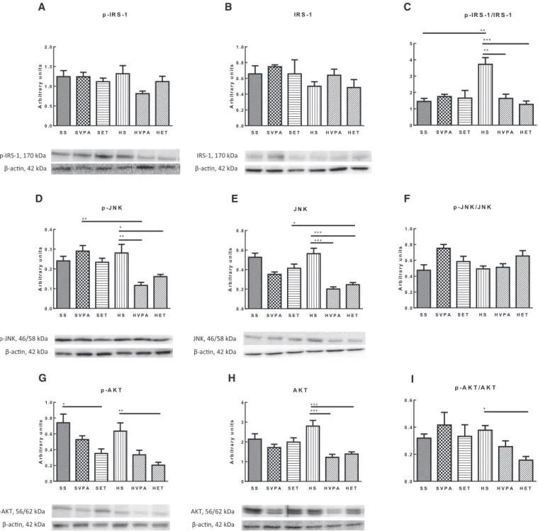
Lipotoxicity activates the c-Jun N-terminal kinase (JNK) and nuclear factor κ-light-chain enhancer of activated B cells (NF-κB) signaling pathways in the liver [21]. Additionally, activation of hepatic JNK (as a consequence of exacerbated oxidative stress in the liver) increases the pro-inflammatory factors TNF-α and interleukin (IL)-6 [5, 67, 127, 128]. Hepatic inflammation mediated by NF-κB, NF-κB inhibitor (IκB) kinase (IKK) and/or JNK pathways, adding to hepatic ectopic lipid deposition and ceramide and diacylglycerol buildup, induces and/or exacerbates hepatic insulin resistance [55, 60, 110, 129-133]. Activation of IKK and JNK, particularly the JNK isoform1, disrupts the insulin signaling pathway through the inhibitory phosphorylation of insulin receptors substrate (IRS) 1 and IRS2 [5, 129, 131, 134, 135].
Additionally, published data suggest that increased expression of protein tyrosine phosphatase 1B (PTP1B) and 11β-hydroxysteroid dehydrogenase type 1 (11β-HSD1) plays an important role in hepatic insulin resistance and inflammation [5, 50, 55, 105, 126, 136-138]. As both enzymes are abundantly located/tethered on the cytosolic surface of the ER [5, 55, 105], the ER functionality is implicated in the onset and/or progression of hepatic insulin resistance and inflammation. Likewise, increased signaling of hepatic transforming growth factor-β (TGF-β) strongly correlates with hepatic insulin resistance and contributes to hepatic inflammation, steatosis and fibrosis, what suggests TGF-β as a crucial player to the onset of NAFLD and the progression of NASH [55, 139-142].
Cross-talk between ER stress, inflammation, insulin resistance, altered redox state and mitochondrial dysfunction
Sustained ER dysfunction and consequent unmitigated activated unfolded protein response (UPR) seem to play an important role in the onset and progression of NASH [5, 12, 60, 143, 144], both in humans and animal models [145-147].
The mechanisms of the complex phenomenon of NASH-related ER stress are not completely understood and most of them are nonexclusive [67, 134, 143, 147-156],
with ER stress-associated hepatic inflammation, insulin resistance, oxidative stress, mitochondrial dysfunction and Ca2+ homeostasis disruption as well as apoptotic signaling involved in this process, in a vicious cycle manner [9, 67, 157-161]. Briefly, the dysfunction of the ER (the principal cellular location involved in secretory and transmembrane protein folding, lipid biogenesis and Ca2+ homeostasis) leads to the activation of an elaborate adaptive UPR [4, 5, 9, 60, 67, 128, 134, 152, 159] driven by three key ER transmembrane proteins [protein kinase RNA-activated (PKR)-like ER kinase (PERK), inositol requiring enzyme (IRE) 1 and activation transcription factor (ATF)], resulting in downstream responses [144, 153] that dynamically adjust the protein folding capacity to the cell needs [162] in order to restore homeostasis and to enhance cell survival. The first stage of UPR activation could be faced as a window of opportunity for the cell to restore homeostasis through expansion of the ER, decrease of protein synthesis, increase of the protein folding capacity and increase of the degradation of ER terminally misfolded proteins [4, 5, 114, 134, 153, 155]. However, prolonged ER stress might trigger and/or increase hepatic inflammation, insulin resistance, oxidative stress, mitochondrial dysfunction, Ca2+ homeostasis disruption and, ultimately, cell death [9, 67, 157-161], all of those accepted as NASH features.
The ER stress-induced unmitigated activation of UPR plays an important role in the inflammatory NASH-related response via phosphorylation of IκB by IKK as well as through the activation JNK/activator protein 1 (AP1) [5, 9, 67, 127, 134, 163, 164]. It is well-recognized that increased activation of the PERK and IRE1 pathways mediate increased expression of pro-inflammatory signaling, such as TNF-α, IL-1β and IL-6, through the activation of JNK, IKK and NF-κB [5, 110, 133]. Nevertheless, it should be noted that increased accumulation of inflammatory cytokines in the liver can also activate UPR [9, 127, 161], suggesting that the relationship between ER stress and inflammation is bidirectional.
Additionally, prolonged ER stress has been involved in the development of hepatic insulin resistance. Although the mechanisms by which it decreases insulin signaling are complex and take place at various and different levels of the insulin signaling pathway, it is widely accepted the involvement of PERK and/or IRE1 pathways activation in this process through the activation of JNK1, IKK and NF-κB [5, 67, 149, 150]. Another mechanism through which prolonged ER stress can induce insulin
resistance is through PTP1B and 11β-HSD1 [5, 105, 136, 165]. Both PTP1B and 11β- HSD1 have been suggested as one of the main negative regulators of insulin sensitivity, being PTP1B involved in dephosphorylation of tyrosine residues in IRS1 and IRS2, while increased glucocorticoid levels (which are regulated by 11β-HSD1) lead to increased phosphorylation of IRS1 at serine307 and, consequently, decreased affinity of IRS1 for the insulin receptor [5, 55, 105, 138, 166, 167]. In fact, the deletion and/or inhibition of 11β-HDS1 is linked with improved insulin sensitivity [55, 168]. Additionally, prolonged ER stress may also induce hepatic insulin resistance through hepatic ectopic lipid deposition [60].
Another major factor that has been implicated in the pathogenesis of NASH is increased oxidative stress [169], which is reinforced by the cross-talk between ER stress and redox state [128, 169]. Because ER and mitochondria are physically and functionally interconnected, and oxidative protein folding occurs in the ER, alterations in the redox state by increased generation of reactive oxygen species (ROS) could, directly and/or indirectly, interfere with ER homeostasis and protein folding capacity as well as influence mitochondrial function by increasing mitochondrial oxidative stress [128]. Moreover, increased mitochondrial ROS generation increases pro- inflammatory signaling [4, 55, 68, 170, 171]. In fact, increased ROS generation and increased pro-inflammatory signaling are closely tangled, with various vicious cycles occurring between them leading to insulin signaling dysfunction and fibrosis development through the production of TGF-β [4, 68, 169-171]. On the other hand, excessive lipid accumulation induces ROS production and lipid peroxidation, both of which are deeply related to hepatic inflammatory reactions [169, 172-174].
Additionally, since ROS generation also occurs during the protein folding process, an increased but untroublesome oxidative stress is a component of the UPR. ROS can be produced both downstream and upstream of UPR suggesting a strong cross-talk between increased ROS production and ER stress [169]. In fact, an increased ROS production as a consequence of mitochondrial dysfunction stimulates an ER stress response that, in turn, aggravates, through a vicious cycle mechanism, mitochondrial dysfunction and ROS production [5, 9, 161, 175]. Simultaneously, an enhanced ROS generation increases mitochondrial dysfunction. Furthermore, both increased oxidative damage and ER stress causes the leakage of Ca2+ from the ER triggering its
accumulation inside the mitochondria. This increases ROS production by the mitochondria what amplifies the ER stress [5]. It is noteworthy that the UPR can act as a binary switch depending on the severity and nature of the ER stress. So, UPR regulates adaptive and death effectors (controlling the fate of the cell: survive or die) [5, 176, 177]. Under conditions such as those observed in NASH, where ER stress in sustained and/or unmitigated, cell death occurs through several mechanisms, including: (I) PERK pathway; (II) IRE1-mediated activation of apoptotic signaling through both apoptosis signaling-regulating kinase 1 (ASK1)/JNK and caspase-12; (III) release of Ca2+ from the ER [5, 177-180].
Overall, in NASH, ER can play an important role in the amplification of the above harmful vicious cycles, as sustained or inadequate ER stress responses per se can lead to or exacerbate hepatic fat accumulation, pro-inflammatory processes, insulin resistance, oxidative stress, mitochondrial dysfunction and apoptosis.
Pharmacological and non-pharmacological therapeutic strategies: the role of physical exercise
Excessive hepatic fat accumulation per se does not lead to liver damage but lays the foundation for others factors, such as increased inflammation, altered redox state (by increased oxidative stress) or increased/prolonged ER stress, to trigger the progression towards NASH [181]. Efforts are being done in order to identify the mechanisms that mediate the development and progression of NASH, with several pharmacological target molecules proposed as therapeutic strategies to fight organelle-associated dysfunction [182-189]. In this regard, and since the ER stress is well-known to play a crucial role in “multiple hits” hypothesis of NASH progression, the ER stress chemical manipulation with exogenous chaperones [compounds with low-molecular-weight such as taurine-conjugated ursodeoxycholic acid (TUDCA), betaine and 4-phenyl butyric acid (PBA)] has been applied to improve the capability of the ER to adapt [151, 185, 186, 188] and, consequently, counteract NASH.
Although it is predicted that NASH may speedily become the most common cause of liver transplantation in many countries in the coming years, a well-established
pharmacological treatment capable to counteract the pathology of such disease is still lacking [20, 21, 29, 53, 83, 189]. Therefore, the core of innovative pharmacological research for the treatment of NASH relates to drugs that targets hepatic lipid metabolism, insulin signaling and inflammation as well as hepatocyte injury, liver fibrosis, bile acid metabolism, gut microbiota, gut permeability, hepatic oxidative stress and hepatic apoptosis, with some of these drugs already in phase II and III trials [4, 189, 190]. The inexistence of approved pharmacological therapies for NASH could be explained by the existence of gaps in the knowledge of the mechanisms underlying the disease as well as the absence of consistent methods for diagnostic and stadiation, except the liver biopsy [189].
For this reason, the pharmacological treatment for NASH consists in the use of different drugs for different therapeutic targets. Nowadays, insulin sensitizers and antioxidants are prescribed for NASH management. The most important classes of drugs currently under investigation are farnesoid X receptor (FXR) agonists, peroxisome proliferator-activated receptors (PPAR) agonists, ASK1 inhibitor, C-C motif chemokine receptor (CCR) 2/CCR5 dual inhibitor, pan-caspase inhibitor, de novo lipogenesis inhibitors, fibroblast growth factor 19 agonist, thyroid hormone receptor agonist, glucagon-like peptide 1 receptor agonists, antidiabetic drugs, anti- inflammatory drugs, anti-apoptotic drugs and anti-fibrotic drugs [36, 52, 189-196].
Unfortunately, only a few of them have had their efficacy confirmed in phase III trials, others haven’t had their superiority confirmed versus placebo, and some others are still under investigation [189, 190].
There is a lack of national guidelines for the treatment of NASH patients and the existent reference guidelines are not widely used [197]. In line, a “Call to Action” has been created by numerous outstanding medical associations to issue guidance strategies for the management of NASH [198, 199].
Considering that the pharmacological agents tested thus far display limited efficacy and NASH is a disease with tremendous pathophysiological complexity, it seems reasonable to consider that, in a near future, drug approach could not be appropriate/enough and lifestyle interventions should be taken into account [4, 5, 189, 194, 200].
The complexity of NASH pathology results from problems caused by “multiple-hits”.
Given that the simultaneity of over nutrition with an inactive lifestyle is the primary cause for the development of NAFLD/NASH [5, 17, 20, 33, 41, 49, 55, 201], in one hand, and the pleiotropic characteristics of the beneficial effects of PE, in another hand, it is conceivable, therefore, that exercise training could be considered as a non- pharmacological cornerstone strategy against NASH and NASH-associated metabolic disorders [5, 202-204]. Although the exact molecular mechanisms responsible for the beneficial effects of physical exercise (PE) against NASH and NASH-associated metabolic disorders are not completely elucidated, the suggested positive modulatory effects may occur at various and distinct levels [5]. In fact, even though there is strong evidence suggesting that a reduction of 5%–10% in baseline body weight, through lifestyle interventions, is needed to improve NASH histological findings [87, 205], PE has been repeatedly shown to improve hepatic steatosis, lipolysis in the adipose tissue and fatty acids flux to the liver, even in the absence of the above-mentioned body weight reduction [84, 85, 87, 205-207]. Likewise, PE also reduces liver fibrosis scores without body weight reduction, highlighting its importance in NASH [87, 207].
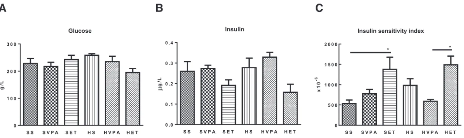
Additionally, regular exercise training improves hepatic insulin sensitivity [55, 208- 211], probable as a result of the favorable modulation of JNK activation (through JNK phosphorylation levels), 11β-HDS1 and PTP1B expression and pro-inflammatory signaling (through NF-κB protein expression, for example) [5, 55, 168, 212-214].
Additionally, anti-inflammatory effects of PE have been described [4, 5, 55, 208, 214, 215].
Given that both increased pro-inflammatory signaling and oxidative stress can be induced by both prolonged ER stress and mitochondria dysfunction, and taking into account that the simultaneity of over nutrition with physical inactivity is the primary cause for the NAFLD/NASH development [5, 49, 55], it will not be a surprise if the protective phenotype induced by PE against the progression of NASH lays on the recovery of ER stress and mitochondrial functionality. Although several studies have analyzed the effects of PE on ER stress response, the response perpetrated by PE still remains obscure with apparent contradictory results (increasing UPR versus decreasing UPR), suggesting that the effects of PE in ER stress are tissue-specific, dynamic and work as a binary switch: suppressing death effectors and increasing the
protective and pro-survival response [4, 5, 99, 208, 216, 217]. The idea that the exercise training could be an efficient therapy against NASH-related ER stress prompt us to hypothesize that exercise training might be able to modulate the protective effects of the existent windows of opportunity in the first phase of UPR.
Regarding the effects of PE on redox state, a variety of studies described PE-induced beneficial redox changes, including improvement of antioxidant defenses, possibly, mediated by nuclear erythroid 2 p45-related factor (Nrf2) [5, 69, 166, 185, 218-220].
Although the preventive and therapeutic effects of exercise training against NASH features and NASH-related ER stress, including its regulatory mechanisms, are only scarcely understood, it is conceivable that the protection provided by exercise training might also result in protection against cell death. Thus, complementing the beneficial impact of PE on redox modulation and mitochondrial and ER function, along with its insulin-like activity and anti-inflammatory role, other positive effects may contribute to the valuable influence upon the apoptotic signaling [5, 97, 221, 222]. Most probably, this could occur by the reduction of the activities of JNK and caspases-8 and -9 rather than by the abrogation of growth arrest and DNA damage inducible protein (GADD34) and caspase-12 expression [4].
References:
1. Zhu, J., et al., Obesity and Dyslipidemia in Chinese Adults: A Cross-Sectional Study in Shanghai, China. Nutrients, 2022. 14(11): p. 2321.
2. Vilalta, A., et al., Adipose tissue measurement in clinical research for obesity, type 2 diabetes and NAFLD/NASH. Endocrinol Diabetes Metab, 2022. 5(3): p. e00335.
3. Stos, K., et al., Prevalence and Sociodemographic Factors Associated with Overweight and Obesity among Adults in Poland: A 2019/2020 Nationwide Cross-Sectional Survey.
Int J Environ Res Public Health, 2022. 19(3): p. 1502.
4. Passos, E., et al., Physical exercise positively modulates nonalcoholic steatohepatitis- related hepatic endoplasmic reticulum stress. J Cell Biochem, 2022. 123(10): p. 1647- 1662.
5. Passos, E., et al., Endoplasmic Reticulum Stress Response in Non-alcoholic Steatohepatitis: The Possible Role of Physical Exercise. Metabolism, 2015. 64(7): p.
780-92.
6. Feldeisen, S.E. and K.L. Tucker, Nutritional strategies in the prevention and treatment of metabolic syndrome. Appl Physiol Nutr Metab, 2007. 32(1): p. 46-60.
7. Hossain, P., B. Kawar, and M. El Nahas, Obesity and diabetes in the developing world- -a growing challenge. N Engl J Med, 2007. 356(3): p. 213-5.
8. Tappy, L. and K.A. Le, Metabolic effects of fructose and the worldwide increase in obesity. Physiol Rev, 2010. 90(1): p. 23-46.
9. Hotamisligil, G.S., Endoplasmic reticulum stress and the inflammatory basis of metabolic disease. Cell, 2010. 140(6): p. 900-17.
10. Hossain, P., B. Kawar, and M. El Nahas, Obesity and diabetes in the developing world- -a growing challenge. The New England journal of medicine, 2007. 356(3): p. 213-5.
11. Ratziu, V., et al., A position statement on NAFLD/NASH based on the EASL 2009 special conference. J Hepatol, 2010. 53(2): p. 372-84.
12. Collison, K.S., et al., Diabetes of the liver: the link between nonalcoholic fatty liver disease and HFCS-55. Obesity (Silver Spring), 2009. 17(11): p. 2003-13.
13. Wei, Y., D. Wang, and M.J. Pagliassotti, Fructose selectively modulates c-jun N- terminal kinase activity and insulin signaling in rat primary hepatocytes. J Nutr, 2005.
135(7): p. 1642-6.
14. Hassen, G., et al., Nonalcoholic Fatty Liver Disease: An Emerging Modern-Day Risk Factor for Cardiovascular Disease. Cureus, 2022. 14(5): p. e25495.
15. Wenzl, F.A., et al., Inflammation in Metabolic Cardiomyopathy. Front Cardiovasc Med, 2021. 8: p. 742178.
16. Gutierrez-Cuevas, J., A. Santos, and J. Armendariz-Borunda, Pathophysiological Molecular Mechanisms of Obesity: A Link between MAFLD and NASH with Cardiovascular Diseases. Int J Mol Sci, 2021. 22(21): p. 11629.
17. Martin-Grau, M., V.G. Marrachelli, and D. Monleon, Rodent models and metabolomics in non-alcoholic fatty liver disease: What can we learn? World J Hepatol, 2022. 14(2):
p. 304-318.
18. Yu, Y., et al., Insights into the Epidemiology, Pathogenesis, and Therapeutics of Nonalcoholic Fatty Liver Diseases. Adv Sci (Weinh), 2019. 6(4): p. 1801585.
19. Younossi, Z., et al., Global burden of NAFLD and NASH: trends, predictions, risk factors and prevention. Nat Rev Gastroenterol Hepatol, 2018. 15(1): p. 11-20.
20. Akbari, G., et al., Characterization of diet based nonalcoholic fatty liver disease/nonalcoholic steatohepatitis in rodent models: Histological and biochemical outcomes. Histol Histopathol, 2022. 37(9): p. 813-824.
21. Chen, Z., et al., A vicious circle between insulin resistance and inflammation in nonalcoholic fatty liver disease. Lipids Health Dis, 2017. 16(1): p. 203.
22. Arguello, G., et al., Recent insights on the role of cholesterol in non-alcoholic fatty liver disease. Biochim Biophys Acta, 2015. 1852(9): p. 1765-78.
23. Nagle, C.A., E.L. Klett, and R.A. Coleman, Hepatic triacylglycerol accumulation and insulin resistance. Journal of lipid research, 2009. 50 Suppl: p. S74-9.
24. Walther, T.C. and R.V. Farese, Jr., Lipid droplets and cellular lipid metabolism. Annual review of biochemistry, 2012. 81: p. 687-714.
25. Ratziu, V., et al., A position statement on NAFLD/NASH based on the EASL 2009 special conference. Journal of hepatology, 2010. 53(2): p. 372-84.
26. Serviddio, G., et al., Effects of dietary fatty acids and cholesterol excess on liver injury:
A lipidomic approach. Redox Biol, 2016. 9: p. 296-305.
27. Noureddin, M., J.M. Mato, and S.C. Lu, Nonalcoholic fatty liver disease: update on pathogenesis, diagnosis, treatment and the role of S-adenosylmethionine. Exp Biol Med (Maywood), 2015. 240(6): p. 809-20.
28. Yu, J., et al., The Pathogenesis of Nonalcoholic Fatty Liver Disease: Interplay between Diet, Gut Microbiota, and Genetic Background. Gastroenterol Res Pract, 2016. 2016:
p. 2862173.
29. Tilg, H., A.R. Moschen, and M. Roden, NAFLD and diabetes mellitus. Nat Rev Gastroenterol Hepatol, 2016. 14(1): p. 32-42.
30. Chen, G., et al., Micronutrient Antioxidants and Nonalcoholic Fatty Liver Disease. Int J Mol Sci, 2016. 17(9): p. 1379.
31. Rosso, C., et al., Crosstalk between adipose tissue insulin resistance and liver macrophages in non-alcoholic fatty liver disease. J Hepatol, 2019. 71(5): p. 1012-1021.
32. Song, Q., et al., Lipidomics Revealed Alteration of the Sphingolipid Metabolism in the Liver of Nonalcoholic Steatohepatitis Mice Treated with Scoparone. ACS Omega, 2022.
7(16): p. 14121-14127.
33. Santos, J., et al., Non-Alcoholic Steatohepatitis (NASH) and Organokines: What Is Now and What Will Be in the Future. Int J Mol Sci, 2022. 23(1): p. 498.
34. Lee, H.H., et al., Non-alcoholic steatohepatitis and progression of carotid atherosclerosis in patients with type 2 diabetes: a Korean cohort study. Cardiovasc Diabetol, 2020. 19(1): p. 81.
35. Steggerda, J.A., et al., Clinical considerations in the management of non-alcoholic steatohepatitis cirrhosis pre- and post-transplant: A multi-system challenge. World J Gastroenterol, 2020. 26(28): p. 4018-4035.
36. Campbell, P., A. Symonds, and A.S.t. Barritt, Therapy for Nonalcoholic Fatty Liver Disease: Current Options and Future Directions. Clin Ther, 2021. 43(3): p. 500-517.
37. Cardoso, A.C., C. de Figueiredo-Mendes, and A.V.-N. C, Current management of NAFLD/NASH. Liver Int, 2021. 41 Suppl 1: p. 89-94.
38. Boland, M.L., et al., Towards a standard diet-induced and biopsy-confirmed mouse model of non-alcoholic steatohepatitis: Impact of dietary fat source. World J Gastroenterol, 2019. 25(33): p. 4904-4920.
39. Miyaoka, Y., et al., A novel hamster nonalcoholic steatohepatitis model induced by a high-fat and high-cholesterol diet. Exp Anim, 2018. 67(2): p. 239-247.
40. Day, C.P. and O.F. James, Steatohepatitis: a tale of two "hits"? Gastroenterology, 1998. 114(4): p. 842-5.
41. Takaki, A., D. Kawai, and K. Yamamoto, Multiple hits, including oxidative stress, as pathogenesis and treatment target in non-alcoholic steatohepatitis (NASH). Int J Mol Sci, 2013. 14(10): p. 20704-28.
42. Takaki, A., D. Kawai, and K. Yamamoto, Molecular mechanisms and new treatment strategies for non-alcoholic steatohepatitis (NASH). Int J Mol Sci, 2014. 15(5): p. 7352- 43. 79. Fang, Y.L., et al., Pathogenesis of non-alcoholic fatty liver disease in children and
adolescence: From "two hit theory" to "multiple hit model". World J Gastroenterol, 2018. 24(27): p. 2974-2983.
44. Buzzetti, E., M. Pinzani, and E.A. Tsochatzis, The multiple-hit pathogenesis of non- alcoholic fatty liver disease (NAFLD). Metabolism, 2016. 65(8): p. 1038-48.
45. Nielsen, T.S., et al., Dissecting adipose tissue lipolysis: molecular regulation and implications for metabolic disease. J Mol Endocrinol, 2014. 52(3): p. R199-222.
46. McCullough, K.D., et al., Gadd153 sensitizes cells to endoplasmic reticulum stress by down-regulating Bcl2 and perturbing the cellular redox state. Molecular and cellular biology, 2001. 21(4): p. 1249-59.
47. Petersen, K.F., et al., Impaired mitochondrial activity in the insulin-resistant offspring of patients with type 2 diabetes. The New England journal of medicine, 2004. 350(7):
p. 664-71.
48. Satapati, S., et al., Partial resistance to peroxisome proliferator-activated receptor- alpha agonists in ZDF rats is associated with defective hepatic mitochondrial metabolism. Diabetes, 2008. 57(8): p. 2012-21.
49. Rector, R.S., et al., Mitochondrial dysfunction precedes insulin resistance and hepatic steatosis and contributes to the natural history of non-alcoholic fatty liver disease in an obese rodent model. Journal of hepatology, 2010. 52(5): p. 727-36.
50. Rosenstock, J., et al., The 11-beta-hydroxysteroid dehydrogenase type 1 inhibitor INCB13739 improves hyperglycemia in patients with type 2 diabetes inadequately controlled by metformin monotherapy. Diabetes care, 2010. 33(7): p. 1516-22.
51. Sanyal, A.J., et al., Nonalcoholic steatohepatitis: association of insulin resistance and mitochondrial abnormalities. Gastroenterology, 2001. 120(5): p. 1183-92.
52. Peng, C., et al., Non-Alcoholic Steatohepatitis: A Review of Its Mechanism, Models and Medical Treatments. Front Pharmacol, 2020. 11: p. 603926.
53. Briand, F., et al., A 3-week nonalcoholic steatohepatitis mouse model shows elafibranor benefits on hepatic inflammation and cell death. Clin Transl Sci, 2020.
13(3): p. 529-538.
54. Goncalves, I.O., et al., Exercise mitigates mitochondrial permeability transition pore and quality control mechanisms alterations in nonalcoholic steatohepatitis. Appl Physiol Nutr Metab, 2016. 41(3): p. 298-306.
55. Passos, E., et al., Role of physical exercise on hepatic insulin, glucocorticoid and inflammatory signaling pathways in an animal model of non-alcoholic steatohepatitis.
Life Sci, 2015. 123: p. 51-60.
56. Li, Z., et al., Mitochondria-Mediated Pathogenesis and Therapeutics for Non-Alcoholic Fatty Liver Disease. Mol Nutr Food Res, 2019. 63(16): p. e1900043.
57. Simoes, I.C.M., et al., Mitochondria in non-alcoholic fatty liver disease. Int J Biochem Cell Biol, 2018. 95: p. 93-99.
58. Ajith, T.A., Role of mitochondria and mitochondria-targeted agents in non-alcoholic fatty liver disease. Clin Exp Pharmacol Physiol, 2018. 45(5): p. 413-421.
59. Xia, S.W., et al., Endoplasmic reticulum stress and protein degradation in chronic liver disease. Pharmacol Res, 2020. 161: p. 105218.
60. Lebeaupin, C., et al., Endoplasmic reticulum stress signalling and the pathogenesis of non-alcoholic fatty liver disease. J Hepatol, 2018. 69(4): p. 927-947.
61. Zhang, X., et al., New insight into inter-organ crosstalk contributing to the pathogenesis of non-alcoholic fatty liver disease (NAFLD). Protein Cell, 2018. 9(2): p.
164-177.
62. Rodrigues, R.M., Y. Guan, and B. Gao, Targeting adipose tissue to tackle NASH:
SPARCL1 as an emerging player. J Clin Invest, 2021. 131(20): p. e153640.
63. Kucukoglu, O., et al., Hepatokines and adipokines in NASH-related hepatocellular carcinoma. J Hepatol, 2021. 74(2): p. 442-457.
64. Ma, D.W., et al., Plasma phospholipids and fatty acid composition differ between liver biopsy-proven nonalcoholic fatty liver disease and healthy subjects. Nutr Diabetes, 2016. 6(7): p. e220.
65. Kartsoli, S., et al., Lipidomics in non-alcoholic fatty liver disease. World J Hepatol, 2020.
12(8): p. 436-450.
66. Mashek, D.G., Hepatic lipid droplets: A balancing act between energy storage and metabolic dysfunction in NAFLD. Mol Metab, 2021. 50: p. 101115.
67. Zhang, K. and R.J. Kaufman, From endoplasmic-reticulum stress to the inflammatory response. Nature, 2008. 454(7203): p. 455-62.
68. Katsiki, N., D.P. Mikhailidis, and C.S. Mantzoros, Non-alcoholic fatty liver disease and dyslipidemia: An update. Metabolism, 2016. 65(8): p. 1109-23.
69. Ascensao, A., et al., Modulation of hepatic redox status and mitochondrial metabolism by exercise: therapeutic strategy for liver diseases. Mitochondrion, 2013. 13(6): p.
862-70.
70. Xu, J. and S. Taubert, Beyond Proteostasis: Lipid Metabolism as a New Player in ER Homeostasis. Metabolites, 2021. 11(1).
71. Wang, L., et al., Endoplasmic Reticulum Stress Related Molecular Mechanisms in Nonalcoholic Fatty Liver Disease (NAFLD). Curr Drug Targets, 2018. 19(9): p. 1087- 1094.
72. Ahmed, L.A., et al., Gut microbiota modulation as a promising therapy with metformin in rats with non-alcoholic steatohepatitis: Role of LPS/TLR4 and autophagy pathways.
Eur J Pharmacol, 2020. 887: p. 173461.
73. Cani, P.D., et al., Changes in gut microbiota control metabolic endotoxemia-induced inflammation in high-fat diet-induced obesity and diabetes in mice. Diabetes, 2008.
57(6): p. 1470-81.
74. Cani, P.D., et al., Changes in gut microbiota control inflammation in obese mice through a mechanism involving GLP-2-driven improvement of gut permeability. Gut, 2009. 58(8): p. 1091-103.
75. Chen, D., et al., The Role of Gut-Derived Microbial Antigens on Liver Fibrosis Initiation and Progression. Cells, 2019. 8(11): p. 1324.
76. Csak, T., et al., Fatty acid and endotoxin activate inflammasomes in mouse hepatocytes that release danger signals to stimulate immune cells. Hepatology, 2011.
54(1): p. 133-44.
77. Endo, H., et al., Butyrate-producing probiotics reduce nonalcoholic fatty liver disease progression in rats: new insight into the probiotics for the gut-liver axis. PLoS One, 2013. 8(5): p. e63388.
78. Farrell, G., et al., Mouse Models of Nonalcoholic Steatohepatitis: Toward Optimization of Their Relevance to Human Nonalcoholic Steatohepatitis. Hepatology, 2019. 69(5):
p. 2241-2257.
79. Iwao, M., et al., Supplementation of branched-chain amino acids decreases fat accumulation in the liver through intestinal microbiota-mediated production of acetic acid. Sci Rep, 2020. 10(1): p. 18768.
80. Raza, S., et al., Current treatment paradigms and emerging therapies for NAFLD/NASH. Front Biosci (Landmark Ed), 2021. 26(2): p. 206-237.
81. Tacke, F. and R. Weiskirchen, Non-alcoholic fatty liver disease (NAFLD)/non-alcoholic steatohepatitis (NASH)-related liver fibrosis: mechanisms, treatment and prevention.
Ann Transl Med, 2021. 9(8): p. 729.
82. Sharpton, S.R., et al., Changes in the gut microbiome associated with liver stiffness improvement in nonalcoholic steatohepatitis. Therap Adv Gastroenterol, 2022. 15: p.
17562848221098243.
83. Stepanova, M., et al., Nonalcoholic steatohepatitis is the most common indication for liver transplantation among the elderly: Data from the United States Scientific Registry of Transplant Recipients. Hepatol Commun, 2022. 6(7): p. 1506-1515.
84. Henkel, J., et al., Reduced Oxidative Stress and Enhanced FGF21 Formation in Livers of Endurance-Exercised Rats with Diet-Induced NASH. Nutrients, 2019. 11(11): p. 2709.
85. Houghton, D., et al., Exercise Reduces Liver Lipids and Visceral Adiposity in Patients With Nonalcoholic Steatohepatitis in a Randomized Controlled Trial. Clin Gastroenterol Hepatol, 2017. 15(1): p. 96-102 e3.
86. Kawanishi, N., et al., Exercise training suppresses scavenger receptor CD36 expression in kupffer cells of nonalcoholic steatohepatitis model mice. Physiol Rep, 2018. 6(23):
p. e13902.
87. Linden, M.A., et al., Aerobic exercise training in the treatment of non-alcoholic fatty liver disease related fibrosis. J Physiol, 2016. 594(18): p. 5271-84.
88. Tsuchida, T., et al., A simple diet- and chemical-induced murine NASH model with rapid progression of steatohepatitis, fibrosis and liver cancer. J Hepatol, 2018. 69(2): p. 385- 395.
89. Ichimura-Shimizu, M., et al., Development of a novel mouse model of diet-induced nonalcoholic steatohepatitis-related progressive bridging fibrosis. Biosci Biotechnol Biochem, 2021. 85(4): p. 941-947.
90. Ichimura, M., et al., Phycocyanin prevents hypertension and low serum adiponectin level in a rat model of metabolic syndrome. Nutr Res, 2013. 33(5): p. 397-405.
91. Diehl, A.M., Lessons from animal models of NASH. Hepatol Res, 2005. 33(2): p. 138- 44.
92. Larter, C.Z. and M.M. Yeh, Animal models of NASH: getting both pathology and metabolic context right. J Gastroenterol Hepatol, 2008. 23(11): p. 1635-48.
93. Leng, Y.R., et al., Pathogenesis of NASH and Promising Natural Products. Chin J Nat Med, 2021. 19(1): p. 12-27.
94. Hebbard, L. and J. George, Animal models of nonalcoholic fatty liver disease. Nat Rev Gastroenterol Hepatol, 2011. 8(1): p. 35-44.
95. Kucera, O. and Z. Cervinkova, Experimental models of non-alcoholic fatty liver disease in rats. World J Gastroenterol, 2014. 20(26): p. 8364-76.
96. Sanches, S.C., et al., Nonalcoholic Steatohepatitis: A Search for Factual Animal Models.
Biomed Res Int, 2015. 2015: p. 574832.
97. Goncalves, I.O., et al., Physical exercise antagonizes clinical and anatomical features characterizing Lieber-DeCarli diet-induced obesity and related metabolic disorders.
Clin Nutr, 2015. 34(2): p. 241-7.
98. Lieber, C.S., et al., Model of nonalcoholic steatohepatitis. Am J Clin Nutr, 2004. 79(3):
p. 502-9.
99. Yang, W., et al., Exercise suppresses NLRP3 inflammasome activation in mice with diet- induced NASH: a plausible role of adropin. Lab Invest, 2021. 101(3): p. 369-380.
100. Ichimura, M., et al., High-fat and high-cholesterol diet rapidly induces non-alcoholic steatohepatitis with advanced fibrosis in Sprague-Dawley rats. Hepatol Res, 2015.
45(4): p. 458-69.
101. Kohli, R. and A.E. Feldstein, NASH animal models: are we there yet? J Hepatol, 2011.
55(4): p. 941-3.
102. Savari, F., et al., A new method to induce nonalcoholic steatohepatitis (NASH) in mice.
BMC Gastroenterol, 2019. 19(1): p. 125.
103. Naiki-Ito, A., et al., A novel model of non-alcoholic steatohepatitis with fibrosis and carcinogenesis in connexin 32 dominant-negative transgenic rats. Arch Toxicol, 2020.
94(12): p. 4085-4097.
104. Wang, D., Y. Wei, and M.J. Pagliassotti, Saturated fatty acids promote endoplasmic reticulum stress and liver injury in rats with hepatic steatosis. Endocrinology, 2006.
147(2): p. 943-51.
105. Pereira, C.D., et al., 11beta-Hydroxysteroid dehydrogenase type 1: relevance of its modulation in the pathophysiology of obesity, the metabolic syndrome and type 2 diabetes mellitus. Diabetes Obes Metab, 2012. 14(10): p. 869-81.
106. Nivala, A.M., et al., Fatty acid-mediated endoplasmic reticulum stress in vivo:
differential response to the infusion of Soybean and Lard Oil in rats. Metabolism, 2013.
62(5): p. 753-60.
107. Zambo, V., et al., Lipotoxicity in the liver. World journal of hepatology, 2013. 5(10): p.
550-557.
108. Park, E.J., et al., Multiple pathways are involved in palmitic acid-induced toxicity. Food Chem Toxicol, 2014. 67: p. 26-34.
109. Osorio, D., et al., Multiple Pathways Involved in Palmitic Acid-Induced Toxicity: A System Biology Approach. Front Neurosci, 2019. 13: p. 1410.
110. Cai, D., et al., Local and systemic insulin resistance resulting from hepatic activation of IKK-beta and NF-kappaB. Nat Med, 2005. 11(2): p. 183-90.