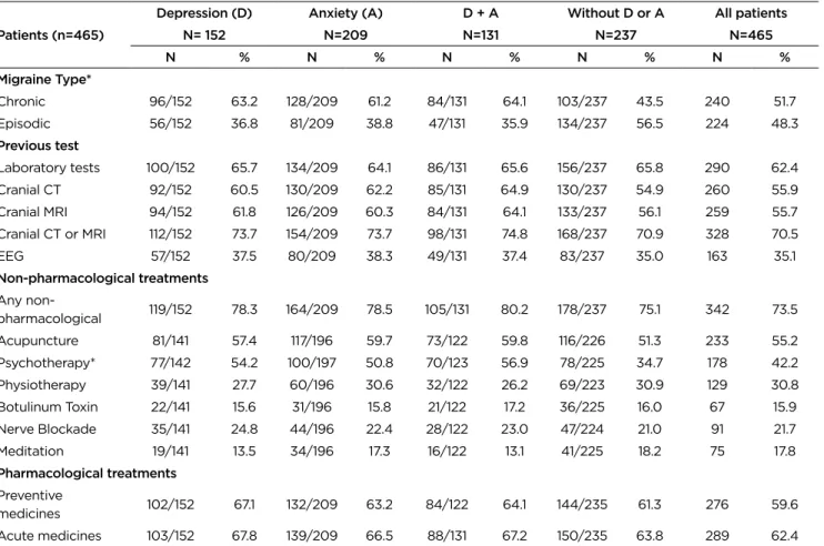
Chronic migraine was more common than episodic migraine among patients with psychiatric comorbidity: 63.2% of depressed patients, 61.2% of anxious patients and 43.5% of patients without any psychiatric disorder. Anxiety and depression influence the journey of patients with migraine before arriving at a tertiary headache centre. Depressed and anxious patients (54.2% and 50.8%, respectively) underwent psychotherapy more often compared to patients with neither depression nor anxiety (34.7%) (Table 2).
DISCUSSION
First, it was an observational cross-sectional study based on medical records and a retrospective self-reported questionnaire, so the associations found may not be due to a cause-and-effect relationship. Thus, our study reflected a specific population and selection bias may have influenced our results.
CONCLUSION
Taking analgesics can occur prior to the onset of headache due to anxiety, and evaluated the reasons for this behavior: 67% of patients reported difficulty coping with pain, 62% feared its occurrence, and 45% used analgesics to reduce anxiety (30).
ABSTRACT
RESUMO
Application of thermal microcautery in migraine management
INTRODUCTION
Sample
Inclusion / exclusion criteria
Technique description
RESULTS
The current study shows promising results with thermal microcautery in the preventive treatment of migraine due to its efficacy and tolerability (13). Possible explanations for our findings are the theory of the disturbed communication within the trigeminocervical complex. A possible pathophysiological mechanism of action is the alteration of the perception of pain by peripheral stimulation (1,14) in the distribution areas of trigeminal and occipital nerves (12,15).
Through the anatomical and functional convergence of these nerve endings, a wider distribution of stimulus is thought to trigger centrifugal pathways that regulate pain (16). It is known that stimulation of the occipital nerves regulates the activity of sensory neurons in the trigeminocervical complex; so stimulation of the trigeminal nerve is supposed to have the same effect. Its branches in the trigeminal divisions and C1 and C2 dermatomes (9) converge with sensory fibers in the dura mater and share the same receptive field.
Thus, it is possible that extracranial stimulation, such as thermal microcautery, also alters dura sensory fiber activity. Thermal microcautery is a promising therapy for migraine, and further randomized clinical trials are needed to confirm its efficacy.
Cefaleia Dialítica Associada à Cefaleia por Privação de Cafeína em Pacientes Submetidos à Hemodiálise
Headaches are classified as primary or secondary, based on the absence or not of underlying structural or metabolic disturbances causing the condition. Headaches are caused by several factors, either intrinsic or extrinsic, as individuals with migraine have lower thresholds for certain exposures, leading to a chain of events and culminating in pain.2. Headache is particularly relevant as a complication of hemodialysis, as this condition increases the discomfort felt by patients undergoing this therapy.
The association between hemodialysis and headache can be observed at the beginning of the dialysis treatment, which can be followed by nausea, vomiting, muscle spasms, disorientation, systemic hypertension and convulsions.3,4. The most frequent precipitating factors for dialysis headache, either mentioned by patients or by the medical team, were arterial hypertension (38%), followed by no identified factor (26%), arterial hypotension (12%) and changes in body weight (6%). The most important symptom of caffeine withdrawal is headache.7 The study by Maia et al.
The objective of this study was to assess the prevalence of headache in patients undergoing hemodialysis sessions, particularly considering dialysis headaches due to caffeine withdrawal.
MATERIAL AND METHODS
CONCLUSION AND DISCUSSION
The International Headache Society (IHS) emphasizes the decrease in serum caffeine as being responsible for headache crises during dialysis sessions. However, there are no controlled studies of prophylactic treatment or abortive treatment of dialysis headache.9. Frontal bilateral pain, characterized as pulsating, without aura, often associated with other symptoms such as scintillating scotomas, nausea, vomiting and photophobia is consistent with the literature found.
PahimLS, Menezes AMB, Rosângela Lima R. Prevalência e fatores associados à migrânea na população adulta de Pelotas, RS.
Prevalence of thunderclap headache in patients with ruptured intracranial aneurysms: series of 60 cases
The character of the headache is not very specific and there is no single pain characteristic that allows to suspect a diagnosis of aneurysm3 except for the presence of headache (TCH), which requires investigation for subarachnoid hemorrhage4. Thunderclap headache” refers to a headache that is very severe and has a sudden onset, reaching maximum intensity in less than 1 minute. A thunderclap headache is usually described by patients as an apoplectic event, an event that is distinctly different from other types of headache they may have previously experienced.
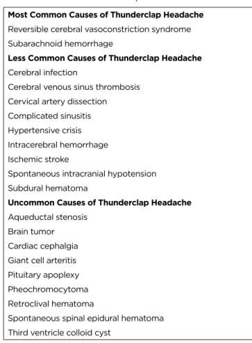
Patients with thunderclap headache often compare the sensation to an explosion in the head or a blow to the head6. We performed a prospective cohort study of consecutive patients with subarachnoid hemorrhage secondary to aneurysm rupture who received EVT. Causes of Thunderclap Headache * Most Common Causes of Thunderclap Headache Reversible Cerebral Vasoconstriction Syndrome Subarachnoid Hemorrhage.
Although the exact incidence of each cause of thunderclap headache is not well defined, certain causes of thunderclap headache are more common than others based on how often they present with the cluster headache and the prevalence of the condition itself. For example, although pituitary apoplexy can often occur with thunderclap headaches, since pituitary apoplexy is an uncommon condition, it is an unlikely cause of a patient's thunderclap headache.
Statistical Analysis
In addition, a recent study identified the presence of migraine as an independent risk factor for the rupture of an intracranial aneurysm5. First, the patients may have overlooked episodes of mild headache or forgotten details of the pain in the 12 months before treatment. In summary, we conclude that almost half of patients with ruptured intracranial aneurysms had thunderclap headache and there is no relationship with aneurysm size and vascular area.
Headache Classification Committee of the International Headache Society (IHS) The International Classification of Headache Disorders, 3. udgave.
Clinical characteristics of headaches attributed to diagnostic and therapeutic procedures
Acute or persistent headache attributed to craniotomy
Post-endarterectomy headache
Headache attributed to carotid or vertebral angioplasty or stenting
However, the relative risk of painful enlargement depends on individual risk factors, such as a history of myocardial infarction.
Headache attributed to cranial venous sinus stenting
Angiography headache
Post-dural puncture headache
Headache attributed to intrathecal injection
Dialysis headache
Headache attributed to radiosurgery of the brain
Post-electroconvulsive therapy headache
Aktan Ç, Özgür Ö, Sindel T, Dora B (2017) Headache characteristics during and after digital subtraction angiography: a critical reappraisal of the ICHD-3 criteria. Goksel BK, Torun D, Karaca S, Karatas M, Tan M, Sezgin N, et al (2006) Is low blood magnesium associated with hemodialysis headache. Roceanu A, Antochi F, Bajenaru O (2015) Symptomatic migraine with aura due to occipital arteriovenous malformation or headache attributed to arteriovenous malformation associated with simple partial seizures in an adolescent – a case report.
Liu Y, Xiao S, Liu M, Li G, Wang D, He J, et al (2000) Analysis of related factors in complications of stereotactic radiosurgery in intracranial tumours. McClelland S, Tendulkar RD, Barnett GH, Neyman G, Suh JH (2006) Long-term outcomes of radiosurgery for refractory cluster headache. Kim JW, Chung HT, Han MH, Kim DG, Paek SH (2016) Cerebral edema after repeated gamma knife radiosurgery for a major arteriovenous malformation: a case report.
Singh VP, Kansai S, Vaishya S, Julka PK, Mehta VS (2000) Early complications after gamma knife radiosurgery for intracranial meningiomas. Li TC, Shiah IS, Sun CJ, Tzang RF, Huang KC, Lee WK (2011) Mirtazapine relieves headaches and nausea after electroconvulsive therapy: a case series and review of the literature.
Telemedicine in the Management of Primary Headaches
A Critical Review
-10) The use of telemedicine in neurology is growing due to the fact that neurological care is still poor around the world. Several studies have shown potential benefits of telemedicine in the treatment of Parkinson's disease, epilepsy, multiple sclerosis, brain and spinal cord injury, and amyotrophic lateral sclerosis.
TELEMEDICINE AND HEADACHE IN THE LITERATURE
-15) However, the availability of healthcare services for headaches is poor worldwide and even greater in developing countries such as Brazil.
Mobile health (mHealth)
In conclusion, the existing evidence favors telemedicine as an alternative in the treatment of primary headache disorders. The use of telemedicine within ethical and compliance parameters by qualified professionals can be incorporated into the treatment of primary headache disorders. The impact of headache disorders in Italy and the public health and policy implications: a population-based study within the Eurolight project.
Acceptability, feasibility and costs of telemedicine for non-acute headaches: a randomized study comparing video and traditional consultations. Telemedicine in the treatment of non-acute headaches: a prospective, open-label, non-inferiority, randomized clinical trial. Added value of an electronic monitoring and alerting system in the management of medication overuse headache: a controlled multicenter study.
Intracranial lipoma manifesting with change in preexisting headache characteristics
Dagiti intracranial lipomas (IL) ket mangbukel ti 0.1% aginggana ti 0.5% kadagiti amin nga intracranial a tumor ken mabirukan a nangruna iti corpus callosum1. Ben Elhend S, Belfquih H, Hammoune N, Athmane EM, Mouhsine A (2019) Lipoma nga addaan iti agenesis ti corpus callosum: 2 a reporta ti kaso ken panagrepaso ti literatura. Piovesan EJ, Tatsui C. E., Kowacs PA, Prazeres R. F., Lange MC, Antoniuk SA, et al (2000), Lipoma ti korpus callosum a nainaig iti hipertropia ti corpus callosum: Maysa a report ti kaso. E) Sagittal nga T1W nga MRI. Ti maysa a sango a median a taba a sugat a mangrukod iti agarup a 6.6 x 4.5 x 3.5 cm a nainaig kadagiti pagilasinan ti dysgenesis ti corpus callosum ken colpocephalic nga aspeto dagiti lateral a bentrikulo.

The neurologist’s hammer O martelo do neurologista
Erb wrote: "If one firmly holds and supports the leg to be examined, slightly bent at the hip and knee joint with all the muscles relaxed, and then lightly and elastically with the finger or with the mallet in the region of the ligamentum patellae taps. [...] each tap is immediately followed by a slight but significant and apparently reflex contraction of the quadriceps; […] and it is extremely difficult to voluntarily suppress this reflex". Westphal wrote that the idea of tendon percussion was given to him by one of his patients who said that when he sat on a chair and tapped lightly on the area below the kneecap of the affected leg, it moved forward with a sudden jerk.New modified versions of the instrument emerged during the last years of the 19th century 6.
The large piece is designed to be used for knee jerks and the small one for bicep jerks. The Queen Square Hospital and Babinski hammers followed, consisting of a rubber disc surrounding a flat metal disc 7. The main difference between these lies in their ease of carrying, as the Queen Square is rigid, while the Babinski is smaller and telescoping with a shorter handle.
Even if some consider them similar, Queen Square is almost 150 grams heavier than Babinski 2. The Rabiner hammer has a rubber disc that can be used parallel to or perpendicular to the handle, as well as an inserted brush and needle for superficial reflexes Figure 1.