Staphylococcus aureus (S. aureus) é uma bactéria Gram-positiva altamente adaptável e versátil que pode causar uma ampla gama de doenças infecciosas em humanos ou animais. Staphylococcus aureus (S. aureus) é uma bactéria Gram-positiva altamente adaptável e versátil que pode causar uma ampla gama de doenças infecciosas em humanos ou animais.
Bibliographic Synthesis
General characteristics of Staphylococcus aureus
- Definition and morphology
- General cultural, biochemical characteristics and genome of S. aureus
- General pathogenesis and clinical diseases
- Pathogenesis
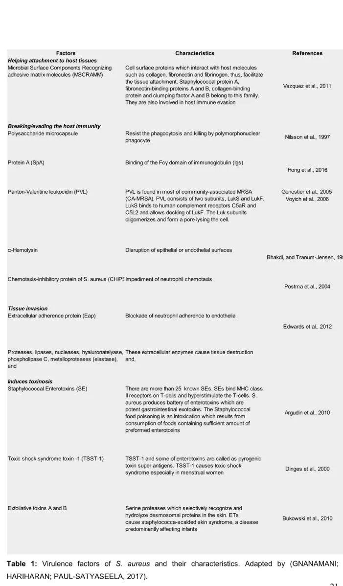
- Virulence factors
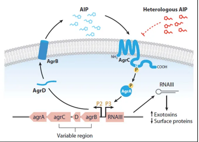
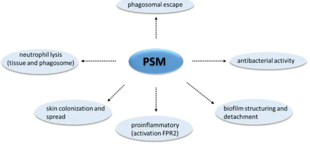
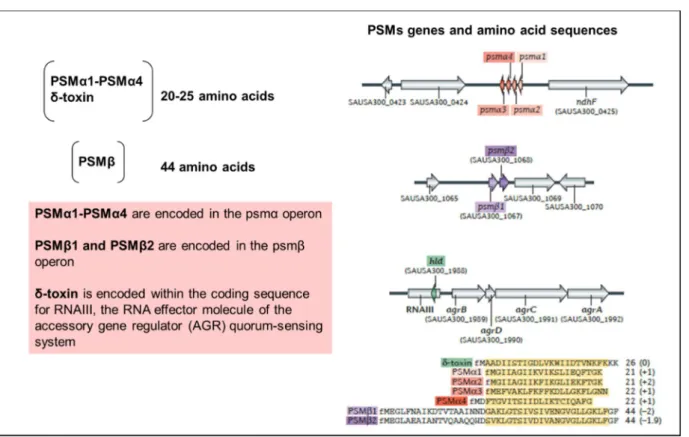
- Phenol-soluble modulins (PSMs)
The ability to induce cellular lysis is the most relevant feature of these PSMs (CHEUNG et al., 2014). Although this group of peptides is linked to the cellular lysis process, only a portion of the PSMs are responsible for this activity (CHEUNG et al., 2014).
The inflammasome, a primordial complex of innate immunity
- Mechanisms of the inflammatory response
- Inducers of the inflammatory response
- Mediators of the inflammatory response
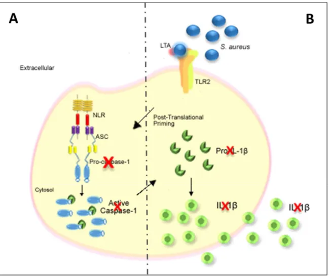
- IL-1β
- The maturation of IL-1β
- Excretion of IL-1β
- The regulation of IL-1β
- Other cytokines
IL-1β participates in most of the events involved in the activation and regulation of the inflammatory response (CHEN et al., 2017a). IL-1β also plays a role in the secretion of vascular endothelial growth factor (VEGF), reactive oxygen species (ROS) and reactive nitrogen species (RNS) (PAPIEWSKA-PAJĄK et al., 2017; ROBERTS et al., 2010 ).
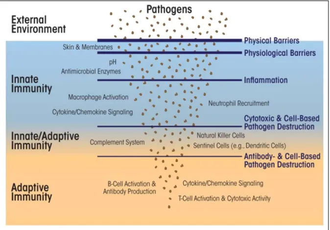
Innate immunity receptors
- Pattern recognition receptors (PRRs)
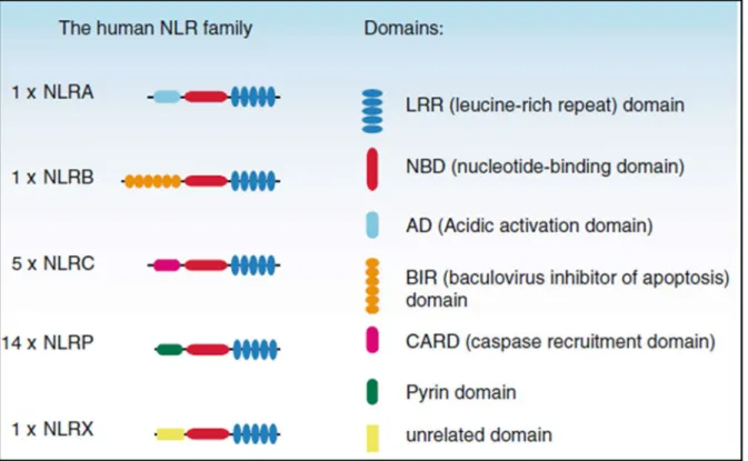
- NOD-like receptors (NLRs)
However, other cell types are able to express PRRs for example epithelial cells (JANG et al., 2015). NLR receptors, together with TLRs, cooperate to recognize and respond to pathogens and to activate pro- and anti-inflammatory mechanisms (CARRILLO et al., 2017).
Molecular platform: The inflammasome
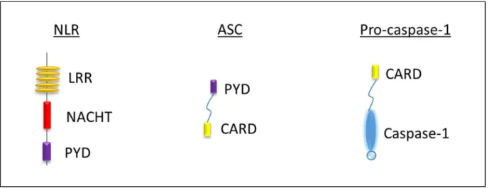
- The Caspase-1 protein
- Non-canonical activation of inflammatory caspases
- The ASC protein
- The NLRP1 inflammasome
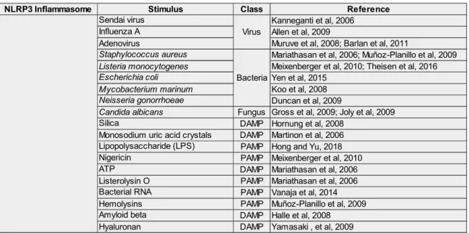
- The NLRP3 inflammasome
- The AIM2 inflammasome
- The NLRC4 inflammasome
- Others inflammasomes
- Inflammasome and pathologies
Caspase-11 (non-canonical) is composed of an N-terminal and C-terminal CARD and can activate the NLRP3 inflammasome by directly binding intracellular lipopolysaccharide (LPS) to the CARD domain in macrophages infected with Escherichia coli, Vibrio cholera, Legionella pneumophila , Salmonella typhimurium and other Gram-negative bacteria that enter the cytosol (murine caspase-11, caspase-4 and caspase-5 are orthologs of caspase-11 in humans) (JOZALA et al., 2013; MUNDAY et al., 1995 ; Yi, 2018). Thus, the decrease in the K+ concentration in the cytosol is proposed as a common trigger for the activation of the NLRP3 inflammasome (HE; HARA; NÚÑEZ, 2016; MUÑOZ-PLANILLO et al., 2013).
Problem statement of the project thesis
This is observed, for example, in diseases such as bone and joint infections (BJI), especially in the presence of orthopedic devices (SAAVEDRA-LOZANO et al., 2017). The increased production of these cytokines then leads to an imbalance in the homeostasis of bone turnover in favor of the activity of osteoclastogenesis and bone resorption (FENG; MCDONALD, 2011; PEETERS et al., 2016; RASIGADE et al., 2013). 34; non-professional phagocytes" has been documented in many cell types, including epithelial cells, endothelial cells, and keratinocytes (KINTARAK et al., 2004; RASIGADE et al., 2013).
Literature review. Strain and cell type-specificity of host cell response to
Introduction
Staphylococcus aureus is a versatile Gram-positive bacterium and an opportunistic pathogen capable of causing a wide range of infections in both humans (Chambers, 1997; Fitzgerald, 2012; . Lowy, 1998) and animals (Peton and Le Loir, 2014). Although these variations are increasingly documented in the literature, to our knowledge they have not been reviewed yet. In this review, we address the variability of strain-specific properties as well as of the host cells, leading to a wide range of cell responses as results of S.
S. aureus, a versatile opportunistic pathogen
These host-specific traits have also been obtained at the genomic level (Ben Zakour et al., 2008) and at the molecular level.
S. aureus adhesion and internalization
61 cytoplasmic domains of β-integrins that occurs as a result of the interaction between α5β1 and the cytoskeleton (Wang et al., 2006). Additionally, the focal adhesion protein tensin, vinculin, and zyxin are recruited to the site of bacterial uptake ( Agerer et al., 2005 ; Wright and Nair, 2010 ). The Alt-dependent internalization involves direct interaction with Hsc70 or an indirect interaction through the Fn coupled to integrin α5β1 ( Hirschhausen et al., 2010 ).
Cell response to S. aureus infection is strain-dependent
It has been shown that when FnBPs interact directly with human heat shock protein 60 (Hsp60) on the membranes of human and bovine epithelial cells, internalization efficiency is maximal (Dziewanowska et al., 2000). The extracellular adhesion protein (Eap) is a multifunctional protein consisting of 4 to 6 tandem-repeated domains that stimulate the adhesion of staphylococci to fibroblasts and endothelial cells (Fraunholz and Sinha, 2012; Hussain, 2002; Palma et al., 1999). However, their host cell-binding partners have not yet been identified, but evidence suggests that a scavenger receptor family is involved in WTA binding (Weidenmaier et al.
Phenotypic modifications also alter the S. aureus-host cell interaction
TLR signaling activates the NF-κB complex of transcription factors, which in turn activates immune gene expression. These clinical outcomes correlate with genetic characteristics such as less toxin production and better adhesion to bMEC in vitro for the strains that cause mild mastitis (Le Maréchal et al., 2011; . Peton et al., 2014). The host's immune response is also different, with a stronger inflammatory response in the early stages of the infection for non-persistent strains, and differential expression of cytokines in the course of the infection (Pereyra et al., 2017).
Strain-specific ability to adhere and internalize
When challenged with the gram-negative pathogen Escherichia coli (E. coli), bMEC strongly expresses a wide variety of immune factors, including cytokines and chemokines, membrane-protective factors, and bactericidal components. Newbould 305 had better adhesion and internalization rates than RF122 and this correlated well with a longer fibronectin binding protein (FnBP) compared to RF122 FnBP, as predicted by genome sequence (Bouchard et al., 2012; Peton et al., 2014).
Strain-specific cytotoxicity
It has been reported that pathogens are able to modulate apoptosis and thus initiate an infection in various types of host cells such as osteoblasts, endothelial and epithelial cells (Bayles et al., 1998; . Fraunholz and Sinha, 2012; Haslinger-Loffler et al. ., 2005; Kubica et al., 2008; Lamkanfi and Dixit, 2014). Cell death of non-prophylactic phagocytes causes activation of caspase-2 due to the efflux of potassium caused by aerolysin or α-toxin ( Imre et al., 2012 ). Next, TRAIL causes the activation of the intrinsic pathway of apoptosis through caspase-9 and the extrinsic pathway through caspase-8, leading to activation of caspase-3 and subsequently to apoptosis (Figure 2) (Alexander and Hudson, 2001; Claro et al. 2001 ; al., 2011; Josse et al.
Cytotoxicity induced by S. aureus extracellular vesicles
In some cases, EV-associated molecules have been shown to be more effective than extracts or soluble proteins in inducing immune responses and cellular cytotoxicity (Hong et al., 2014; Kim et al., 2019). These findings suggest that EV cargo and integrity can influence the host cell response and that even non-cytotoxic EVs can promote immunomodulation. EVs can also induce the production of cell adhesion molecules such as E-selectin, ICAM1, and VCAM1 and subsequent monocyte recruitment (THP1) (Kim et al., 2019), which contributes to immune cell infiltration.
Impact on the host cell cycle
In addition to these PSMs and SElOs, also known as cyclomodulins (El-Aouar Filho et al., 2017), “lipoprotein-like” proteins (lpl) in S. Like lpl genes are carried by strain-specific genomic islands (νSaα) ( Baba et al., 2008 ), this Lpl-associated phenotype is also strain-dependent. Impact on innate immune response and inflammatory response Certain bacterial traits are associated with a higher inflammatory response (Chen et al., 2018).
Impact on the innate immune response and on the inflammatory
Impact on the epigenetic modifications in host cells
Transcriptome analysis in mammary tissue from infected mice revealed a set of upregulated genes that significantly correlated with promoter-specific histone H3K14 acetylation. Another strain suppressed the inflammatory response, which could result in long-term infection of the host mammary tissue (Modak et al., 2014).
Cell types and S. aureus invasion
Primary endothelial cells and epithelial cells are known to absorb a large number of bacteria and can ingest S. These differences between cell types (primary cells and cell lines) with respect to bacterial degradation and host inflammatory processes have been reviewed (Seidl et al., 2012; Tuchscherr et al., 2011). Persistence induces bacterial phenotypic diversity, including SCV phenotypes, accompanied by changes in virulence factor expression (Tuchscherr et al., 2019).
Conclusion
Factors affecting the internalization of Staphylococcus aureus and impact on the course of infection in humans. Inhibition of Staphylococcus aureus invasion into bovine mammary epithelial cells by contact with live Lactobacillus casei. Gram-positive bacteria produce membrane vesicles: Proteomics-based characterization of Staphylococcus aureus-derived membrane vesicles.
Original article - Involvement of caspase-1 in inflammasomes activation and
Results
- Caspase-1 activation and IL-1β release triggered by inflammasomes activators
- Deletion of caspase-1 gene in human osteoblasts MG-63 using the
- Inflammasomes involvement in caspase-1 dependent IL-1β release by S
- S. aureus clearance by osteoblasts depends on caspase-1
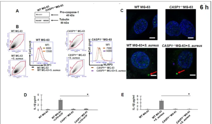
Therefore, we analyzed immunofluorescent staining for the adapter ASC in WT and CASP1–/–MG-63 cells. To elucidate the possible role of human osteoblasts in IL-1β production, we analyzed the kinetics (from 2 to 11 days) of IL-1β production by WT and CASP1–/–MG-63 cells exposed to S., IL-1β was not detected in supernatants from infected CASP1–/–MG-63 cells during the tested period.
Discussion
Moreover, a low level of IL-1β is produced at the beginning of the infection, so that the monitoring of IL-1β depends on the sensitivity of the detection method. The absence of IL-1β production by cells exposed to heat-killed bacteria indicates that factors associated with viable bacteria are involved in the activation of the NLRP3 inflammasome. Nevertheless, recent studies indicate that, in addition to the processing of IL-1β and IL-18, caspase-1 regulates unconventional protein secretion [46], activates lipid metabolic pathways [47], limits pathogen replication in professional phagocytes [48], [ 49 ].
Materials and Methods
- Maintenance of eukaryotic cells lines
- Deletion of the caspase-1 gene in human osteoblast-like MG-63 cells using
- S. aureus strains description
- Cell culture infection
- Western blot analysis
- Flow cytometry analysis
- Confocal microscopy
- IL-1β quantification by ELISA
- Statistical analysis
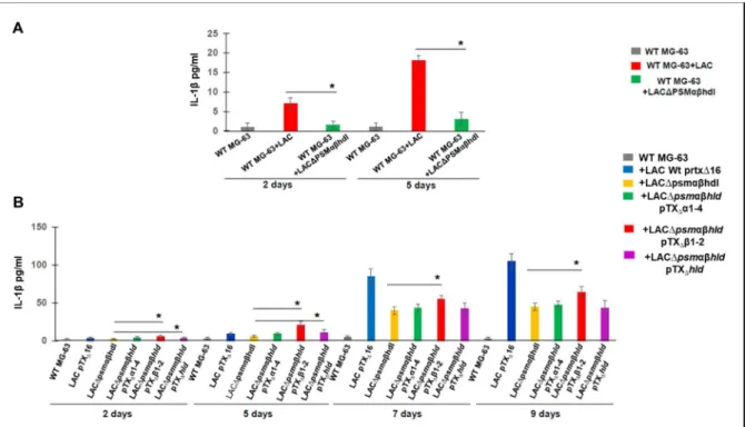
Detection of 45-kDa pro-caspase-1 in MG-63 cells by Western blot analysis was performed using anti-caspase-1 antibody (AdipoGen) as described in Materials and Methods. WT MG-63 cells were exposed to wild type LAC (USA300) and its isogenic mutant LAC. MG-63 cells were exposed to USA300 LAC (pTX∆16), which carries the control plasmid, the deletion mutant LAC∆psmαβhld (pTX∆16) and the complemented strains carrying the.
Results and general discussion
Caspase-1 activation and IL-1β release triggered by inflammasome activators
After different times after infection (2h, 6h, 2 days, 5 days, 7 days and 9 days), cell supernatants were collected and the level of IL-1β was determined by a commercial sandwich-ELISA (Invitrogen, France) and the activation of caspase-1 in MG cells -63 was measured by Western blot. In contrast, IL-1β was not detected 2 h after treatment, whereas only 4 pg/ml IL-1β was detected in MG-63 cells 6 h after LPS+ATP exposure. IL-1β production by MG-63 cells started later and the level of IL-1β was much lower than IL-1β production by ThP1 cells, whose level of IL-1β production corresponds to the findings of other investigators (GRAHAMES et al., 1999). ).

Deletion of caspase-1 gene in human osteoblasts MG-63 using the
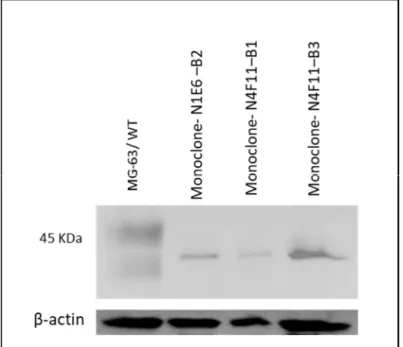
Deletion of the caspase-1 gene in human MG-63 osteoblasts using the CRISPR/Cas9 gene editing system. Therefore, after determining the method for isolation of monoclonals, MG-63 cells were transduced and selected with 0.2 μg/ml puromycin for 3 days. Monoclone G2 (CASP1–/– MG-63 cells) (Figure 19-B), B9 and C3 (data not shown), which were identified as a result of limiting dilution of puromycin-treated cells, were tested by Western blotting for lack the 45 kDa band corresponding to pro-caspase-1.
Inflammasome involvement in caspase-1 dependent IL-1β release by S. aureus
MG-63 cells by Western blot analysis using anti-caspase-1 antibody (AdipoGen) was performed as described in Material and Methods. To verify whether IL-1β release by infected osteoblasts was strain dependent, we analyzed the kinetics of IL-1β release by MG-63 exposed to different S. IL-1β was not detected in the supernatants of MG-63 cells exposed to killed bacteria of one of three strains, suggesting that factors associated with viable bacteria are involved in inflammation activation.
Pivotal role of S. aureus PSM toxins in stimulation of IL-1β release by infected
23-A, the level of IL-1β was significantly decreased in the supernatants of WT MG-63 cells exposed to Δpsmαβhld LAC compared to WT MG-63 cells exposed to wild-type LAC at days 5 and 9 post-infection. Analysis of the kinetics of IL-1β release from WT MG-63 cells exposed to either six clinical isolates or S. Moreover, we found different levels of IL-1β production induced by clinical strains collected from three patients .
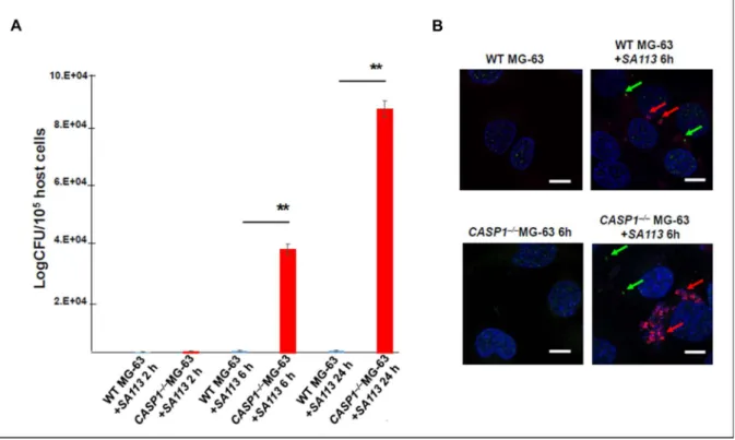
S. aureus clearance by osteoblasts depends on caspase-1
Consequently, we analyzed whether the absence of caspase-1 in CASP1–/–MG-63 cells is associated with the failure to control intracellular replication of S. Six and 24 h post-infection, significantly higher numbers of viable bacteria were found by CASP1 –/– MG-63 cells compared to WT MG- 63 cells (Fig. 25-A). WT MG-63 or CASP1–/– MG-63 cells were grown on slides of 12-well plates overnight, then the cells were exposed to a fluorescent derivative of strain S .
General conclusion and perspectives of the work performed during PhD
Distribution and regulation of the mobile genetic element-encoded phenol-soluble modulin PSM-mec in methicillin-resistant Staphylococcus aureus. Understanding the rise of the superbug: investigating the evolution and genomic variation of Staphylococcus aureus.