Die langsamen Wellen während des Schlafs sind im Zusammenhang mit CSWS interessant, da die Bildung langsamer Wellen und Spike-Wave-Komplexe gemeinsame Mechanismen haben [14]. Folglich kann die Steigung der langsamen Wellen als indirekter, aber zuverlässiger elektrophysiologischer Marker für die synaptische Stärke verwendet werden. Dies gilt auch für internationale, multizentrische „Investigator Initiated Trails“, die bereits in anderen europäischen Ländern etabliert sind.
Gesichtsfelddefekte nach Vigabatrintherapie bei West-Syndrom – Internationale mul- tizentrische Langzeitbeobachtung
Im Deutschen wird dieses Epilepsiesyndrom auch als BNS-Epilepsie („Blitz-Nick-Salaam-Anfälle“) bezeichnet, in der englischen Literatur werden die klinischen Symptome als „infantile Anfälle“ klassifiziert. Für die Therapieentscheidung zur Behandlung des West-Syndroms ist es wichtig zu wissen, ob bei der Behandlung mit VGB im Kindesalter das Risiko irreversibler Gesichtsfeldausfälle besteht. Lachen ohne Lachen – morphometrische MRT-Analyse in der präoperativen Beurteilung von MRT-negativer Epilepsie im Kindesalter mit elastischen Anfällen.
Lachen ohne Heiterkeit – Morphometrische MRT-Analyse in der prächirurgischen Ab- klärung einer MRT-negativen kindlichen Epilepsie mit gelastischen Anfällen
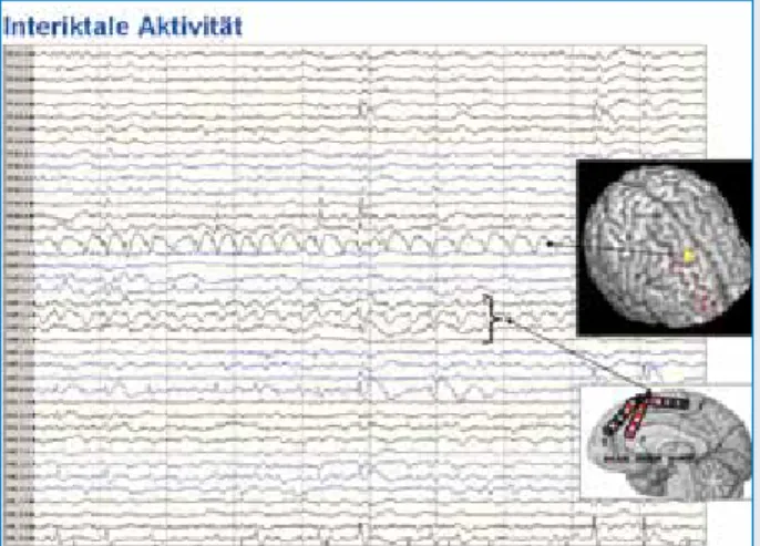
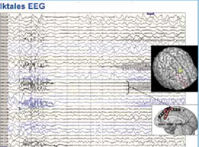
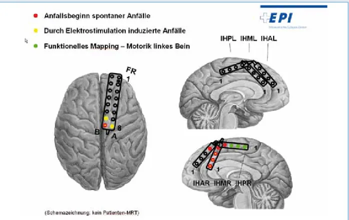
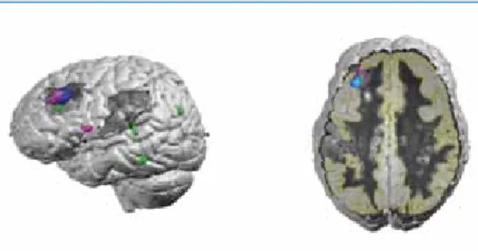
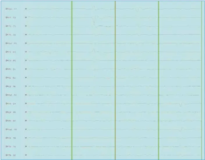
Interiktale, kontinuierliche Spike-Wave-Aktivität war bei einem einzelnen Elektrodenkontakt über der rechten frontalen Konvexität direkt über der mutmaßlichen Läsion sowie bei mehreren ipsilateralen interhemisphärischen Kontakten erkennbar (Abbildung 3). Interiktale, kontinuierliche Spike-Wave-Aktivität war bei einem einzelnen Elektrodenkontakt über der rechten frontalen Konvexität direkt über der mutmaßlichen Läsion sowie bei mehreren ipsilateralen interhemisphärischen Kontakten erkennbar. Typische Anfälle könnten durch die Stimulierung der rechten, aber nicht der linken interhemisphärischen Elektrodenkontakte und durch die Stimulierung von drei Kontakten auf der rechten frontalen Konvexität direkt über der Läsion induziert werden.
Localisation of Focal Epileptic Activity in Children Using High Density EEG Source Imaging*
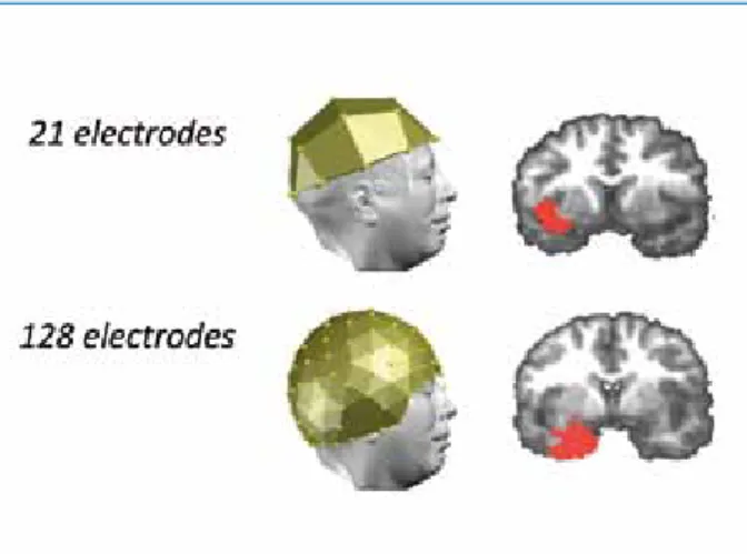
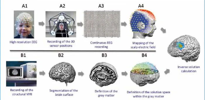
In children with epilepsy, a better understanding of the brain networks involved in epileptic activity is of great clinical and scientific importance. Noninvasive epileptic focus mapping can be performed using EEG source imaging (ESI), a technique based on scalp EEG recordings to assess the localization of intracerebral electrical sources. EEG Source Imaging (ESI) is a technique that enables 3-dimensional reconstruction of electrically active areas in the brain based on surface-recorded EEG.
The inverse solution is then performed to locate the generators of the peak in the solution space. The exact position of the electrodes on the head can be determined using a photogrammetry system (Electrical Geodesic Inc). Interictal spikes are identified by visual inspection of the spurs using a standard bipolar mount.
The exact position of the electrodes on the head can be determined using a photogrammetric system (Electrical Geodesic Inc). The consequences of insufficient coverage of the inferior temporal regions in a patient with a temporal lobe focus are shown in Figure 2. ESI is a reliable noninvasive imaging tool that allows for some patient movement during recording.
In vivo measurement of brain and skull resistivity using an EIT-based method and the combined analysis of SEF/SEP data.
Status Epilepticus in Children and SCN1A Gene
SE can occur in the context of an acute brain injury, such as encephalitis, ischemia, or hemorrhage. Hypotheses to explain the occurrence of SE include persistence of seizure-initiating factors and/or a defect in the mechanisms responsible for seizure arrest. The exact role these mutations play in the genesis of these epilepsies remains to be understood.
Myoclonus and severe developmental delay appear later in the course (in the classic form). Our work aims to know whether genetic variants of SCN1A play a role in the occurrence of SE in children. In the longer term, the goal is to improve the management of SE in children by adapting treatment to the patient's genotype.
We also expect to find more recurrences of status epilepticus and MRI abnormalities in the case group. So far, mutations or deletions have been identified in 11 children, most of whom had episodes of SE in the context of Dravet syndrome or GEFS+. These initial partial results suggest that SCN1a may indeed play a role in seizure duration.
Clinical correlates of mutations in the SCN1A gene: from febrile seizures to severe myoclonic epilepsy in infants.
Abnormal Autoantibodies in Pediatric Refractory Epilepsies
Therefore, we plan to collaborate with physicians of Pediatric Neurology Units at Swiss Children's Hospitals for case reporting, most of whom have already agreed to participate in principle. Most published reports concern adult patients, and the frequency of these antibodies in the pediatric epileptic population is unknown. Antibodies against NR1-NR2 heteromers of the NMDA receptor have been reported in a form of acute reversible encephalitis in adults and children [2, 11-14].
Glutamic acid decarboxylase (GAD) is involved in the anabolism of γ-aminobutyric acid (GABA), the most important cerebral inhibitory neurotransmitter. However, as stated by Liimatainen et al., “intrathecal synthesis of anti-GAD antibodies may be a marker of ongoing immune response and. The EEG and MRI of these patients, performed as part of standard management in such situations, will be analyzed in detail and repeated according to clinical developments.
Antibodies to the GABA(B) receptor in limbic encephalitis with seizures: case series and characterization of the antigen. Childhood idiopathic epilepsies usually appear at distinct, characteristic ages and usually tend to disappear in most cases or respond well to antiepileptic treatment without impairing the child's development – this is why they can be considered to have a good prognosis . 2 main studies by the UKBB team of pediatric neurology and development are presented, which are dedicated to this particular topic.
The case-control study No. 2 on children with benign epilepsy with centro-temporal spikes (BECTS) and control children should show the influence of several parameters, such as the onset, duration and severity of epilepsy and the exact localization of epilepsy. the epileptic focus on language and working memory network and its reorganization using fMRI, source EEG and neuropsychological examination.
Epilepsy and Cognition in Childhood*
Decreased fractional anisotropy in the middle cerebellar peduncle in children with epilepsy and/or ADHD. Neuropsychological and attention-induced cerebral hemodynamic effects in children with epilepsy and attention-deficit/hyperactivity disorder. Attention problems are one of the most common cognitive problems in children with epilepsy.
Therefore, we examined structural features in children with developmental ADHD and ADHD associated with epilepsy. Working memory is one of the most common and most reported executive function disorders in children with ADHD. To investigate this question, we examined behavioral differences in working memory performance and functional brain organization in children with epilepsy.
A good indicator to demonstrate the ability of reorganization in children with BECTS is the laterality index (LI). Reduced fractional anisotropy in the middle cerebellar peduncle in children with epilepsy and/or attention-deficit/hyperactivity disorder: a preliminary study. Effects of methylphenidate on working memory functioning in children with attention deficit/hyperactivity disorder.
Language reorganization in children with incipient left hemisphere lesions: an fMRI study.
Déficit en transporteur cérébral du glucose de type 1 dans les épilepsies généralisées idiopathiques et cryptogéniques de l‘enfant
Déficit cérébral en transporteur de glucose de type 1 dans les épilepsies généralisées idiopathiques et cryptogéniques de l'enfant. Comme la maladie est traitable, le diagnostic est important, basé sur la détection d'un rapport LCR/plasma à jeun < et d'une mutation du gène SLC2A1. La résistance aux médicaments, la survenue de difficultés d'apprentissage avec une intelligence limite et la fluctuation des symptômes liés aux repas ont conduit au diagnostic de déficit en GLUT1 [10].
Ces découvertes ont ensuite été retrouvées dans les formes familiales d'épilepsie : en 2009, Mullen et al. Le modèle d'épilepsie dans GLUT1DS a donc évolué au fil du temps d'un tableau d'encéphalopathie avec épilepsie sévère vers des formes familiales d'EGI ou d'autres syndromes d'épilepsie infantile, en particulier le syndrome de Doose [12]. La ponction lombaire (LP) doit être réalisée à jeun et précédée d'une mesure de la glycémie veineuse pour éviter une hyperglycémie de stress réactif [2, 4].
Diagnostic d'épilepsie généralisée (absence et/ou myoclonies et/ou crises généralisées tonico-cloniques et/ou crises toniques et/ou atoniques), idiopathique (développement normal, rythme de fond EEG normal, pas de résistance aux médicaments) ou cryptogéalique (retard de développement et/ou rythme de fond EEG anormal et/ou résistance aux médicaments). Les résultats positifs (présence d'un changement dans SLC2A1) seront confirmés par une deuxième amplification et séquençage à partir d'une nouvelle dilution de la solution d'ADN du patient. Le formulaire de consentement ainsi qu'une fiche d'information expliquant les objectifs de l'étude, les implications diagnostiques et thérapeutiques et les modalités pratiques seront remis à chaque famille lors d'un entretien avec l'investigateur ou un collaborateur lors du recrutement des patients.
La reconnaissance d'un GLUT1DS est d'une grande importance en termes de traitement et de pronostic, un traitement par un régime cétogène peut être proposé pour les cas réfractaires aux traitements conventionnels.
Im Schatten des Wolfes”
L’ombre du loup”
Postfach 1084
Epilepsie-Preise
Epilepsie-Liga-Mitteilungen
Johannes Lemke von der Universitätsklinik für Pädiatrie, Abteilung Mensgenetik, Inselspital Bern, erkundigte sich nach der Schweizer Anti-Epilepsie-Liga. Mai 2011 in Luzern, Margitta Seeck, Genf, in Urs Sennhauser, Hettlingen, bis in die letzten Jahre. Ausschreibung – Forschungsunterstützung Förderung der wissenschaftlichen Forschung im Bereich Epilepsie (insbesondere in Form von Ersthilfe) durch die Schweizerische Liga gegen Epilepsie (Liga gegen Epilepsie).
La Ligue contre l'épilepsie soutient des projets scientifiques dans le domaine de l'épileptologie avec un montant total de. En revanche, la prise en charge des frais de déplacement et d'hébergement (sans salaire) est possible pour des séjours de courte durée (quelques semaines au maximum) lorsque ce séjour est utilisé pour apprendre des méthodes appliquées dans le cadre d'un projet bénéficiant d'un accompagnement en Suisse. La Ligue suisse contre l'épilepsie (Ligue contre l'épilepsie) décerne tous les 3 ans un prix d'un montant de.
Tous les domaines professionnels et groupes professionnels couvrant des disciplines fondamentales ou cliniques sont invités à postuler. Le jury qui décerne le prix est composé de trois membres du comité directeur de la Ligue contre l'épilepsie. Le prix est toujours décerné l'année suivante dans le cadre de l'assemblée annuelle ou générale de la Ligue contre l'épilepsie.
Les candidatures doivent parvenir au secrétariat de la Ligue contre l'épilepsie (Seefeldstrasse 84, case postale Zurich) par.
Informations de la Ligue contre l’Epilepsie
Les bourses ne sont pas accordées pour une formation initiale ou continue ou pour des séjours à l'étranger. Si le demandeur a déjà demandé une aide ailleurs, veuillez nous en informer en indiquant où et avec quel résultat.
Kongresskalender
Jahrestagung der Deutschen Gesellschaft für Epileptologie e.V. (DGfE)