Across Europe, the number of patients with neurological diseases in the emergency department is increasing, both due to demographic changes and specialty development [ 1 - 3 ]. Before the puncture, the patient is informed about the painful and traumatic nature of the procedure. One of the most important characteristics that helps distinguish between migraine aura and epileptic seizures is the speed of the "march" of symptoms.
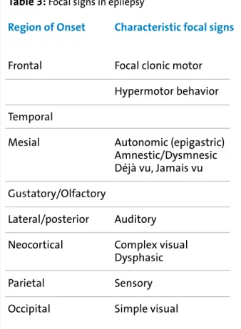
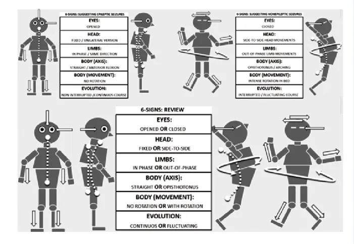
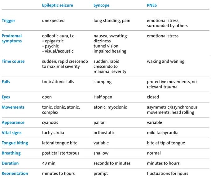
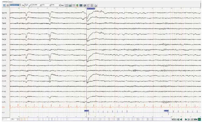
Duration of spells (minutes in cataplexy) and history (provocation of emotional triggers and excessive daytime sleepiness in cataplexy) can be helpful in differentiating atonic seizures from cataplexy events. When the patient arrived, the on-call neurologist and anesthesiologist were called, and they were ready to intubate the patient. Some of the most important signs that are easy to evaluate are summarized in Figure 2 and Table 1.
Simply communicating the diagnosis to the patient early in the course of the disease has been shown to be effective [55].
Yield of EEG After a First Unprovoked Seizure
- Routine EEG in first seizure
- First seizure and epilepsy: current definition and epidemiology
- Sleep EEG
- Relevance of ictal recordings in the 1st EEG It is possible that the first seizure which comes to
- Risk of relapse
Two factors are associated with an increased risk of relapse: the presence of a cerebral lesion and epileptiform abnormalities (EA) in the electroencephalogram (EEG). In this paper we focus on the risk of relapse after a first unprovoked attack; we review the yield of standard and sleep EEG to identify EA and/or abnormal but nonspecific slowing. Standard EEG carries valuable information regarding the underlying syndrome and risk of relapse.
This number is based on the risk of relapse after two unprovoked seizures, which is about 60% at 2 years and 70 - 75% at 5 years of follow-up. As we discuss below (“Risk of relapse”), two factors are consistently associated with an increased risk of relapse: the presence of cerebral lesions and epileptiform abnormalities on the EEG. In this case, the risk of repeated seizures, at least during the first 2 years, is significantly reduced by an average of 34% [8].
In 2009, Hesdorffer showed that a patient who experiences a single unprovoked seizure after a distant brain injury, such as a stroke, tumor, central nervous system infection, or trauma, is at high risk for a second unprovoked seizure. The risk of relapse was higher in patients with an abnormal EEG than with an abnormal picture, as not all epilepsy syndromes are related to cerebral lesions [24]. Although interictal EA have been associated with a higher risk of relapse, their diagnostic value has long been unclear.
However, by five years, the cumulative risk of recurrence was higher in older adults (75 versus 61 percent). Psychiatric and neuropsychological comorbidities are associated with a lower response to drug treatment and a higher risk of remission failure. Risk of seizure recurrence after a first unprovoked afebrile seizure in childhood: an extended follow-up.
Brain Imaging After a First Seizure
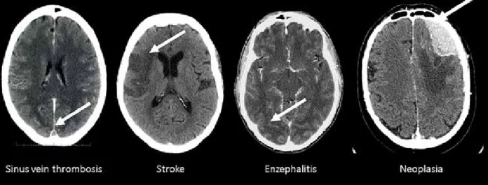
Whether the patient's baseline imaging is associated with a seizure or a mimicking condition (ie, a seizure-like episode). Is there significant brain damage that explains the first seizure and affects the prognosis - or is it just a chance finding. For discussion, we refer to the guidelines of the German Society of Neurology (DGN, the British National Institute for Health and Care Excellence (NICE) guidelines [8] and the American Academy of Neurology (AAN) imaging guidelines (2015). ) [3].
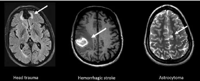
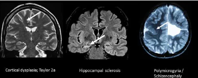
Is there a substantial lesion of the brain that explains the first seizure and affects the prognosis - or is it just a coincidental finding. 2nd image left: Acute stroke in the right medial territory visualized by hypodense delineation of parenchyma on non-enhanced CT. It is striking that almost 50% of the brain lesions discovered during the effects of a first seizure can now be regarded as incidental.
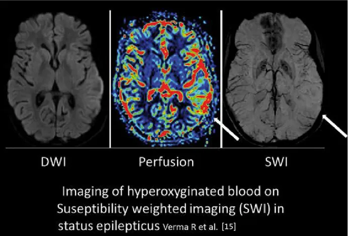
Currently, knowledge of the diagnostic benefits of these techniques is based on case studies. Brain perfusion measurements on CT or MRI are part of the emergency imaging protocols in most hospitals in Switzerland. Imaging techniques based on brain perfusion changes such as perfusion measurements themselves, SWI or EEG/fMRI rely on measuring indirect effects of epileptic neuronal activity.
In most cases the site of seizure initiation was located in the activation lobe in the NCI. Diffusion-weighted imaging (DWI) (left image) reveals a diffusion restriction within the symptomatogenic area as defined by perfusion imaging of the current status epilepticus. Epileptology of first seizure presentation: a clinical, electroencephalographic and magnetic resonance imaging study of 300 consecutive patients.
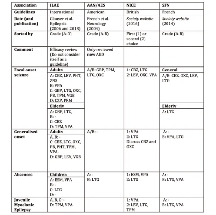
First-Line Antiepileptic Drugs in Adults: From Guidelines to Personalized Medicine
Furthermore, they are limited to the inclusion criteria of the corresponding studies, which rarely include patients with significant somatic or psychiatric comorbidities. From a review of the guidelines, we will present the individual characteristics of the AED and how they differ from each other. Considering this fictitious patient's situation, we will discuss what are likely to be the best AEDs in this case.
The differences between guidelines are due to the date of redaction and methodological differences in grading and the evaluation of the available literature [6]. We will now discuss five of these studies [7 - 11] to illustrate this point and understand the rationale of the published guidelines. Tables 3 and 4 summarize important aspects in the selection of the first line AED with 11 of the most common.
Beyond the most important AEDs from the guidelines, this table adds two of the more recent AEDs lacosamide (LCM) and perampanel (PER), whose prescription rate is likely to increase in daily practice (e.g. (LCM was approved for monotherapy in the US in 2014). Titration and dosing schedule is another important aspect of choosing first-line AEDs. Liver enzyme induction is also problematic in this case: CBZ, PHT, PB and primidone (PRM) can decrease the efficacy of the chemotherapy agent.
It is generally agreed that AEDs should be continued during pregnancy because of the potentially severe consequences of repeated seizures for both mother and child. The goal of treatment should not be to impair cognition or motor skills and to avoid causing behavioral problems. Efficacy and tolerability of new antiepileptic drugs I: treatment of new-onset epilepsy: report of the Therapeutics and Technology Assessment and Quality Standards Subcommittee of the American Academy of Neurology and the American Epilepsy Society.
Neu: Flyer „Nichtepileptische Anfälle“
CH 8008 Zürich
Ligue suisse contre l'épilepsie Schweizerische Epilepsie-Liga Lega Svizzera contre Epilessia Ligue suisse contre l'épilepsie. Il n'est pas facile de faire la différence entre les deux, et même les neurologues expérimentés confondent parfois les crises épileptiques et non épileptiques. Il s'adresse en priorité aux personnes en crise physiologique ou psychogène et à leurs proches, mais intéresse également les professionnels.
Son auteur est le vice-président de la Ligue contre l'épilepsie, le Dr. Andrea Rossetti, guide privé. J'ai souffert de crises récurrentes depuis l'âge de 17 ans mais je n'ai trouvé de soulagement qu'à l'âge de 49 ans. Et je sais qu'il existe un remède, que je peux comprendre la maladie et peut-être même la laisser derrière moi.
Nouveau : le dépliant « Crises non épileptiques »
Epilepsie-Liga-Mitteilungen
Dezember 2016
8008 Zürich
Since 2004, the Swiss League Against Epilepsy (SLAE) annually awards a research recognition award (Forschungsförderungspreis) to promote experimental and clinical research in epileptology. With a prize money of CHF 25,000, the Research Recognition Award is the highest award given by SLAE. The most important criteria for awarding the research recognition award are excellent scientific quality, the opportunity to study new methods and techniques and to establish or consolidate international collaborations, as well as the overall feasibility of the project [1].
Fritschy is one of the brightest stars in preclinical epilepsy research in Switzerland. Several clinical features and neuropathological changes of temporal lobe epilepsy associated with hippocampal sclerosis can be reproduced experimentally by intrahippocampal injection of kainic acid in adult mice, the animal model used in this project. However, it has not yet been established whether such epileptiform activity is merely a manifestation of the functional changes induced by kainic acid or actually the driver of epileptogenesis.
The main goal of the funded project is to investigate how epileptic discharges during the latent phase of the kainic acid model are involved in the formation of an epileptic focus. The technique used here makes use of light-sensitive proteins called opsins, which are expressed in ion channels of specific neuronal populations in the hippocampus. By using this technique, it is now possible to perform targeted manipulations of neuronal function on demand during the phase of epileptogenesis in the kainic acid model, which has never been done before.
This will make it possible to better understand how epileptic discharges during epileptogenesis are involved in the development of spontaneous recurrent seizures and the formation of epileptic foci. In 1996, he completed his habilitation at the Faculty of Medicine of the University of Zurich on the subject of GABAA receptor subtypes in the brain and shortly thereafter was awarded the "Georg Friedrich Götze-Preis". Since 2004 he has been professor of pharmacology at the Department of Pharmacology and Toxicology at the University of Zurich and since 2010 director of the Neuroscience Center Zurich.
8008 Zurich
Promotion de la recherche scientifique dans le domaine de l'épilepsie (principalement sous forme d'aide initiale) par la Ligue suisse contre l'épilepsie. La Ligue contre l'épilepsie soutient des projets scientifiques dans le domaine de l'épileptologie pour le montant total. En revanche, la prise en charge des frais de déplacement et des per diem (sans salaire) est possible pour des séjours de courte durée (quelques semaines au maximum) lorsque ces séjours servent à apprendre les méthodes utilisées dans le cadre d'un projet soutenu en Suisse. .
Si le demandeur a déjà demandé une aide ailleurs, veuillez nous indiquer où et avec quel résultat.
Informations de la Ligue contre l’Epilepsie
Kongresskalender
Fachtagung für Neurophysiologie und angrenzende Gebiete
Dreiländertagung der Österreichischen und Deutschen Gesellschaften für Epileptologie und der
Autorenverzeichnis Jahrgang 33 | 2016
Inhaltsverzeichnis Jahrgang 33 | 2016
- April 2016
- Juni 2016
- Oktober 2016
- Dezember 2016 First Seizure: What’s Next?
Quantitative EEG in the intensive care unit Frédéric Zubler, Mojtaba Bandarabadi, Rebekka Kurmann, Andreas Steimer,.
Schlüsselwörter/Mots clés/Key words Jahrgang 33 | 2016
Postictal 56