Long-term potentiation of synaptic transmission in the dentate gyrus of the anesthetized rabbit after perforant path stimulation. Intact sister cultures received TrkB blocking antibody to control for antibody effects. Addition of receptor-specific antibodies to the BDNF and NT-4/5 neurotrophin receptor, i.e., TrkB antibodies for 14 days in uninjured cultures did not lead to the formation of GAP-43 immunopositive fibers.
This suggests that activation of the TrkB receptor is required for upregulation of GAP-43 and axonal reorganization after brain injury. The lesion of the Schaffer collaterals after the incision led to axonal sprouting in CA3 (red) but not to replacement of the Schaffer collaterals. Therefore, the increase in the connectivity of the CA3 pyramidal cells, as seen in the paired recordings, could explain the observed increase in excitability.
The time required for the preparation of these new axonal collaterals and the formation of the new synapses may underlie the delay between the initial injury and the development of posttraumatic damage. Lesion-induced sprouting and hyperexcitability in the hippocampus in vitro: implications for the genesis of posttraumatic epilepsy.

Circuits in a Dish: A Rodent Slice Preparation as Model for the Development of Epileptiform Activity in the Temporal Lobe
- Interictal-like bursting in a slice preparation of the hippocampus
- A new slice model for burst spreading in the temporal lobe
- Whole-cell patch clamping to establish changes on the cellular level
- Changes in both excitation and inhibition lead to persistent bursting
Circuits in a dish: a rodent slice preparation as a model for the development of epileptiform activity in the temporal lobe. The hippocampus, entorhinal cortex, and amygdala are structures in the temporal lobe of the brain that are extensively and directly connected to each other [6]. After a longer exposure to bicuculline, we also saw the development of a bursting activity that started spontaneously in the CA3 region of the hippocampus.
In the entorhinal cortex and amygdala, individual bursts had a lower frequency than in the hippocampus (see Figure 2B). 16 Epileptologie 2004 Circuits in a Dish: A Rodent Slice Preparation as Model for Development of ..| Ron Stoop Figure 2: Preparation and spread of interictal-like activity in the rodent horizontal slice preparation. White circles indicate the usual recording sites in the hippocampus (CA3/2), the entorhinal cortex (EC) and the lateral amygdala (LA).
Previous studies have shown that the connections between cells in the pyramidal cell layer can undergo long-term potentiation, i.e. 18 Epileptologie 2004 Circuits in a Dish: A Rodent Slice Preparation as Model for the Development of ..| Ron Stoop Figure 3: A) Effects of bicuculline application and washout on spontaneous bursting activity in the hippocampus and entorhinal cortex (EC). Bottom graph: Mean frequencies of bursting activity in hippocampus and entorhinal cortex before and after their dissociation (as indicated at top) followed by bicuculline washout (n = 15 slices).
At the same time, we found that the projections sent by the excitatory cells in the pyramidal layer to the inhibitory interneurons, i.e., in conclusion, the persistent bursting that occurs in the preparation of the horizontal slice can be used for several purposes. Requirement of basolateral amygdala neuron activity for the induction of long-term potentiation in the dentate gyrus in vivo.
The role of excitatory amino acid receptors in the propagation of epileptiform discharges from the entorhinal cortex to the dentate gyrus in vitro.

A New Animal Model of Temporal Lobe Epilepsy*
It is characterized by extensive loss of neurons and gliosis, most severe in the CA1 and CA3 regions of the hippocampus and in the hilus of the dentate gyrus [4]. The neurodegeneration also affects granule cells in the dentate gyrus and CA2 pyramidal cells to varying degrees. Histologically, these insults resemble hippocampal sclerosis, indicating a selective vulnerability of certain types of hippocampal neurons, particularly in the hilus of the dentate gyrus.
KA treatment causes severe neuronal loss in the hippocampal formation, particularly in the hilus of the dentate gyrus and in the CA3 area. The second phase lasts about 2 weeks and corresponds to the latent phase of the pilocarpine model. During this phase, there is a slow progression of the lesion in CA1, evidenced by increasing neuronal loss in the pyramidal cell layer (figure 1).
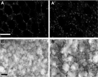
A diagram (adapted from [17]) showing the main types of neurons and hippocampal circuitry is provided for reference. They are therefore likely to be functionally altered during the chronic phase of the model. TLE with hippocampal sclerosis offers the advantage, as mentioned in the introduction, that late stages of the disease can be studied in tissue resected from patients suffering from intractable seizures.
Such seizures were observed with variable frequencies (up to a few per hour), mainly during the resting phase of the mice. This marker labels their cell body, dendrites and axons that form a dense plexus in the granule cell layer of the dentate gyrus and in the CA3-CA1 pyramidal cell layer. Excitatory granule cells of the dentate gyrus show a dual inhibitory neurochemical content following intrahippocampal administration of kainate in adult mice.
Neurodegenerative and morphogenic changes in a mouse model of temporal lobe epilepsy do not depend on the expression of the calcium-binding proteins parvalbumin, calbindin, or calretinin.

Adenosin-Zelltherapie der Epilepsie
- Aktivierung von Adenosin-Rezeptoren
- Anfallsunterdrückung in pharmakoresistenter Epilepsie
- Rolle der Adenosin-Kinase in der Epilepsie Die extrazellulären Adenosin-Spiegel hängen direkt
- Anfallsunterdrückung durch lokale Zell-Implan- tate
- Stammzellen – ein neuer Weg zur lokalen Adenosin-Freisetzung?
Eines der größten Probleme der Epilepsieforschung ist heute die Entwicklung verbesserter antikonvulsiver Therapien. Neben den krampflösenden Eigenschaften von Adenosin sind auch seine neuroprotektiven Eigenschaften bei der Behandlung von Epilepsie von großer Bedeutung. Es wurde gezeigt, dass weder Carbamazepin noch Valproat noch Phenytoin die chronische Anfallsaktivität bei diesen Tieren unterdrücken konnten [19].
Um periphere Nebenwirkungen zu vermeiden, scheint auch die lokale Applikation von Adenosin in der Nähe eines epileptischen Herdes mittels implantierter Adenosin-freisetzender Zellen zur Unterdrückung medikamentenresistenter Anfälle geeignet zu sein. Pharmacocore-resistente Anfälle im Maus-Kainat-Modell der chronischen Temporallappenepilepsie werden durch Carbamazepin nicht beeinflusst (oben). Die Rolle der Adenosinkinase bei Epilepsie Die extrazellulären Adenosinspiegel stehen in direktem Zusammenhang mit dem Aktivitätszustand neuronaler Schaltkreise.
Die zentrale Rolle der Adenosinkinase bei der Regulierung des endogenen Adenosinspiegels wirft daher die Frage auf, welche Rolle dieses Enzym bei der Epileptogenese spielt. Basierend auf dem oben Gesagten sollte es möglich sein, epileptische Anfälle durch lokale Adenosinfreisetzung in der Nähe eines epileptischen Herdes zu unterdrücken. Nun gilt es, diejenigen Zellen zu finden und gezielt zu vermehren, die einen Defekt in der Adenosinkinase aufweisen.
Zwei Möglichkeiten, die derzeit intensiv evaluiert werden, sind der Einsatz von verkapselten Muskelzellen, die Adenosin freisetzen, und der Einsatz von Stammzellen mit einem genetischen „Knockout“ der Adenosinkinase. Allerdings lässt sich experimentell zeigen, dass zumindest einige dieser Zellen in der Lage sind, differenzierte Zellen aus einer anderen Keimbahn zu produzieren. Die lokale Freisetzung von Adenosin aus Zellen in der Nähe eines epileptischen Herdes reicht aus, um die krampfartige Krampfaktivität nahezu vollständig zu unterdrücken.
Das iktale N-Acetylaspartat (NAS) ist durch eine anfängliche Anstiegs- und eine späte Abfallphase gekennzeichnet.

MR-Spektroskopie: Eine non-invasive Methode zur Untersuchung der neurochemischen Veränderungen im epileptischen Gehirn
Der interiktale Fokus ist normalerweise auch durch eine Verringerung des NAA und einen Anstieg des Pi gekennzeichnet. MR-Spektroskopie: eine nicht-invasive Methode zur Untersuchung neurochemischer Veränderungen im epileptischen Gehirn. Im interiktalen Zustand ist der epileptische Fokus durch eine verringerte NAA und einen erhöhten Pi gekennzeichnet.
M0 kann nun abgelehnt werden oder Das MR-Signal enthält somit das gesamte Frequenzspektrum aller in der Probe vorhandenen 1H-haltigen Metaboliten. Abschließend ist noch zu beachten, dass die gemessenen Konzentrationen jeweils der Gesamtkonzentration in der Probe entsprechen, d.h.
In einem typischen In-vivo-31P-Spektrum, das auf einem klinischen Gerät gemessen wird, sind die Peaks der Phosphomonoestergruppe (PME), die hauptsächlich direkte Bestimmung der N-Acetyl-L-Aspartat-Syntheserate im menschlichen Gehirn durch 13CMRS- und [1-13C]-Glukoseinfusion sind . Gehirnenergiezustand und Laktatstoffwechsel während des Status epilepticus beim neugeborenen Hund: In-vivo-31P- und 1H-Kernspinresonanzstudie.
Proton magnetic resonance spectroscopy characteristics of a focal cortical dysgenesis during status epilepticus and in the interictal state. Pierre Gloor worked closely with the clinical and research teams at the Montreal Neurological Institute in the treatment of epilepsy. Pierre Gloor was a key figure in the training of graduate students in neuroscience and of young doctors in neurology.
Pierre Gloor was truly a star in McGill's crown and in international neurophysiology and epileptology.

Abschied von Professor Pierre Gloor
Professor Gloor was born in Basel, Switzerland and began his career in neuroscience with Professor Boranger in Colmar. During his training with Penfield and Jasper in the early 1950s, he developed a lifelong passion for clinical neurophysiology, EEG and, in particular, epilepsy. He was as concerned with human heredity and personality as he was with his work on the electroencephalogram.
Pierre Gloor began lecturing at McGill in 1954 as an assistant professor, became full professor in 1968 in the Department of Neurology and Neurosurgery, and McGill Emeritus Professor in 1998. Gloor was a teacher-clinician-scientist-researcher able to combine these activities into an incredible degree. He was a consummate researcher, a wonderful teacher and he had extraordinary clinical judgment and observation.
Pierre Gloor was a tremendous worker and, after his retirement from administration, it took three people to handle his workload. Loaded with honors and admired by his many disciples, he received the American Epilepsy Society's Epilepsy Research Award, the Michael West Award. He was president of the Canadian and American Electroencephalographic Societies, the Eastern Society of Electroencephalographers, and the American Epilepsy Society.
Lectureships in his honor were established by the American EEG Society, the Eastern Society of Electroencephalographers, and the University of Chicago. This resulted in his magnum opus, The Temporal Lobe and the Limbic System, published by Oxford University Press, a culmination of his life's interest in the anatomy and physiology of the temporal lobe and its disorders. He was sensitive and generous to his employees, to his friends and especially to his Swiss colleagues who sought advice and always helped with any of their problems.
Geboren auf einem Bauernhof in der Lüneburger Heide, aufgewachsen in Hamburg, Westfalen und im Rheinland, studierte er Allgemeine Naturwissenschaften mit Schwerpunkt Biochemie und Analysis in Köln, Tübingen, Wien, Bonn und Karlsruhe.
Ausschreibung - Forschungsförderung
Oktober 2004
Dezember 2003 überreichte Dr. Claude Kahn den Forschungspreis der Hugo Kahn Jubiläumsstiftung
- Arbeitstagung des Deutsch-Österreichischen- Schweizer Arbeitskreises (Dach-AK) Epilepsie
Kongresskalender
Jahrestagung der Deutschen Gesellschaft für klini- sche Neurophysiologie und funktionelle Bildgebung
Impressum
![Figure 2: Traces from intrahippocampal EEG recordings taken at various stages after KA treatment with a bipolar electrode implanted in the hippocampus (see [9] for details)](https://thumb-eu.123doks.com/thumbv2/pubpdfco/325672.41404/27.892.87.423.97.404/figure-traces-intrahippocampal-recordings-treatment-electrode-implanted-hippocampus.webp)