Hana Zemková, CSc, Head of the Department of Cellular and Molecular Neuroendocrinology, Institute of Physiology, Czech Academy of Sciences. Circadian ATP release in organotypic cultures of the rat suprachiasmatic nucleus is dependent on P2X7 and P2Y receptors.
Introduction
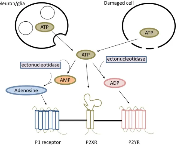
Purinergic signaling
- ATP release and extracellular ATP accumulation
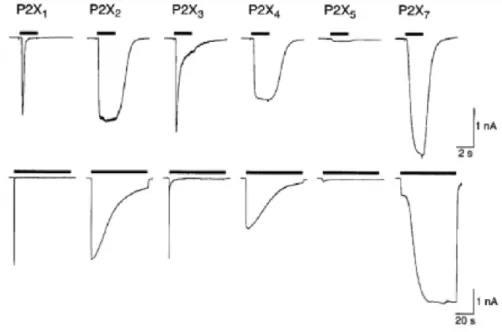
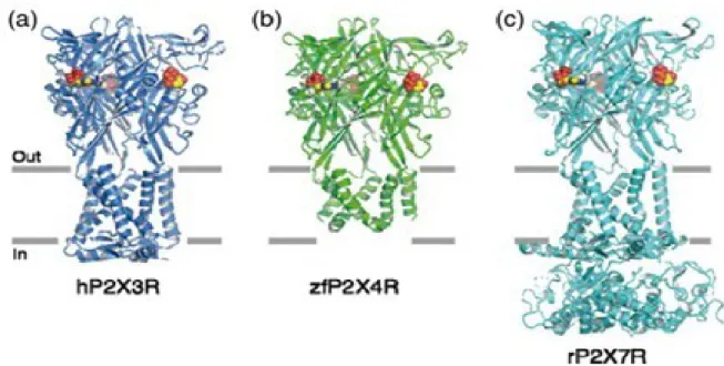
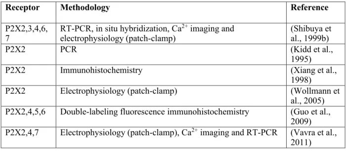
- Purinergic P2X receptors
- Specific properties of P2X2
- Specific properties of P2X4
- Specific properties of P2X7
- Purinergic P2Y receptors
P2X4 exhibits higher sensitivity to some other related antagonists, such as 5-BDBD (Donnelly-Roberts et al., 2008). The P2X7 has a number of features that distinguish it from the other P2X receptors (Surprenant et al., 1996).
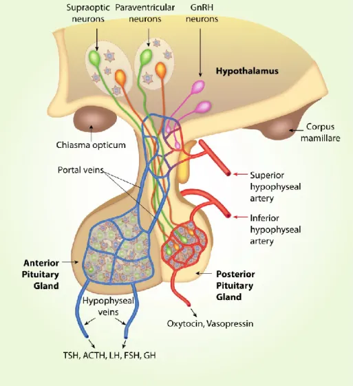
Hypothalamo-neurohypophyseal system
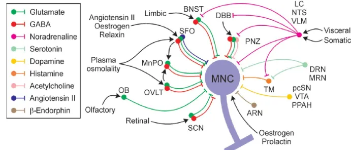
- SON, synaptic inputs and outputs
- PVN and SCN, cell composition, synaptic inputs and outputs
- Characteristics of magnocellular neurosecretory neurons
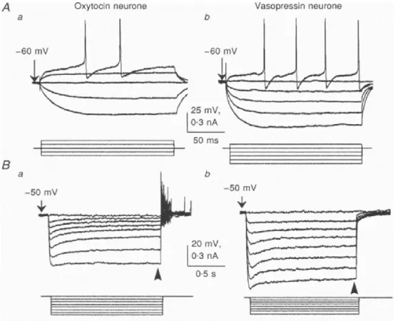
- Electrophysiological properties of magnocellular neurosecretory neurons
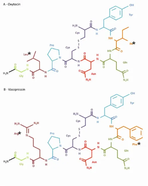
- Neurohypophyseal hormones
- Vasopressin
- Oxytocin
The perinuclear zone also contains cholinergic neurons that are believed to innervate the SON (Wang et al., 2015). The light-induced suppression of food intake was largely offset by blockade of the oxytocin receptor in the brain (Santoso et al., 2018).
Purinergic signaling in hypothalamus
- Expression and function of P2X in hypothalamus
- P2Y distribution and function in SON and SCN
- Circadian ATP release from astrocytes
This was the first evidence that P2X receptors in the PVN can modulate the sympathetic nervous system (Ferreira-Neto et al., 2013). In the SON, activation of somatic receptors induces depolarizing currents and extracellular Ca 2+ entry ( Shibuya et al., 1999a ). ATP application induced increases in cytosolic calcium and peptide release from nerve terminals (Song et al., 2006; Gomes et al., 2009).
Contribution of both P2X2 and P2X4 receptors to ATP-induced somatic current has been reported in SON neurons ( Vavra et al., 2011 ). Another study showed that ATP potentiates AVP secretion in nerve terminals through P2X2, P2X3, P2X4 and possibly P2X7 (Lemos et al., 2012). Consistent with this, ATP-induced somatic current was observed only in a subpopulation (62%) of unidentified SON neurons ( Vavra et al., 2011 ).
Purinergic receptors are also involved in astrocytic modulation of magnocellular neurosecretory cells (Espallergues et al., 2007).
Experimental protocols used to stimulate synthesis and release of hormones
- Lactation
- Sweetened condensed milk gavage
- Salt-loading
- Refeeding after fasting
ATP release from cultured cortical astrocytes also follows the circadian rhythm ( Marpegan et al., 2011 ), indicating that oscillations in ATP release are intrinsic properties of astrocytes. Acute osmotic stimuli activate oxytocin and vasopressin cells to a similar extent, and chronic dehydration or salt loading produces similar depletion of pituitary stores of both hormones (Leng et al., 1999). In hyponatremic rats, oxytocin and vasopressin cells also responded linearly to intravenous infusions of hypertonic saline, but with much lower slopes ( Leng et al., 2001 ).
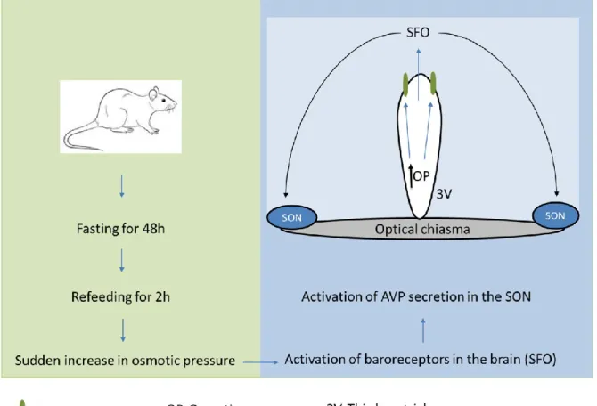
Food deprivation for 48 h causes a reliable decrease in AVP level in the SON, while little change in oxytocin concentration was detected (Burlet et al., 1992). Increased plasma osmolality upon food intake (after 48 h of fasting) stimulates baroreceptors in neurons of the subfornical organ of the brain projecting to the SON (Fig. 8) and induces increases in plasma AVP (Lucio-Oliveira et al., 2015) and oxytocin (Lucio -Olivira and Franci, 2012). It has been shown that 2–4 h of refeeding after 48 h of fasting significantly increases mRNA levels of both oxytocin and AVP in mouse hypothalamus compared to normally fed mice ( Poplawski et al., 2010 ).
An increased number and percentage of Fos-positive oxytocin neurons in the SON, with no significant alteration in oxytocin mRNA expression, was also found after refeeding (Uchoa et al., 2009).
Aims of the study
Materials and methods
Animals, tissue slices and primary cultures
- Animals
- Slice preparation
- Organotypic culture preparation
- Primary cultures of SCN astrocytes
Coronal sections of the hypothalamus (~350 μm thick) were cut from approximately 1x1 mm blocks of tissue containing the SCN using a vibratome. Slices were cultured in neurobasal medium for 7 days before starting the ATP accumulation assays to allow the slices to stabilize ( Svobodova et al., 2003 ). At this time, organotypic cultures of the SCN maintain a clear organization of SCN cells along the dorsoventral axis (Figure 12).
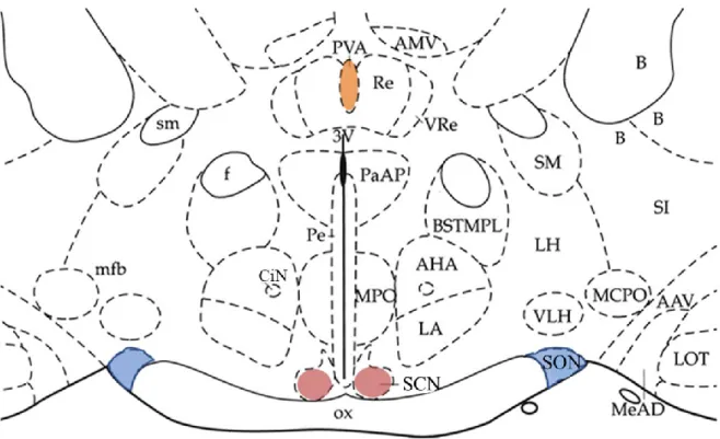
The immunopositive signal predominates in the dorsomedial subdivision of the SCN (experiments performed by Zdena Bendova, Faculty of Science, Charles University). B) Acute isolated hypothalamic slice containing SCN stained with anti-vasopressin antibody (Abcam - ab39363). The AVP immunoreactivity (red) is present at the dorsomedial subdivision of the SON. C) Illusory borders showing dorsomedial SCN (DM-SCN), ventrolateral SCN (VL-SCN), optic chiasm (OC) and the third ventricle (3V). SCN regions were dissected from ∼600-μm-thick hypothalamic slices, and cells were dissociated after treatment with trypsin according to published methods ( Watanabe et al., 1993 ; Svobodova et al., 2003 ).
Next, cells were purified on a discontinuous protein gradient, and approximately 100,000 cells were plated on coverslips coated with a 1% poly-L-lysine solution (Sigma) in 35 mm culture dishes (BD Falcon) and cultured in Neurobasal A medium with 2 % B27 supplement and 0.5 mM L-Glutamine in a humidified CO2-containing atmosphere at 37°C until use (14-21 days).

Experimental assays and techniques
- Patch-clamp recordings
- ATP luminescence assay
- Calcium imaging
- Quantitative Real-Time PCR
- Immunohistochemistry
- Drug application by rapid solution changer
- Solutions
- Chemicals
Each drug tested was added at 8:00 AM and cultures were exposed to these drugs for an incubation period of 24-48 hours. In control experiments, the protocol was the same and the medium was replaced with fresh medium without drugs every 4 h. Images of the emitted light at 515 nm were acquired with a d 40 x 0.9 NA objective and the light emission intensity was measured.
Briefly, samples were homogenized in the lysis buffer provided with the kit using ceramic beads (MagNA Lyser Green Beads) and a MagNA Lyser homogenizer (Roche Diagnostics GmbH, Germany). RNA was then extracted from the tissue homogenate by acid-phenol:chloroform extraction and subsequent column purification with a glass fiber filter containing the columns according to the manufacturer's instructions. First-strand cDNA was synthesized from up to 1 mg of isolated RNA using the SuperScriptTM VILOTTM cDNA Synthesis Kit (Invitrogen, Thermo Fisher Scientific, United States) in a 20-mL reaction volume using the random primers provided with the kit according to the manufacturer's protocol.
The patch electrodes used for whole-cell recording were filled with an intracellular solution containing (in mM): 140 KCl, 3 MgCl2, 0.5 CaCl2, 10 HEPES, and 5 EGTA, and the pH was adjusted to 7.2 with KOH.
Statistics and data analysis
- Electrophysiological data analysis
- Statistical analysis
Comparisons between two groups were performed by Student's unpaired t-test, and for multiple group comparisons, significant differences were determined by two-way analysis of variance (ANOVA) and Tukey's post hoc test using SigmaPlot v10.01 with p < and p < 0.05 . .
Results
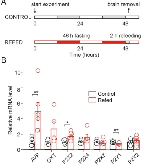
The effect of refeeding after fasting on P2X expression and function in the rat SON (Study I)
- Upregulation of AVP and P2X2 mRNA and decrease of P2Y1 mRNA in fasted/refed rats
- Basal electrophysiological properties of SON neurons in slices
- Increased amplitude of ATP-induced somatic current in fasted/refed rats
- Increased ATP-induced potentiation of GABA release in the P2X-expressing SON neurons 55
We used acutely isolated hypothalamic slices to determine the fundamental electrophysiological properties of SON neurons from fasted/refed and normally fed rats. Application of 100 μM ATP to some of the SON neurons with a voltage clamp of -60 mV caused an inward somatic current. We first examined the effect of ATP on mIPSC frequency and amplitude in SON neurons showing ATP-evoked somatic current, most likely AVP neurons (Fig. 15C and D).
Effect of ATP on a Subpopulation of SON Neurons Not Expressing Somatic P2X Next, we investigated the effect of ATP on GABAergic synaptic transmission in SON neurons that do not exhibit somatic ATP-evoked current (Figs. 17 and 18), most likely neurons oxytocin (Troadec et. al., 1998; Lack of somatic and presynaptic effects of ATP in a subpopulation of SON neurons Lack of effect of ATP on the frequency of mIPSCs in control (A) and refed (B) animals.
These data reveal that fasting/refeeding has no effect on presynaptic ATP-induced responses in SON neurons that do not express somatic P2X receptors, presumably oxytocin neurons.
Involement of P2X7 and P2Y in circadian ATP release in organotypic SCN cultures (Study II) . 59
- Dependence of extracellular ATP accumulation on P2X7 activity
- Role of pannexin-1 hemichannel in ATP release
- Dependence of extracellular ATP accumulation on P2Y1 and P2Y2
- Ca 2+ signals mediated by P2X7 and P2Y in primary cultured SCN astrocytes
- Lack of an effect of P2X7 inhibitors and mitochondrial blocker on SCN neuronal
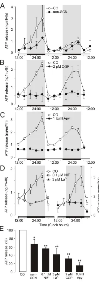
Inhibition of ATP release by the L-type Ca2+ channel blocker nifedipine (D, left panel) and non-selective voltage-gated Ca2+. Summary histogram showing cumulative ATP release in control cultures (open column) and experimental cultures (black columns) (E). ATP release was spontaneous, with a circadian rhythm that is a continuation of the endogenous rhythm in the SCN in vivo.
We examined the effects of several selective P2X7 antagonists to examine the role of P2X7 in ATP release from SCN organotypic cultures (Fig. 20). Summary histogram comparing the effects of different concentrations of AZ10606120 (E) and positive and negative modulators (J) on cumulative ATP release. These results demonstrated that P2X7 receptors play an important role in the circadian accumulation of extracellular ATP in SCN organotypic cultures, and astrocytes represent the source of P2X7-dependent circadian ATP release.
These results demonstrate that the inhibitory effects of P2X7 and mitochondrial blockers on ATP release from astrocytes are not associated with inhibition of neuronal activity.
Increased expression and function of P2X2 in SON of fasted/refed rats (Study I)
Increased expression and function of P2X2 in SON from fasted/refed rats (Study I) We found significantly increased AVP and P2X2 expression and decreased P2Y1 expression, . Sladek, 2006; Vavra et al., 2011). P2X2 activation increases AVP release from explants of the hypothalamic-neurohypophyseal system (Gomes et al., 2009; Troadec et al., 1998; Song and Sladek, 2006) and elicits somatic current in the SON neurons of hypothalamic slices (Vavra et al., 2011). Second, ATP endogenous from the posterior pituitary gland during electrical stimulation depolarizes nerve endings and enhances AVP secretion (Knott et al., 2008).
P2X2 has also been shown to be expressed on presynaptic nerve terminals in hypothalamic slices and its activation facilitates glutamate and GABA release in a subpopulation of SON neurons (Vavra et al., 2011). On the other hand, electron microscopy observations have shown that not all axons in the rat SON show P2X2 immunoreactivity (Loesch et al., 1999). P2Y receptors couple to the phospholipase C (PLC) pathway, the activation of which results in an increase in [Ca2+]i due to the release of Ca2+ from intracellular stores and stimulation of a Ca2+-dependent K+ current (Schicker et al., 2010).
GABA innervation appears to play a role in patterning the pulsatile discharge of oxytocin neurons (Voisin et al., 1995; Moos, 1995; Brussaard and Kits, 1999).
Role of P2X7 and P2Y in extracelllar ATP rhythm in the SCN (Study II)
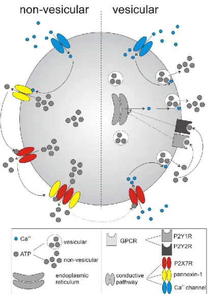
These results may also explain why circadian ATP release in SCN2.2 cells is independent of changes in intracellular Ca2+ concentrations (Burkeen et al., 2011) and occurs in cultured mouse cortical astrocytes even after disruption of the vesicular release mechanism ( Marpegan et al., 2011). However, astrocytic [Ca2+]i is high at night in SCN slices (Brancaccio et al., 2017), indicating that Ca2+-dependent release of vesicular ATP could also contribute to circadian ATP release (Fig. 26, right). ). This effect has also been shown to be related to the P2X7-dependent and Ca2+-dependent release of vesicular ATP (Ballerini et al., 1996; Suadicani et al., 2012) and glutamate (Cervetto et al., 2013) in astrocytes.
Finally, increases in [Ca2+]i can activate pannexin-1 hemichannels (Stout et al., 2002), which are important for ATP release via conductance mechanisms. The involvement of P2Y1 in ATP-triggered Ca2+ signaling and Ca2+-dependent release of vesicular gliotransmitters is also well established in astrocytes (Fumagalli et al., 2003; Verkhratsky et al., 2009). We previously found that ATP application modulates the synaptic activity of SCN neurons and potentiates GABA release via presynaptic P2X2 receptors ( Bhattacharya et al., 2013 ).
Surprisingly, the glutamate-evoked increase in astrocytic [Ca2+]i in the intact optic nerve is significantly reduced in P2X7 knockout mice (Hamilton et al., 2008).
Conclusions
Ali AAH, Avakian GA, Gall CV (2020) The role of purinergic receptors in the circadian system. Lemos JR, Wang G (2000) Excitatory versus inhibitory modulation by ATP of neurohypophysial terminal activity in the rat. Loesch A, Miah S, Burnstock G (1999) Ultrastructural localization of ATP-gated P2X2 receptor immunoreactivity in the rat hypothalamic-neurohypophysial system.
Ludwig M, Leng G (2000) GABAergic projection from the arcuate nucleus to the supraoptic nucleus in the rat. Tasker JG, Dudek FE (1991) Electrophysiological properties of neurons in the region of the paraventricular nucleus in slices of rat hypothalamus. Theodosis DT, Paut L, Tappaz ML (1986) Immunocytochemical analysis of the GABAergic innervation of oxytocin- and vasopressin-secreting neurons in the rat supraoptic nucleus.
Xiang Z, Bo X, Oglesby I, Ford A, Burnstock G (1998) Localization of ATP-gated P2X2 receptor immunoreactivity in the rat hypothalamus.