The liver dysfunction and fibrosis in DDC mice were confirmed by the significant increase in the level of ALT, TBIL and an increased ratio of liver to body weight. However, TRC105 treatment itself has no significant effect on Eng expression in liver fibrosis in the DDC diet.
Introduction
Carotuximab (Tracon Pharmaceuticals Inc), also known as TRC105 (chimeric IgG1), is a monoclonal antibody (mAb) that affects Eng expression, signaling and sEng level by binding to Eng. This thesis focuses on the activity of this novel mAb on Eng expression and sEng as a circulating biomarker of liver injury following a 3,5-diethoxycarbonyl-1,4-dihydrocollidine (DDC) diet.
Theoretical part
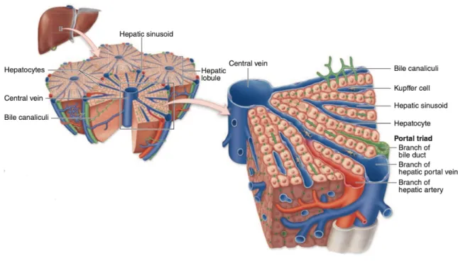
The Liver
- Liver Histology
- Liver Function
Non-alcoholic fatty liver disease (NAFLD)
- NAFLD and type 2 diabetes
- Liver fibrosis in NAFLD
NAFLD is caused by fat accumulation in the liver when there is no or little alcohol consumption, which is associated with inflammation. If scar tissue begins to replace a large portion of the normal liver tissue over time, liver function is compromised.
Endoglin (Eng)
- Structure and Function
- Endoglin role in liver fibrosis
- Soluble endoglin role in liver disorders
To summarize, short endoglin inhibits endothelial cell migration, proliferation and angiogenesis, while long endoglin stimulates them (Vicen et al., 2021). As a result, ALK1 activation stimulates cell proliferation and migration, whereas ALK5 activation inhibits these responses (Goumans et al., 2002; Lebrin et al., 2005). TGF-β is one of the most potent profibrotic cytokines, and its role in fibrotic conditions has been established ( Biernacka et al., 2011 ).
This is necessary to activate TGF-β stores in close proximity to the fibroblasts ( Walton et al., 2017 ). There are two types of the latent complexes, small and large (Taipale et al., 1996). A disulfide bond connects a small latent complex to a protein called latent TGF- β binding protein (LTBP) (Murphy-Ullrich & Poczatek, 2000). Both matrix and non-matrix proteins can be degraded by this enzyme in the extracellular environment (Nagase et al., 2006).
However, sEng is not cleaved at position 586 in preeclampsia and possibly other diseases (Rathouska et al., 2015). The elevation of sEng in preeclamptic women increased the severity of the disease and accelerated its progression.
Liver fibrosis animal models
NAPQI increases the mRNA expression of various factors and leads to the phosphorylation of extracellular signal-regulated kinase 1/2 (ERK1/2) which controls a wide range of stimulated cellular processes, mainly proliferation, differentiation and stress response (Bai et al., 2017). Toll-like receptor 4 (TLR4) signaling is activated by chronic alcohol administration and high-fat diet-induced NASH, resulting in fibrosis (Gäbele et al., 2011). Chronic feeding of DDC to rats was originally established to study Mallory bodies, which are cytoplasmic body inclusions of hepatocytes associated with NASH as well as other metabolic liver diseases (Denk et al., 2000; Fickert et al., 2003).
Glutathione and phospholipid excretion has also been shown to decrease significantly over time (Fickert et al., 2007). In the same study, the DDC diet induced a novel xenobiotic model in mice for sclerosing cholangitis, which is caused by inflammation, and biliary fibrosis leading to bile duct scarring (Ishida et al., 2016). Overall, they found that DDC causes renal insufficiency through tubular cell damage, resulting in chronic kidney disease (Ishida et al., 2016). Oval cells, which have an oval nucleus, appear in the damaged liver after toxins inhibit hepatocyte proliferation (Yamazaki et al., 2011).
DDC inhibits mitochondrial and microsomal heme synthesis in vivo by inhibiting ferrochelatase activity (J. Y. Kim et al., 2017). In conclusion, the DDC diet is useful for testing various liver and kidney pathologies in vivo.
Carotuximab
- Structure, function, pharmacodynamic
- Carotoximab treatment
Not only does sEng have an antagonistic effect on Eng, but it also binds directly to BMP-9, inhibiting angiogenesis ( Kumar et al., 2014 ). This was discovered in a study which showed that sEng binds specifically to BMP9 and BMP10, leading to the suppression of tumor growth and blood vessel formation (Castonguay et al., 2011). In conclusion, increased levels of sEng inhibit TGF-β1 binding to its membrane receptors on endothelial cells, preventing Smad2/3 activation, inhibiting eNOS activation and eNOS-dependent vasodilation (Karzai et al., 2015; Venkatesha et al., 2006).
It was originally developed for use in oncology, aiming to target proliferating endothelial cells in the vasculature of solid tumors (Liu et al., 2014; Rosen et al., 2014). The overall outcome of TRC105 treatment was angiogenesis suppression with an increase in sEng. The treatment would be safe, with clinical activity indications, and served as a basis for further clinical development (Liu et al., 2021). It was subsequently discovered that the combination of carotuximab and the vascular endothelial growth factor (VEGF) antibody bevacizumab has sustained activity in a VEGF inhibitor-refractory patient population with a reduction in tumor density, as confirmed by CT scan (Gordon et al., 2014).
Combination therapy was found to be more effective than monotherapy in inhibiting vascular leakage (W. Shen et al., 2018).

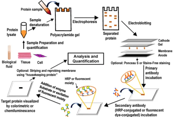
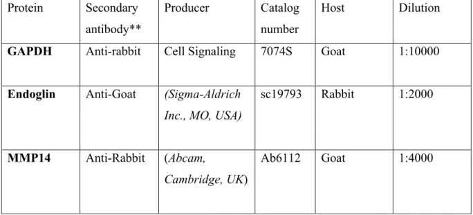
Western blot analysis
- The principle of the method
- Sample preparation
- Gel electrophoresis
- Blotting
- Blocking and antibodies
- Chemiluminescence detection
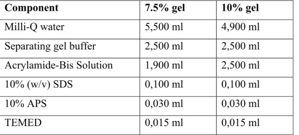
The proteins we are looking for can be found in different parts of the cells, such as the cell membrane, mitochondria or the nucleus. The protein concentration of the samples must be determined after protein extraction, and this step is extremely critical to calculate the exact amount of protein mass to be applied to each well of the gel. The only differences between these two gels are the pH of the separation and stacking buffers and also the concentration of the acrylamide; The pH of the stacking gel is 6.8 and also has a lower concentration of the acrylamide, therefore it is extremely porous, while the pH of the separating gel is 8.8 with a higher concentration of acrylamide, which causes protein distribution on the gel according to their molecular weight.
After electrophoresis, the current of electricity drives the negatively charged proteins from the gel to the membrane. The membrane is placed between the gel and the positive electrode, and therefore, traveling proteins can bind to the membrane. By masking the non-specific binding of antibodies to the membrane, the blocking agent reduces the chance of a false positive.
After the blocking step, the membrane is incubated in a certain concentration of the primary antibody to bind to the protein of interest.
Aim
Experimental part
Method
- Animals
- Western blot
- Sample preparation
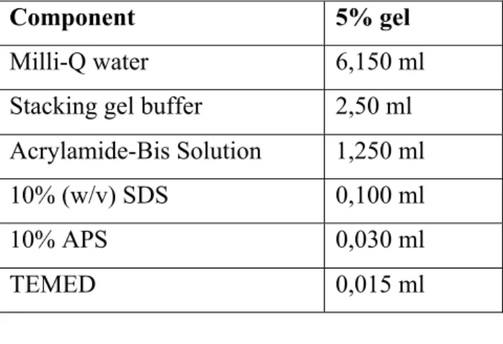
- Gel preparation
- Applications of samples on gels
- Gel electrophoresis
- Blotting
- Blocking antibody application
- Chemiluminescence detection
- ELISA
- Biochemical analysis
- Statistical analysis
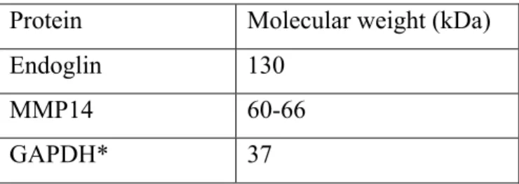
Based on the molecular weight of the desired protein, different percentages of separation gel should be made. At the end, the remaining space of the tank was filled up to the mark of the 2 gels with running buffer. While the instrument was warming up during the gel electrophoresis, two ice blocks were placed on both sides of the electrophoresis tank during the electrophoresis process.
Two soaked black sponges and a soaked white filter paper were placed on the black side of the cassette to assemble the so-called sandwich. The black side of the cassette represented the negative pole and the transparent side the positive pole. The glass slides containing the gel were carefully opened, the gel was removed and transferred to the center of the thick filter paper followed by placing the activated membrane on top.
According to the expected molecular weight of the protein of interest, the membranes were cut into strips and marked with a pencil.
Results
DDC diet induces liver impairment in mice
- The effect of DDC on the ratio of liver weight to the body
- The effect of DDC on liver enzyme
- The impact of DDC on total bilirubin
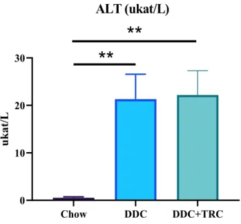
To confirm liver damage in rats fed the DDC diet, plasma ALT activity was measured. Compared with the control group, there was a significant increase in the ALT level in the plasma of rats fed the DDC diet, implying that the DDC diet causes liver damage (Figure 6). DDC-fed rats had significantly higher TBIL levels than chow-fed rats, indicating liver damage in DDC-fed rats.
Although the DDC diet successfully induced liver injury, carotuximab did not significantly affect liver weight-to-body weight ratio, ALT activity, and total plasma bilirubin ( figure 7 ).
The effect of DDC and TRC105 on Eng expression, signalling and sEng
- DDC modulates Eng expression in liver
- The impact of DDC and carotuximab on MMP14 expression
- The impact of DDC and caotuximab on the level of sEng
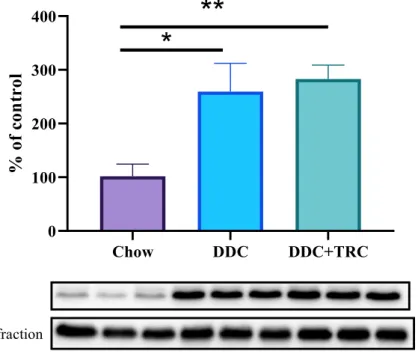
The impact of DDC and carotuximab on MMP14 expression Western blot analysis showed a significant increase in MMP14 protein expression in DDC Western blot analysis reported a significant increase in MMP14 protein expression in DDC-fed mice compared to the control group, implying liver fibrosis and Eng cleavage in DDC-fed mice. On the other hand, MMP14 protein expression did not reach statistical significance between DDC and TRC-treated groups with respect to protein expression of MMP14 (Figure 9). As expected, the significantly higher level of sEng was detected in the plasma of the DDC group mice compared to the control group, suggesting the cleavage of liver tissue Eng via MMP14 protein and release of sEng into the bloodstream (Figure 10).
Discussion
In our study, the levels of sEng in the bloodstream and Eng expression were discussed in relation to liver fibrosis (in animal model of liver cholestasis and fibrosis). Conversely, cholestatic effects of DDC depend on its ability to stimulate bile porphyrin secretion, leading to the generation of intraductal pigment plugs after 4 weeks of treatment. Surprisingly, the Western blot analysis for the samples from the three groups revealed a significant decrease in Eng protein expression in DDC-fed mice compared to the control group.
These data are contradictory to our previous work where we showed increased expression of endoglin in the liver in the NASH animal model compared to control rats. The explanation may be related to the different pathological stimulus here from the DDC diet (cholestasis model) and the NASH diet. After that, a significant increase in MMP14 protein expression in DDC-fed mice compared to the control group was confirmed by western blot, implying liver fibrosis and Eng breakdown in DDC-fed mice in which they did not differ significantly between DDC and TRC105-treated groups.
As expected, significantly higher levels of sEng were detected in the plasma of DDC group mice compared to the control group, implying Eng cleavage via MMP14 protein and sEng release into the bloodstream.
Conclusion
Endoglin is an accessory protein that interacts with the signaling receptor complex of several members of the transforming growth factor-β superfamily. Identification and expression of two forms of the human transforming growth factor-β-binding protein endoglin with distinct cytoplasmic regions. Increased expression of the transforming growth factor-beta signaling pathway, endoglin, and early growth response-1 in stable plaques.
Endoglin is a component of the transforming growth factor-beta receptor system in human endothelial cells. Identification of BMP9 and BMP10 as functional activators of the orphan activin receptor-like kinase 1 (ALK1) in endothelial cells. Key role of the Endothelial TGF-β/ALK1/Endoglin signaling pathway in human and rodent pulmonary hypertension.
Induction and maintenance of the neuronal cholinergic phenotype in the central nervous system by BMP-9.