Roles of the TRAPP-II Complex and the
Exocyst in Membrane Deposition during
Fission Yeast Cytokinesis
Ning Wang1, I-Ju Lee1, Galen Rask1, Jian-Qiu Wu1,2*
1Department of Molecular Genetics, The Ohio State University, Columbus, Ohio, United States of America, 2Department of Biological Chemistry and Pharmacology, The Ohio State University, Columbus, Ohio, United States of America
*wu.620@osu.edu
Abstract
The cleavage-furrow tip adjacent to the actomyosin contractile ring is believed to be the pre-dominant site for plasma-membrane insertion through exocyst-tethered vesicles during cyto-kinesis. Here we found that most secretory vesicles are delivered by myosin-V on linear actin cables in fission yeast cytokinesis. Surprisingly, by tracking individual exocytic and endocytic events, we found that vesicles with new membrane are deposited to the cleavage furrow relatively evenly during contractile-ring constriction, but the rim of the cleavage furrow is the main site for endocytosis. Fusion of vesicles with the plasma membrane requires vesi-cle tethers. Our data suggest that the transport partivesi-cle protein II (TRAPP-II) complex and Rab11 GTPase Ypt3 help to tether secretory vesicles or tubulovesicular structures along the cleavage furrow while the exocyst tethers vesicles at the rim of the division plane. We con-clude that the exocyst and TRAPP-II complex have distinct localizations at the division site, but both are important for membrane expansion and exocytosis during cytokinesis.
Author Summary
Cytokinesis partitions a mother cell into two daughter cells at the end of each cell-division cycle. A significant amount of new plasma membrane is needed at the cleavage furrow during cytokinesis in many cell types. Membrane expansion is achieved through the bal-ance of exocytosis and endocytosis. It is poorly understood where and when the mem-brane is deposited and retrieved during cytokinesis. By tracking individual vesicles with high spatiotemporal resolution and using electron microscopy, we found that new mem-brane is deposited relatively evenly along the cleavage furrow in fission yeast, while the rim of the division plane is the predominant site for endocytosis. The secretory vesicles/ compartments carrying new membrane are mainly delivered along formin-nucleated actin cables by myosin-V motors. Surprisingly, we find that both exocytosis and endocytosis at the division site are ramped up before contractile-ring constriction and last until daugh-ter-cell separation. We discovered that two putative vesicle tethers, the exocyst and TRAPP-II complexes, localize to different sites at the cleavage furrow to promote tethering OPEN ACCESS
Citation:Wang N, Lee I-J, Rask G, Wu J-Q (2016) Roles of the TRAPP-II Complex and the Exocyst in Membrane Deposition during Fission Yeast Cytokinesis. PLoS Biol 14(4): e1002437. doi:10.1371/ journal.pbio.1002437
Academic Editor:Frederick Hughson, Princeton University, UNITED STATES
Received:October 21, 2015
Accepted:March 15, 2016
Published:April 15, 2016
Copyright:© 2016 Wang et al. This is an open access article distributed under the terms of the
Creative Commons Attribution License, which permits
unrestricted use, distribution, and reproduction in any medium, provided the original author and source are credited.
Data Availability Statement:All relevant data are within the paper and its Supporting Information files.
Funding:This work was supported by an OSU Presidential Fellowship to NW (http://www.gradsch.
osu.edu/presidential-fellows-autumn-2014.html) and
the National Institute of General Medical Sciences of NIH grants GM086546 and GM118746 to JQW
(https://www.nigms.nih.gov/Pages/default.aspx). The
of different, yet overlapping, classes of secretory vesicles/compartments for exocytosis and new membrane deposition.
Introduction
Cytokinesis partitions a mother cell into two daughter cells following chromosome segregation. In most eukaryotes, except plants, cytokinesis relies on an actomyosin contractile ring, the con-striction of which in coordination with plasma-membrane invagination forms the cleavage fur-row [1,2]. A significant amount of new plasma membrane is needed for cytokinesis in many cell types [3–5]. Membrane expansion is under sophisticated regulation of the exocytic and endocytic pathways [6–8].
During exocytosis, post-Golgi secretory vesicles mostly travel on actin or microtubule cyto-skeleton to their destination [9,10]. Once vesicles approach the target membrane, a series of reactions trigger the vesicle-membrane fusion: tethering, docking, priming, SNARE complex assembly, and fusion [11]. Tethering determines the sites and specificity of vesicle fusion. Vesi-cle tethers physically attach vesiVesi-cles to the target membrane over a distance and promote the subsequent fusion processes [12,13]. The pairing between v-SNARE on the vesicle and t-SNARE on the plasma membrane provides the force for the vesicle-membrane fusion [14,15]. On the other hand, branched actin filaments nucleated by the Arp2/3 complex provide the force for membrane invagination during clathrin-mediated endocytosis [16–18]. Given that endocytosis removes membrane from the plasma membrane, it is intriguing that cytokinesis is inhibited or delayed when endocytosis is blocked by drug treatment or in endocytic mutants [19–21]. In mammalian cells, recycling endosomes, which have irregular tubulovesicular shapes [22], deliver retrieved membrane from endocytosis back to the cell surface for reuse [23,24]. Therefore, close investigation of exocytic and endocytic events during cytokinesis is of great interest.
Although studies from different systems have not reached a consensus, it is believed that new plasma-membrane insertion in most animal cells and budding yeast is biased towards the leading edge of the cleavage furrow and that the exocyst complex tethers vesicles delivering the new membrane for cytokinesis [7,25–27]. These are consistent with the unified view of cytoki-nesis that the contractile ring guides and coordinates membrane invagination [28]. However, these paradigms have not been rigorously tested using live-cell imaging with high spatiotempo-ral resolution.
The octameric exocyst is the main tether of vesicles at the plasma membrane in all eukary-otes [29–31] and the only known vesicle tether functioning during cytokinesis in animal cells and fungi [32–34]. Besides the exocyst, many other vesicle tethers in two groups are involved in intracellular vesicle trafficking: elongated coiled-coil tethers and a variety of multisubunit tethering complexes (MTCs) including the transport protein particle (TRAPP) complexes [13,35,36]. Initially discovered in budding yeast, the three forms of TRAPP complex (TRAPP-I, -II, and -III) share six core components but have distinct functions [37,38]. The TRAPP-II complex is proposed to regulate intra-Golgi, endosome-Golgi, and Golgi-exit trafficking in budding yeast and plant cells [37,39,40] but has not been shown to function in cytokinesis in yeast or mammalian cells. Interestingly, TRAPP-II is required for cleavage furrow ingression in male meioticDrosophilacells [41], and TRAPP-II and exocyst complexes play sequential and overlapping roles in plant cytokinesis [42]. However, it was untested in these studies whether the TRAPP-II affects vesicle tethering in cytokinesis. Furthermore, the specificity of the vesicles that TRAPP-II potentially tethers or helps to tether to the cleavage furrow was unknown. Competing Interests:The authors have declared
that no competing interests exist.
The fission yeastSchizosaccharomyces pombeis an excellent model organism for studying cytokinesis because the principle mechanisms and the proteins involved are largely conserved from fission yeast to humans [2,43]. InS.pombe, the contractile ring is assembled through the condensation of cytokinesis nodes, the ring precursors at the cell equator, into a compact ring [44–47]. The compact ring matures by recruiting more cytokinesis proteins during anaphase B and begins constricting after chromosome segregation [44]. Ring constriction is coupled with invagination of the plasma membrane and formation of a trilaminar septum (primary septum sandwiched by two secondary septa). Digestion of the primary septum leads to cell separation after septum matures.
Rod-shapedS.pombecells have a diameter of ~3.5μm and must add ~19μm2of new
mem-brane to make a cleavage furrow during cytokinesis. A typical secretory vesicle in yeast has a surface area of ~0.02 to 0.03μm2[48,49]. Thus, new membrane from>600 secretory vesicles is needed for cytokinesis if assuming secretory vesicles are the only membrane source and not considering membrane loss due to endocytosis. How this immense number of vesicles are brought to the division site is unknown. In fission yeast, the exocyst localizes to the rim of the cleavage furrow throughout cytokinesis, thus it is separated from the constricting ring [33,50– 52]. It is known that the exocyst targets the secretory vesicles carrying the hydrolytic glucanases to the division site for daughter cell separation [33,50]. However, it remains untested whether vesicle fusion and membrane deposition are limited to the rim of the cleavage furrow by the localization of exocyst.
Here we find that secretory vesicles/compartments are delivered to the cleavage furrow evenly during contractile-ring constriction by actin cytoskeleton and myosin-V motor, rather than predominantly to the leading edge adjacent to the ring or to the rim of the division plane as predicted. In contrast, endocytosis is more active near the rim of the cleavage furrow. In addition, our data also indicate that the exocyst and the TRAPP-II complex have distinct locali-zations at the division site but both affect vesicle tethering for plasma-membrane expansion and cargo delivery during cytokinesis.
Results
Secretory Vesicles Start to Deliver New Membrane to the Future
Division Site before Contractile-Ring Constriction
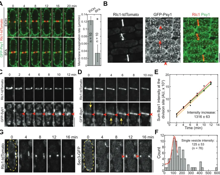
Consistent with previous studies on exocytosis in fission yeast [33,50], we found that exocytosis is critical for plasma-membrane invagination and contractile-ring constriction during cytoki-nesis. Brefeldin A (BFA; blocking endoplasmic reticulum [ER] to Golgi trafficking) treatment of wild-type (wt)S.pombecells and mutation in exocyst subunit Sec8 [33] both significantly slowed down plasma-membrane invagination (marked with GFP tagged t-SNARE Psy1 [53]) and ring constriction (marked with tdTomato tagged myosin regulatory light chain Rlc1) (Figs 1A,S1A and S1B).
intensity of Bgs1 puncta that moved to the division site in a fast and directional manner (Fig 1D, arrows;S1D Fig), we calculated that11 Bgs1 containing vesicles were delivered to the division site per minute during ring maturation (Fig 1E and 1F). Since secretory vesicles with other cargos are delivered to the division site during this time [57], these data suggest that vesi-cle tethering and deposition begin before vesi-cleavage-furrow ingression. Indeed, the exocyst, the only known vesicle tether at the plasma membrane in fungi (seeIntroduction), localized to the division site right after node condensation into a compact ring (Fig 1G).
A
B
E
F
0 2 4 6 8 10 12 min
Rlc1-tdT
o
mato
GFP-Syb1
C
Rlc1-tdTomato GFP-Psy1 Rlc1 Psy1
Sum Bgs1 intensity at the division site (AU, x 10
3) 0 2 4 6 8 10 12
0 100 200 300 400 500 600 Intensity/Bgs1 punctum (AU)
Single vesicle intensity: 125 ± 53 (n = 76)
Count
0 4 8 12 16 20 min
BF A GFP-Psy1 Rlc1-tdT omato 0 0.02 0.04 0.06 0.08 0.1 0.12 0.14
Membrane invagination rate (µm/min)
EtOHBFA
n = 10 n = 10
D
Rlc1-tdT
o
mato
GFP-Bgs1
0 2 4 6 8 10 min
EtOH
G
0 4 8 12 16 min
Rlc1-tdT o mato 0 5 20
0 2 4 6 8 10 12 14 15
10
Time (min) Intensity increase:
1316 ± 63
*
0 4 8 12 16 min
Sec3-GFP
Fig 1. Exocytosis is important for cytokinesis, and vesicles are delivered to the division site during contractile-ring maturation.(A) Time course in min (middle focal plane; left panels) and quantification (right panel) of ring constriction and membrane invagination in cells expressing GFP-Psy1 and Rlc1-tdTomato (strain JW2402, seeS1 Tablefor details) treated with either EtOH or BFA (seeMaterials and Methods). Time 0 marks the start of the movie.
*,p<0.05 compared with the control from two-tailedttest in this and other graphs. (B) Psy1 (green) concentrates to the medial cortex during ring (red) maturation, during which the diameter of a compact ring has no changes (indicated by arrowheads, compared to the cell in ring-assembly stage marked by an arrow). (C) Time course of the appearance and localization (arrowheads) of the v-SNARE Syb1 at the division site (sum projection) during ring maturation. (D) Time course (sum projection) and (E) plot (of sum intensity) of accumulation ofβ-glucan synthase Bgs1 at the division site during ring maturation. Arrowheads mark division-site localization and arrows mark Bgs1 vesicles in (D). Intensity increase rate (mean±standard deviation [SD]) in min is indicated (n= 3 cells). (F) Histogram showing the intensity of single Bgs1 vesicles imaged with the same setting as for the cells in (D, E). Mean±SD of the first peak (possibly a single vesicle) after fitting with a Gaussian distribution is shown. (G) Time course showing localization of the exocyst subunit Sec3 to the division site during ring maturation (marks with Rlc1 in the same cell) in the middle focal plane. The cell boundaries are marked with broken lines in this and some other figures. Bars, 5μm.
New Membrane Deposition Sites at the Cleavage Furrow Are Not
Biased by the Locations of the Exocyst or the Contractile Ring
We next examined where new membrane is deposited at the cleavage furrow. As reported [33,50–52], the exocyst localized to non-constricting rings at the rim of the cleavage furrow during membrane invagination and ring constriction (Fig 2A), similar to septins [58], which seems to contradict the prevailing model in animal cells that new membrane is predominantly inserted at the leading edge of the cleavage furrow, behind the constricting ring (see Introduc-tion). To elucidate the discrepancy, we tracked the directional movement of v-SNARE Syb1 labeled post-Golgi vesicles towards the division site during ring constriction (Fig 2B–2G;S1 Video, middle panels). Surprisingly, individual punctum could travel to the regions close to the constricting ring (Fig 2B and 2C), to the rim of the division plane where the exocyst localizes, or to other locations along the cleavage furrow (S1 Video, middle panels). Given the heteroge-neous shapes and sizes of Syb1 puncta (Fig 2B), Syb1 might label multiple secretory compart-ments including but not limited to the typical spherical secretory vesicles.
To investigate quantitatively the docking sites for incoming secretory vesicles and/or other Syb1 labeled secretory compartments that travel to the division site, we bleached the Syb1 sig-nal around the division site on a single focal plane and tracked the incoming Syb1 puncta (Fig 2D–2F;S2 Video). Once at the division site, the Syb1 signal often remained still for a few sec-onds before spreading out (Fig 2D, yellow arrowheads), which may indicate vesicle tethering/ fusion. We detected no obvious lateral movement of Syb1 signal along the division plane (S2 Video), indicating that the last tractable location of vesicle was likely the final docking and fusion site. The Syb1 labeled secretory vesicles/compartments were deposited relatively evenly between the constricting ring and the rim of the division plane (Fig 2F), suggesting that the vesicle docking sites at the cleavage furrow are not biased by the locations of exocyst or con-tractile ring.
We were able to track ~11 Syb1 puncta travelling to the division site per minute on a single focal plane when the division-site Syb1 signal was unperturbed and eight when bleached (Fig 2G). Typical spherical secretory vesicles in wt and exocyst mutantsec8-1had similar diameters (90 ± 10 nm) under electron microscopy (EM) (Fig 2H). Their calculated surface area is ~0.025μm2. Thus,0.25μm2new membrane from the secretory vesicles is delivered to the
division site within single focal plane during ring constriction if we assume all the travelling Syb1 puncta are spherical secretory vesicles. We also detected elongated, tubulovesicular mem-brane structures close to the plasma memmem-brane in EM thin sections (S2A Fig, arrowheads), which resemble recycling endosomes in mammalian cells [22] and the late-Golgi cisternae in budding yeast [59]. The heterogeneity of the shape, size, and intensity of delivered Syb1 puncta (Fig 2B) suggest these tubulovesicular structures also contribute to cytokinesis.
main source of membrane inS.pombecytokinesis. Thus, our Psy1 data also support that new membrane is inserted evenly at the cleavage furrow.
We also tracked Syb1 vesicle movement and Psy1 dynamics during septum maturation, the stage that the ring has fully constricted and disassembled but before daughter-cell separation (S2B–S2D Fig). Interestingly, a large amount of post-Golgi vesicles/compartments still deliv-ered relatively evenly to the division plane (S2B and S2C Fig). Consistently, Psy1 signal recov-ered evenly after the division-site signal was bleached (S2D Fig). Together, vesicles are delivered to the division site throughout cytokinesis until cell separation.
EM images of wt cells were also consistent with the even deposition of new membrane dur-ing cytokinesis. Secretory vesicles or the tubulovesicular structures appeared to be tethered to the plasma membrane (distance of vesicles to plasma membrane was within ~100 nm) along the whole cleavage furrow instead of exclusively at the leading edge or at the rim (S2A Fig, arrows for vesicles and arrowheads for tubulovesicular structures). Together, our data indicate that new membrane deposition sites at the cleavage furrow are not biased by the locations of the exocyst or the contractile ring.
For3-Nucleated Actin Filaments and the Myosin-V Myo52 Are Important
for Vesicle Trafficking to the Division Site
The fast and directional movements of Syb1 and Bgs1 vesicles (Figs1,2,S1andS2) suggest that they are transported by motors on cytoskeletal tracks. In animal and plant cells, vesicles delivered on microtubules are an important source for new plasma membrane during cytokine-sis [25,68]. In budding yeast, myosin-V delivers secretory vesicles on actin cables to the bud for polarized growth and to bud neck for cytokinesis [69–71]. In fission yeast, although both actin filaments and microtubules are involved in polarity establishment and maintenance [72–74], it is unknown which one is more important for vesicle transport to the division site for mem-brane expansion during cytokinesis.
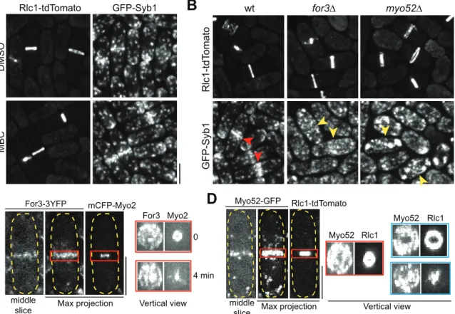
We first investigated whether microtubules affect the delivery of Syb1 vesicles during cell division (Fig 3A). Treatment of wt cells with methyl benzimidazole-2-yl carbamate (MBC) to disrupt microtubules had no detectable effects on Syb1 localization at the division site (Fig 3A). In contrast, Syb1 did not concentrate at the division site during ring constriction in ~93% cells with a deletion of the formin For3 (Fig 3B, yellow arrowheads;n= 58 cells), which nucleates actin filaments in actin cables [74]. The myosin-V motor Myo52 is critical for transportingβ -glucan synthase Bgs1 and other cargos to cell tips in interphase and to the division site in divid-ing cells [75,76]. As expected, Syb1 concentration at the division site was abolished in ~90% myo52Δcells during ring constriction (Fig 3B, yellow arrowheads;n= 67 cells), indicating that Myo52 is a motor that drives the actin-dependent movement of secretory vesicles/compart-ments to the division site. The fact thatfor3Δcells still finish cytokinesis with a delay in ring
Trafficking of Syb1 vesicles to the division site in a single focal plane after photobleaching Syb1 signal within the white-boxed region in a cell with a partial septa (DIC) and a constricting Rlc1 ring. Two examples of traveling vesicles from the same cell are marked with arrowheads. The yellow arrowheads mark a vesicle remaining still for 1.5 s before signal spreading out, suggesting vesicle tethering/fusion. (E) Enlarged red- and blue-boxed regions in (D). (F) Distribution of final tractable deposition sites of Syb1 vesicles relative to the position of constricting ring (at the beginning of the movie) from 7 cells (color coded) in 2-min movies after Syb1 signal at the division site was bleached as in (D).X- andy-axes are along the division site and cell long axis, respectively. The ring position (the diameter marked by the color lines) is displaced along they-axis away from its real position aty= 0 for clarity. (G) Numbers of tractable Syb1 vesicles per min at the middle focal plane when the division site Syb1 signal is unperturbed or bleached. (H) EM images (left) and diameters (right) of secretory vesicles (arrows) in wt andsec8-1mutant. (I-K) Dynamics of Psy1 on the plasma membrane revealed by FRAP. Red boxes mark the bleached regions, which were bleached at time 0. (I) Psy1 dynamics at the non-growing side of an interphase cell (left panel) or the division site of a cell with
constricting ring (right panel), which is marked by Rlc1 taken at -1 min. (J) Recovery of Psy1 intensity after photobleaching at time 0. Mean±standard error of
the mean (SEM) is plotted. (K) The center of the bleached region (arrows) at the division site (enlarged on the right) does not move in any directions in cells with a constricting ring. Bars, 5μm.
constriction [77] andmyo52Δcells displayed prolonged ring constriction and septum matura-tion (S3A and S3B Fig) is consistent with the finding that vesicles can reach their destination by actin-independent random walk [56,78]. Thus, For3 nucleated actin cables are critical for efficient delivery of secretory vesicles/compartments by myosin-V motors to the division site.
Next we observed For3 and Myo52 distribution at the division site. We and others found that both For3 and Myo52 dynamically localized to the division plane during cytokinesis [72,75,77,79], forming a disk structure, although the signals were not smooth and continuous (Fig 3C and 3D). This motivated us to track Myo52 movements towards the division plane at different cytokinesis stages. Myo52 puncta docked evenly along the division plane during ring constriction and septum maturation (S3C–S3E Fig;S4 Video, middle and right panels), resem-bling v-SNARE Syb1 (Figs2FandS2C). Together, these data support our hypothesis that vesi-cle tethering and fusion with plasma membrane happen all over the division plane.
Endocytosis Is More Active at the Rim of the Cleavage Furrow during
Cytokinesis
The balance between exocytosis and endocytosis controls plasma-membrane expansion during polarized growth and cytokinesis (seeIntroduction). Thus, we investigated how endocytosis contributes to cytokinesis, which was unclear partly due to insufficient investigation into the
A
B
C
D
for3∆ myo52∆
wt
Rlc1-tdT
o
mato
GFP-Syb1
Vertical view For3 Myo2
For3-3YFP mCFP-Myo2
middle
slice Max projection
0
4 min
middle
slice Max projection
Myo52-GFP Rlc1-tdTomato
Vertical view Myo52 Rlc1
Myo52 Rlc1
DMSO
Rlc1-tdTomato GFP-Syb1
MBC
Fig 3. Actin filaments nucleated by the formin For3 and myosin-V motor Myo52 are important for vesicle delivery to the division site.(A) Syb1 localization at the division site does not depend on microtubules revealed by MBC treatment. (B) Syb1 localization at the division site depends on For3 and Myo52. Yellow arrowheads mark examples of greatly reduced Syb1 signal at the division site in mutant cells compared with wt cells (red arrowheads). (C, D) For3 and Myo52 localize to the whole cleavage furrow as a washer or disk during ring constriction (labeled with myosin-II heavy chain Myo2 or light chain Rlc1). Localization of For3 or Myo52 in the middle focal plane, maximum-intensity projection of a z-stack spaced at 0.2μm, or vertical view of the red-boxed regions of the same cell (and 4 min later for For3) or from another two Myo52 cells with different extent of ring constriction (blue boxes). Bars, 5μm.
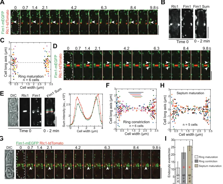
precise locations of endocytosis during cell division. InS.pombe, active endocytic sites are at growing cell tips during interphase and at the cell equator during cytokinesis [80]. The appear-ance of actin cross-linker fimbin Fim1 at endocytic patches represents the growth of actin fila-ments surrounding the endocytic pits [81,82], which can be used to mark the endocytic sites. Using Rlc1 to label cytokinesis nodes and the contractile ring, we found that endocytic patches started to assemble at the cell equator during ring maturation (Fig 4A–4CandS5 Video), which is consistent with the timing of the relocation of other proteins involved in endocytosis from cell tips to the division site [80,83–85].
0 1 2 3 4
Cell width (µm) 1.5
2 2.5 3
Sum intensity (au., x10
6) -3 -2 -1 0 1 2 3
0 0.5 1 1.5 2 2.5 3 3.5 Cell width (µm)
Cell long axis (µm)
A
B
G
C
D
E
F
H
Rlc1 Fim1 Fim1 Sum
Time 0 0 - 2 min
0 0.7 1.4 2.1 4.2 6.3 8.4 9.8S
Fim1-mEGFP Rlc1-tdT
o
mato
0 0.7 1.4 2.1 4.2 6.3 8.4 9.8S
Fim1-mEGFP Rlc1-tdT
o
mato
DIC Rlc1 Fim1 Fim1 Sum
Time 0 0 - 2 min
Ring maturation -3 -2 -1 0 1 2 3
0 0.5 1 1.5 2 2.5 3 3.5 Cell width (µm) Cell long axis (µm) Ring constriction
n = 6 cells n = 6 cells
0 0.7 1.4 2.1 4.2 6.3 8.4 9.8S
Fim1-mEGFP Rlc1-tdTomato DIC -3 -2 -1 0 1 2 3
0 0.5 1 1.5 2 2.5 3 3.5
Cell long axis (µm)
Cell width (µm) Septum maturation
n = 5 cells
I
Ring maturation Ring contriction Septum maturation 0 5 10 15 20 25 30 Endocytic events/minn = 6 n = 6 n = 6
* *
Fig 4. Sites of endocytosis at different stages of cytokinesis.Formation of endocytic patches labeled with fimbrin Fim1 at the division site during ring (Rlc1) maturation (A–C), ring constriction (D–F), and septum maturation (G–H). (A, D, G) Montages showing endocytic patch assembly (arrowheads) viewed
in the middle focal plane. (B, E) Sum intensity projection of 498 Fim1 images in the middle focal plane in a 2-min movie along with Rlc1, Fim1, and DIC images at time 0. The right panel in E: line scans (color-coded) of Fim1 intensity as marked on the sum image to the left. (C, F, H) Distribution of endocytic patch assembly sites from multiple cells (color-coded) during ring maturation, ring constriction, and septum maturation. The dashed line aty= 0 marks the division site. (F) The ring (the diameter marked by the color lines) is displaced along they-axis away from its real position aty= 0 for clarity. (I) Numbers of assembled Fim1 patches in the middle focal plane per minute at different stages of cell division.*,p<0.05 compared with ring maturation stage. Bars, 5μm.
Next we examined the distribution of endocytic sites during cell division using the emerging locations of Fim1 patches. During ring maturation, the predominant site for endocytic activity appeared as a broad band on the plasma membrane at the cell equator around the contractile ring (Fig 4B and 4C). During ring constriction, Fim1 emerged from both the cortex adjacent to the division plane (within ~2μm) and the cleavage furrow (Fig 4D–4F). However, more
endo-cytosis occurred on or near the rim of the cleavage furrow (Fig 4E and 4F). Endocytosis was even more active in later stage of cytokinesis when a full septum has formed (Fig 4G and 4H; S5 Video). During this stage, more endocytic events were detected (Fig 4I) and slightly more endocytic patches formed at the interior of the division plane (Fig 4H). Together, frequencies and locations of endocytic events appeared to be temporally regulated.
Putative Vesicle Tether TRAPP-II Complex Localizes to Trans- and
Post-Golgi Secretory Vesicles/Compartments and the Division Site
during Cytokinesis
Our finding that new membrane is deposited along the whole cleavage furrow (Figs2andS2) but the exocyst concentrates at the rim of the division plane (Fig 2A; [33,50,51,56]) suggests that other vesicle tethers or tethering factors contribute to cytokinesis besides the exocyst. The TRAPP-II complex has been proposed to regulate cytokinesis in plants andDrosophila[41,42]. Thus, we tested whether TRAPP-II is involved in cytokinesis to help tethering vesicles in fission yeast. Trs120, a specific component of the TRAPP-II complex [86], localized to punctate struc-tures (Fig 5A–5D) and was slightly concentrated at the growing cell tips in interphase cells (Fig 5A, arrowheads). In late anaphase B, a fraction of Trs120 puncta concentrated to cell equator, the future division site (Fig 5A, arrow;S6 Video). During ring constriction and septum forma-tion, Trs120 puncta localized along the division plane (Fig 5A, vertical views), similar to For3 and Myo52 (Fig 3C and 3D).
Trs120 puncta resemble secretory vesicles, we reasoned that they might participate in vesi-cle trafficking. Indeed, Trs120 puncta partially co-localized with the trans-Golgi network marker Sec72 [87] and secretory vesicles/compartments (β-glucan synthase Bgs4, a vesicle cargo [57];Fig 5B, white arrows;Fig 5E), but did not overlap with cis-Golgi (Golgi mannosyl-transferase complex component Anp1 [87];Fig 5B and 5E) or transitional-ER (COPII vesicle coat protein Sec24 [87]). ~94% of Trs120 puncta also colocalized with Syb1 labeled trans- or post-Golgi vesicles/compartments (Fig 5C, arrows;Fig 5E) and traveled with Syb1 to the divi-sion site during cytokinesis (Fig 5C, arrowheads) although only ~50% Syb1 puncta contained Trs120 (n= 179 puncta). Trs120 may also be involved in cargo delivery to cell tips during interphase, given that Trs120 and Bgs4 moved together on some puncta to cell tips (Fig 5D, arrowheads). The division-site accumulation of Trs120 largely depended on the formin For3 (Fig 5F).
A
B
C
G
H
0 0.5 1 1.5 2 2.5 s
Rlc1 Trs120-3GFP for3∆ DIC Trs120-3YFP Trs120-3YFP wt DIC
Cell width (µm)
Cell long axis (µm)-2
-1 0 1 2
0 0.5 1 1.5 2 2.5 3 3.5 n = 8 cells
Septum maturation
0 0.5 1 1.5 2 2.5 s Trs120-3GFP
Rlc1
Trs120-tdTomato GFP Trs120 GFP
Sec72-GFP T rs120-tdT o mato trans-Golgi GFP-Bgs4 T rs120-tdT o mato vesicle cargo Anp1-GFP T rs120-tdT o mato cis-Golgi GFP-Syb1 T rs120-tdT omato Syb1 T rs120
0 1 2 3 4 s
0 20 40 60 80 100 T
rs120 colocalizing puncta (%)
w/ Anp1w/ Sec72w/ Syb1
0 1 2
GFP-Bgs4 T rs120-tdT o mato Bgs4 T rs120
3 4 5 s
D
F
E
1 2 3
1 2 3 Middle DIC Sad1 -mCFP T rs120-3YFP Max Vertical view
Interphase G2/M Anaphase B Ring constriction maturationSeptum
0 20 40 60 80 100
Septating cells with
T
rs120
signal at the division site (%)
wtfor3∆
n = 106 n = 1
18 n = 204 n = 186 n = 165
Cell long axis (µm)
Ring Constriction -2 -1 0 1 2
0 0.5 1 1.5 2 2.5 3 3.5 Cell width (µm)
n = 9 cells
Fig 5. The TRAPP-II complex colocalizes with post-Golgi vesicles and travels to the division site during cytokinesis.(A) Trs120 localization to cell tips (arrowheads) and division site (arrow) during the cell cycle marked with spindle pole body protein Sad1. Images of DIC, max intensity projection, and middle focal plane of cells are shown along with vertical views on the right of the regions marked by short red lines on the numbered cells. (B) Trs120 colocalizes with Sec72 and Bgs4 (examples marked by white arrows) but not with Anp1 labeled cis-Golgi structures. Red arrows point out the division-site signal of Trs120. (C) Trs120 colocalizes with Syb1 labeled vesicles/compartments (arrows) and travels to the division site with Syb1 (arrowheads) during cytokinesis. (D) A Trs120 punctum travels with Bgs4 to the cell tip in an interphase cell. (E) Percentage of Trs120 puncta containing Anp1, Sec72, or Syb1. (F) Trs120 accumulation at the division site (arrows) is dramatically reduced or undetectable infor3Δ. Quantification of septating cells (with partially or fully formed septa) with detectable Trs120 concentration at the division site is shown on the right. (G) Trs120 puncta (arrowheads) move to the division site during ring maturation (left) and constriction (right). The dashed horizontal line is to aid tracking Trs120. (H) Distribution of final tractable docking sites of Trs120 labeled puncta in 2-min movies during ring constriction (top) and septum maturation (bottom). The data are plotted as inFig 4F and 4H. Bars, 5μm.
TRAPP-II Mutants Display Cytokinesis Defects and Synthetic Genetic
Interactions with Exocyst Mutants
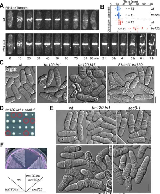
To further study the function of TRAPP-II complex in cytokinesis, we examined the phenotype oftrs120mutants. Sincetrs120is an essential gene [88], we generated a homozygous diploid strain expressing integrated Rlc1-tdTomato and then deleted one copy oftrs120. We per-formed tetrad fluorescence microscopy [77,89,90] to observe the growth and division of wt and trs120Δhaploid cells after thetrs120+/trs120Δdiploid underwent meiosis and sporulation. Haploid cells withtrs120deleted usually died after two to four generations.trs120Δcells had no obvious defects in contractile-ring assembly (Fig 6A and 6B), but often failed (Fig 6A, n= 12 cells) or had very slow (Fig 6B,n= 11 cells) ring constriction and plasma-membrane invagination, which led to premature ring disassembly, incomplete membrane insertion, and/ or cell lysis. This indicates that the TRAPP-II is involved in later stages of cytokinesis.
To facilitate rapid inactivation of TRAPP-II complex in vivo, we generated temperature-sensitivetrs120mutants using a marker reconstitution mutagenesis method [89,91]. Both trs120-ts1andtrs120-M1mutants had cytokinesis defects at 36°C with increased septation indices, formation of multi-septated cells, and cell lysis (Fig 6C).trs120depletion mutant 81nmt-trs120showed similar phenotype at 36°C (Fig 6C). Consistent with our hypothesis that the exocyst and TRAPP-II complexes have overlapping functions in vesicle trafficking, exocyst mutantsec8-1[33,49] was synthetic lethal with81nmt-trs120andtrs120-M1(Fig 6D).
trs120-ts1 sec8-1double mutant, although viable, displayed severe cytokinesis defects such as multi-septated cells at 25°C (Fig 6E) and stopped growing at 30°C (S4D Fig).trs120-ts1, trs120-M1, and81nmt-trs120also displayed additive growth defects with exocyst mutant exo70Δ(Fig 6F). Given that the two complexes had different localizations during ring constric-tion, septum formation and maturation (Figs2A,5A,S4A and S4B;S6andS7Videos), it is not surprising that Trs120 localization appeared normal in exocyst mutantsec8-1(S4E Fig) and the localization of exocyst component Sec3 was not perturbed bytrs120mutations (S4F and S4G Fig). Together, the TRAPP-II and exocyst are independent for localization to the division site but have overlapping functions during late stages of cytokinesis.
TRAPP-II and Exocyst Mutants Affect the Delivery of Cargo Proteins to
the Division Site in Distinct Patterns
To test whether TRAPP-II mutants are defective in exocytosis, we first examined the localiza-tion of v-SNARE Syb1 and the secrelocaliza-tion of acid phosphatase. Similar tosec8-1,trs120-ts1 mutant accumulated Syb1 in proximity to the division plane in dividing cells and at the cell tips in interphase cells (Fig 7A). The secretion of acid phosphatase was reduced in twotrs120 mutants although not as dramatic as insec8-1(S4H Fig), suggesting that TRAPP-II functions in exocytosis.
secretory pathway [37,39,40,86], which may explain the detection of various vesicular mem-brane structures in thetrs120mutant.
Given the different localization of exocyst and TRAPP-II at the division site (Figs2A,5A, S4A and S4B) and the accumulation of distinct secretory vesicles as well as the tubulovesicular
Fig 6. Phenotypes of TRAPP-II mutants and synthetic interactions between TRAPP-II and exocyst mutations.(A, B) Time courses (A) and quantification (B) of the contractile-ring assembly (from the appearance of cytokinesis nodes to formation of a compact ring without lagging nodes) and constriction (from the start of ring contraction until it constricts to a dot with the highest Rlc1 intensity) in wt andtrs120Δcells observed with tetrad
fluorescence microscopy. (B) The rings of ~50%trs120Δcells did not or only partially constricted during the 14-h movies, so the quantification includes only those fully constricted ring. (C) Morphological defects of threetrs120mutants in YE5S medium at 36°C. Wt,trs120-ts1,trs120-M1, and81nmt1-trs120cells were cultured at 36°C for 6, 4, 2, and 6 h, respectively. (D, E) Synthetic genetic interactions between TRAPP-II and exocyst mutations. (D) The predicted trs120-M1 sec8-1mutant (circles) cannot form colonies on YE5S plate at 25°C. (E) DIC images showing the synthetic interaction betweentrs120-ts1and sec8-1mutations at 25°C. (F) Growth oftrs120-ts1,exo70Δ, and the double mutant at 32°C on YE5S medium with phloxin B (PB), which accumulates in dead cells [131]. Bars, 5μm.
membrane structures in their mutants (Figs2Hand7C), the exocyst and TRAPP-II complex may affect recruiting vesicles with different cargos to distinct sites on the plasma membrane. To test this idea, we observed the localization of the glucanase Eng1 and the glucan synthase Bgs1, two post-Golgi cargos [55,92], insec8-1andtrs120-M1mutants. Eng1 localizes to the rim of the division plane as a ring and to the center as a bright spot (arrowheads) in wt cells during septum maturation (Fig 7E; [93]). As reported [50], Eng1 signal spread throughout the division plane with less signal at the rim insec8-1mutant (Fig 7E), although total Eng1
Fig 7. Exocyst and TRAPP-II mutants affect cargo delivery to the division site differently.(A) Syb1 accumulates at the division site or cell tips in trs120-ts1andsec8-1mutants grown in YE5S medium at 36°C for 2 h. (B–D) EM images (B, C) and quantification (D) showing abnormal accumulation of
vesicles and other secretory compartments at the division site or cell tips intrs120-M1orypt3-i5cells grown at 36°C for 4 h. (D) Quantification of secretory vesicles or round-shaped vesicle-like structures (diameter<150 nm without engulfed membrane-bound materials) accumulated in each longitude EM thin section. (E) Localization of the glucanase Eng1 to the division site intrs120-M1andsec8-1cells. Arrowheads mark the center localization of Eng1 at the division plane in wt cells. Arrows mark the remaining Eng1 at the rim intrs120-M1. Vertical views of the boxed regions are shown at the bottom corner. (F, G) Localization of glucan synthase Bgs1 in the middle focal plane at the division site intrs120-M1andsec8-1cells during septum maturation. (G) Line scans of Bgs1 intensity along the division plane (marked by arrows in F). Top, Mean±SD from multiple cells. Bottom, individual wt andtrs120-M1cells. Bars in A, E, F, 5μm.
intensity at the division site appeared normal. Intrs120-M1mutant, the Eng1 intensity at the division plane seemed to be lower than in wt andsec8-1and the central spot localization was lost in 93% cells during septum maturation (Fig 7E;n= 41 cells). These data suggested that TRAPP-II is more important for cargo delivery to the center, whereas the exocyst is more important for delivery to the rim of the division plane. Consistently,trs120-M1mutation decreased Bgs1 intensity at the center of the cleavage furrow dramatically, whereassec8-1 com-promised Bgs1 localization at the rim (Fig 7F and 7G). Together, our data indicate that the exo-cyst and TRAPP-II complexes may function at distinct spatial locations to tether (or help tether) vesicles for membrane and cargo delivery during cytokinesis.
The TRAPP-II Complex Works Preferentially with the Rab11 GTPase
Ypt3 in Cytokinesis
TRAPP complexes have been proposed to work with Rab GTPases to tether vesicles by func-tioning as their guanine nucleotide exchange factors (GEFs) [37,94,95]. Recently, the TRAPP-II was shown to activate Rab11 as a GEF inAspergillus nidulans[40]. Mammalian Rab11 GTPase is a marker for recycling endosomes that are critical for transporting internalized proteins from endocytosis back to cell surface [96,97]. In budding yeast, the recycling traffic to cell sur-face is through late Golgi [98–100], and Rab11 GTPases Ypt31/32 label late-Golgi cisternae and the secretory vesicles emerging from late Golgi in a signaling cascade to activate Sec4, the Rab8 GTPase on secretory vesicles for polarized exocytosis [101,102].
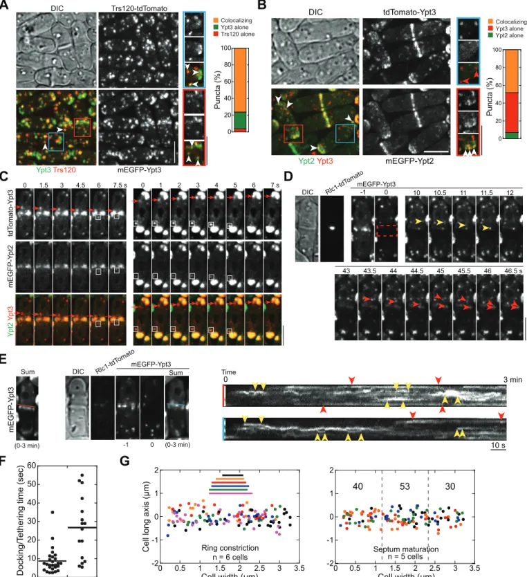
We tested whether the TRAPP-II complex works with Rab11 homologue Ypt3 [103] or Rab8 homologue Ypt2 [104] inS.pombe. Trs120 and Ypt3 co-localized in ~80% cytoplasmic puncta (n= 439 puncta from 8 cells) although more Ypt3 was on the plasma membrane (Fig 8A). However, only 14% of Trs120 puncta contained with Ypt2 (S5A Fig, arrowheads;n= 159 from 8 cells). Interestingly, Ypt2-containing puncta in the cytoplasm seemed to be more homo-geneous than Ypt3 puncta (Fig 8B). Ypt2 and Ypt3 colocalized in ~48% of the punctate struc-tures, mainly at cell tips or the division site (Fig 8B, white arrowheads), suggesting that they act in a similar signaling cascade on the secretory vesicles as in budding yeast [101]. However, ~45% of puncta contained only Ypt3 but no Ypt2 (Fig 8B, red arrowheads), suggesting that Ypt3 also functions independent of Ypt2 GTPase. Indeed, those Ypt3-only puncta also traveled to the division site during cytokinesis and to the growing cell tips during interphase (Fig 8C andS5B Fig, arrows). Given that ~70% of Ypt3 puncta (derived fromS5C Fig) colocalized with the cargoβ-glucan synthase Bgs4 (S5C Fig, arrowheads), these Ypt3-only secretory compart-ments may also be important for cargo delivery and membrane traffic, and likely resembled the tubulovesicular structures detected in EM images (FigsS2Aand7C). Together, our data sug-gest that the TRAPP-II mainly works on Rab11 Ypt3-labeled secretory compartments regard-less of whether Ypt2 is present.
yeast (~18 s; [105]). Ypt3-labeled secretory vesicles or recycling endosome equivalents were delivered to the whole division plane likely with a small bias towards the center of the maturing septum (Fig 8G;S9 Video), similar to Trs120 (Fig 5G and 5H). Thus, TRAPP-II may specifi-cally recognize Ypt3 containing secretory structures throughout the cleavage furrow.
Additional lines of genetic and cellular evidence further support that the TRAPP-II complex and Rab11 GTPase Ypt3 work together in cytokinesis. First, similar to its homolog inA. nidu-lans[40], TRAPP-II is important for Ypt3 localization since cell tip and division-site localiza-tion of Ypt3 was dramatically reduced intrs120mutants, although Ypt3 global level was not affected by thetrs120mutation (S6A Fig). Second, Trs120 was more concentrated at the divi-sion site (red arrow) and cell tips (yellow arrow) inypt3-i5mutant (S6B Fig), possibly due to delayed fusion of TRAPP-II containing vesicles to the plasma membrane. Third,ypt3-i5 mutant accumulated GFP-Syb1 at the active growth sites (S6C Fig, arrows), similar totrs120 mutant (Fig 7A). Fourth, liketrs120mutant during cytokinesis,ypt3-i5cells accumulated vesi-cles (arrowheads) and tubulovesicular structures (arrows) at the division site (Fig 7C and 7D; S6D Fig), with similar distance to the plasma membrane (S6D Fig, yellow arrows and arrow-heads), suggesting that the events after tethering, likely the fusion step, was affected byypt3 mutation. Lastly,trs120mutations displayed synthetic genetic interactions withypt3-i5 muta-tion (S6E and S6F Fig). Collectively, our data indicate that the TRAPP-II complex works with Rab11 GTPase Ypt3 for membrane deposition along the cleavage furrow during cytokinesis.
Ectopic Targeting of Secretory Vesicles and Tubulovesicular Membrane
Structures to Mitochondria by the TRAPP-II Complex
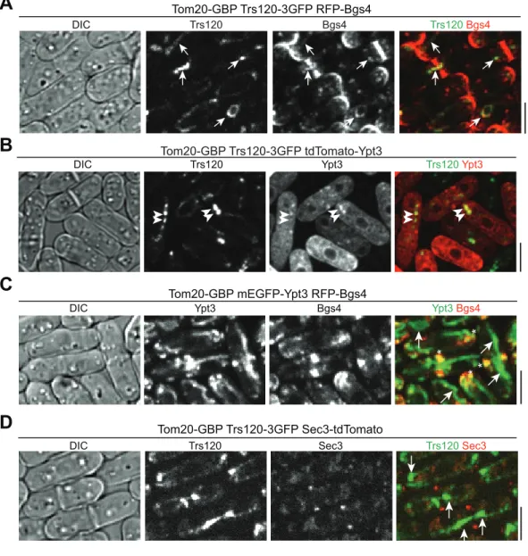
Although direct evidence from in vitro reconstitution that the exocyst tethers secretory vesicles to the plasma membrane is missing, it has been shown in budding yeast that ectopic targeting of Sec3 to mitochondria or peroxisomes results in the recruitment of secretory vesicles to these surrogate organelles [106]. To test whether TRAPP-II complex can target and/or tether Golgi-derived secretory vesicles/compartments, we mislocalized Trs120-3GFP to mitochondrial sur-face using the GFP-binding protein (GBP; [107]). We tagged the mitochondrial outer mem-brane protein Tom20 with GBP at its cytosol-exposed COOH-terminus [106]. GBP recognizes most GFP variants but not RFP or Tomato-tagged fluorescence proteins (S7A and S7B Fig; [107]). We used RFP or tdTomato tagged proteins to check whether other components of TRAPP-II complex or cargos of secretory vesicles/compartments are mistargeted by misloca-lized Trs120-3GFP. Consistent with the reported central role of Trs120 in TRAPP-II assembly [108], mislocalized Trs120 could recruit other components of TRAPP-II complex, such as Trs130, to mitochondria (S7A Fig). Importantly,β-glucan synthase Bgs4 labeled secretory vesi-cles/compartments colocalized with Trs120 on mitochondria (arrows) in 75% oftom20-GBP trs120-3GFPcells (n= 324 cells;Fig 9A), suggesting that the TRAPP-II complex can recruit and possibly tether vesicles.
Consistent with our conclusion that the TRAPP-II complex works preferentially with the Rab11 GTPase Ypt3, we detected mitochondrial localization of Ypt3 in 80% oftom20-GBP trs120-3GFPcells (n= 211 cells;Fig 9B, arrowheads). In contrast, mislocalized Trs8502 (the
furrow, respectively. (E) Left, sum projection of the 3-min movie for the cell in (D). Middle panels, sum projection of Ypt3 signal for another cell during septum maturation along with Rlc1 and DIC images at time 0 and Ypt3 images at time -1 and 0 min. Right panels, kymographs along the color-coded lines marked on the sum projections on the left (to avoid blocking Ypt3 signal, the lines were moved upward slightly) showing different docking/tethering times for Ypt3 puncta at the leading edge (or center) or the rim of the division site. Pairs of red and yellow arrowheads mark Ypt3 with long and short dwell time, respectively. (F) Docking/tethering time for Ypt3 puncta at the center or the rim of the division site of 10 cells during late stage of ring constriction or early stage of septum maturation. (G) Distribution of final tractable docking sites of Ypt3 labeled puncta in 3-min movies during ring constriction (left) and septum maturation (right, numbers of vesicles/compartments delivered to each region are shown). The data are plotted as inFig 4F and 4H. Bars, 5μm.
TRAPP-III component Trs85) did not lead to mitochondrial localization of Ypt3 (S7C Fig, arrows). Ectopically targeted Ypt3 did not recruit Bgs4 to mitochondria (Fig 9C), suggesting that the Rab GTPase itself was not sufficient to tether vesicles. In addition, the exocyst was not mistargeted by ectopically localized TRAPP-II (Fig 9D), although TRAPP-II and exocyst com-plexes physically interact in plant cells [42]. Together, these data suggest that the TRAPP-II complex works together with Ypt3 to promote vesicles/compartments tethering, although whether TRAPP-II functions as a GEF to activate Ypt3-dependent tethering activities or as a direct tether itself is still unclear.
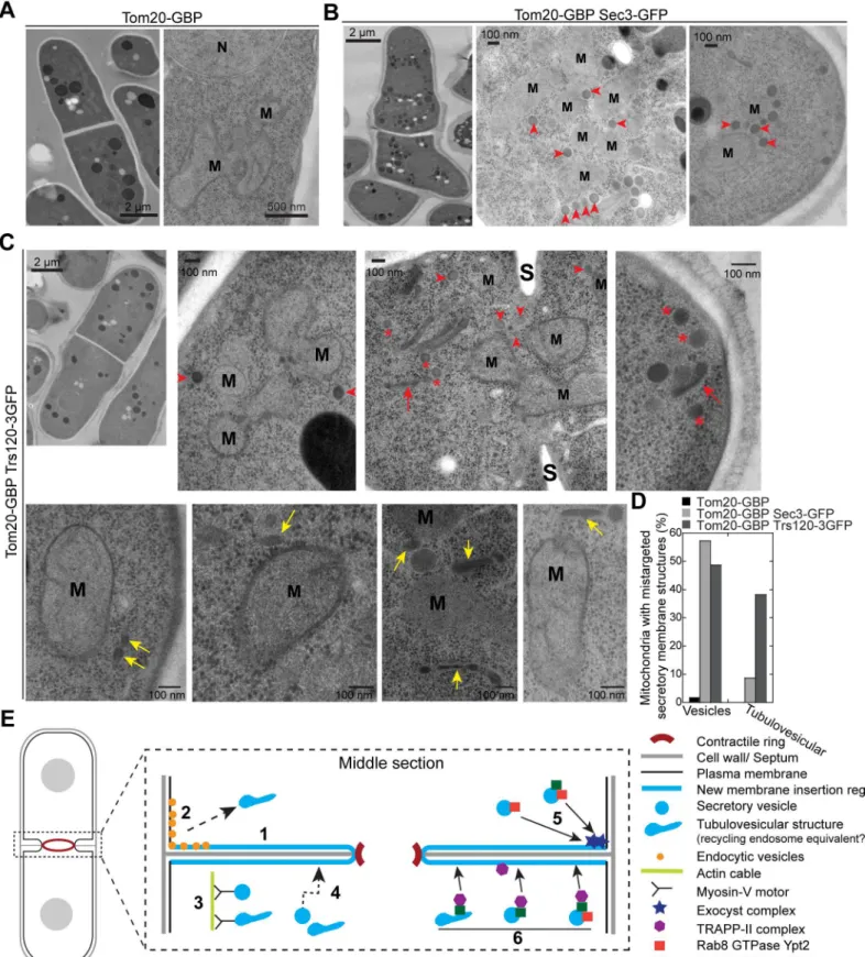
To confirm that secretory vesicles/compartments are tethered to mitochondria in
tom20-GBP trs120-3GFPcells, EM was performed as described previously [89]. In cells express-ing Tom20-GBP only, vesicles were not detectable at mitochondria (distance of vesicles to
A
DIC
Tom20-GBP Trs120-3GFP RFP-Bgs4
Trs120 Bgs4 Trs120 Bgs4
B
C
DIC Trs120 Ypt3 Trs120Ypt3
Tom20-GBP Trs120-3GFP tdTomato-Ypt3
Tom20-GBP Trs120-3GFP Sec3-tdTomato
Trs120 Sec3 Trs120Sec3
DIC
Ypt3 Bgs4
*
*
*
*
Ypt3Bgs4 DIC
Tom20-GBP mEGFP-Ypt3 RFP-Bgs4
D
Fig 9. Mislocalized TRAPP-II complex but not Ypt3 can ectopically target vesicle cargos to mitochondria.(A) Micrographs of cells expressing Tom20-GBP Trs120-3GFP RFP-Bgs4. Colocalization of Trs120 with vesicle cargo Bgs4 on some mitochondrial structure is marked by arrows. (B) Mislocalization of Ypt3 to mitochondrial structure (arrowheads) by Trs120. (C) Mistargeted Ypt3 cannot recruit Bgs4 to mitochondria (arrows), although Ypt3 at its native location still colocalizes with Bgs4 at the division site and cell tips (asterisks). (D) Mistargeted Trs120 does not ectopically target the exocyst to mitochondria (arrows). Bars, 5μm.
mitochondria<100 nm) in EM thin sections (n= 12 cells;Fig 10A and 10D). As a positive con-trol, in cells expressing Tom20-GBP Sec3-GFP, we observed attachment of secretory vesicles with a diameter of ~90 nm to ~60% mitochondria from all imaged cells with 3.0 ± 0.9 vesicles per mitochondria (n= 12 cells;Fig 10B and 10D, arrowheads). Similar to the budding yeast strain carrying comparable constructs [106], mitochondria were clustered in this mutant (Fig 10B). Similar secretory vesicles were also detected to associate with mitochondria in ~70% cells expressing Tom20-GBP Trs120-3GFP (n= 14 cells;Fig 10C, arrowheads), with ~1.5 ± 0.4 vesi-cles per mitochondria detected in ~50% mitochondria (Fig 10C and 10D). In addition, ~40% of mitochondria had attached tubulovesicular membrane structures (Fig 10C, yellow arrows; Fig 10D), suggesting that secretory compartments resembling recycling endosomes were also mistargeted. However, only ~9% of mitochondria in tom20-GBP sec3-GFPcells attached such structures (Fig 10D), indicating exocyst complex prefers to tether spherical vesicles. In addi-tion, some vesicles (Fig 10C, asterisks) and tubulovesicular structures (Fig 10C, red arrows) were either free in the cytoplasm or accumulated at the active growth site, possibly caused by the depletion of TRAPP-II from its native localizations. Together, secretory vesicles and the tubulovesicular structures can be retargeted and tethered to mitochondria by mislocalized TRAPP-II complex. Together, our data indicate that the TRAPP-II complex is critical for teth-ering Ypt3-containing vesicles or other secretory compartments to the plasma membrane.
Discussion
Previous studies found that new plasma membrane was predominantly inserted at the leading edge of the cleavage furrow during cytokinesis in animal cells [7,25–27]. In this study, we chal-lenged the universality of the model with our finding that post-Golgi secretory vesicles and compartments are delivered to everywhere along the cleavage furrow for membrane insertion during fission yeast cytokinesis (Figs2and3). The involvement of exocyst complex as a vesicle tether in cytokinesis was confirmed (Fig 10B). However, the fact that the exocyst localizes to the rim of the division plane but vesicle fusion and membrane insertion do not happen exclu-sively at the rim indicates that other vesicle tether or tethering factor is involved. Indeed, we find that the putative vesicle tether TRAPP-II complex localizes to the cleavage furrow and is also involved in vesicle tethering and membrane insertion during cytokinesis (Figs5–10; see the proposed model inFig 10E).
Where Is the New Membrane Inserted during Furrow Ingression in
Cytokinesis?
Fig 10. Mislocalized TRAPP-II complex can ectopically target vesicles and tubulovesicular membrane structures to mitochondria.(A–D) EM
images (A–C) and quantification (D) showing post-Golgi secretory vesicles (arrowheads) or elongated tubulovesicular membrane structures (yellow arrows,
whole cleavage furrow instead of just to the furrow tip. Spreading vesicle insertion sites may avoid crowding of vesicles and the vesicle fusion machineries, especially in cell types that require a large amount of new membrane during cytokinesis. Consistently, new membrane addition in a broader region along the cleavage furrow have been detected inXenopusembryos and zebrafish blastomeres [19,112].
Our discovery that new membrane from secretory vesicles/compartments is not predomi-nantly inserted adjacent to the contractile ring raises an interesting question on the coordina-tion between membrane deposicoordina-tion and ring constriccoordina-tion during cytokinesis. It has been proposed that the contractile ring recruits secretory compartments to the division site and the F-BAR protein Cdc15 in the ring helps to deliver Bgs1, a vesicle cargo, to the cleavage furrow [55,87]. Our data are not incompatible with that proposal, although these pioneer studies do not have enough spatiotemporal resolutions on the exocytic events. Cdc15 may mainly help recruit Bgs1 to the division site during ring maturation when they colocalize but not during ring constriction. Ring constriction may help to generate more vesicle insertion sites at the cleavage furrow. The distribution of the formin For3 and myosin-V motor Myo52 at the divi-sion site (Fig 3) is consistent with this possibility. Alternatively, ring constriction may generate tension on the existing plasma membrane in the cleavage furrow and this tension guides mem-brane expansion by facilitating exocytosis and inhibiting endocytosis. Similar hypotheses or models have been proposed for membrane translocation during cell motility [113,114]. It will be interesting to test these possibilities in future studies.
The Function and Coordination of the Exocyst and TRAPP-II Complexes
during Cytokinesis
The exocyst complex is the best known presumptive vesicle tether at the plasma membrane during cell polarization, cell motility, cytokinesis, and many other cellular processes
[30,31,115]. However, direct evidence of exocyst’s vesicle tethering activity from in vitro recon-stitution is missing [31]. We found that fission yeast exocyst can be mistargeted to mitochon-dria by its component Sec3 and then recruits post-Golgi secretory vesicles to mitochonmitochon-dria (Fig 10B), which strongly supports its vesicle tethering ability inS.pombe.
The TRAPP complex was initially indicated as a tethering factor for ER-derived vesicles in budding yeast [116]. The TRAPP-I complex plays an essential role in ER-to-Golgi transport [117]. TRAPP-II is proposed to affect intra- and post-Golgi traffic [37,39,40]. TRAPP-III is for autophagosome formation [37]. Like the exocyst, vesicle tethering ability and specificity of the TRAPP complexes have not been reconstituted in vitro [118]. Instead, their potential functions as the GEFs for Rab/Ypt GTPases have been studied in several different systems [37,94,95]. Although our data did not unambiguously assign TRAPP-II as a genuine MTC, they do indi-cate that TRAPP-II can recognize some secretory vesicles/compartment and promote vesicle tethering and fusion to the plasma membrane at the division site during cytokinesis.
the cytoplasm or near the active growth sites were marked. N = Nucleus, S = septum. (D) Quantification of the mitochondria associated with mis-targeted secretory vesicles or tubulovesicular structures. (E) A working model for the roles of the exocyst and TRAPP-II complexes in vesicle trafficking and
membrane deposition at the cleavage furrow during cytokinesis. The events 1–6 are symmetrical at the division site and omitted at some locations for clarity.
1. New membrane is deposited throughout the cleavage furrow. 2. Endocytic vesicles are mostly generated at the rim of the division plane or the adjacent regions and may become tubulovesicular structures like recycling endosomes. 3. Myosin-Vs transport secretory vesicles or recycling endosome equivalents along actin cables to the division plane. 4. Secretory vesicles can also reach the division site by actin-independent random walk. 5. Exocyst complexes localize to the rim of the cleavage furrow and preferentially tether 90-nm secretory vesicles probably through interaction with Rab8 GTPase Ypt2. 6. TRAPP-II complexes localize along the cleavage furrow (slightly biased to the leading edge) to directly tether or indirectly promote the tethering of Rab11 GTPase Ypt3-labeled recycling endosome equivalents or 90-nm secretory vesicles.
Given the different localizations of the TRAPP-II and exocyst complexes at the division site, we favor a model that the TRAPP-II and the exocyst function at different locations, although both regulate membrane addition and dynamics at the division site (Fig 10E). This model is further supported by the observation that localization of the glucan synthase Bgs1 and gluca-nase Eng1 was affected differently intrs120andsec8mutants (Fig 7E–7G). Our data suggest that TRAPP-II may preferentially tether (or help to tether) Ypt3-labeled secretory vesicles as well as the tubulovesicular structures, whose identity is still unclear at this moment, along the cleavage furrow (Fig 10E). It is of great interest to examine the contribution of TRAPP-II’s potential GEF activity to vesicle tethering in the future. In contrast, it appears that the exocyst prefers to tether the typical spherical secretory vesicles together with Rab8 GTPase Ypt2 at the rim of the division plane (Fig 10E), given that the exocyst is an effector of the Rab8 GTPase Sec4 in budding yeast [119], which primarily locates on the surface of the secretory vesicles [120]. Because a portion of exocyst complexes are transported to the cell tips together with secretory vesicles by random walk [56], therefore we cannot rule out that a small fraction of exocyst may contribute to vesicle tether along the cleavage furrow. Exocyst and TRAPP-II may have overlapping localization during ring maturation given that they both start to concentrate at the cell equator during this stage (Figs1Gand5A;S6 Video). However, the localization inde-pendence between the two complexes (S4E–S4G Fig) and the lack of mis-targeting of Sec3 by Trs120 (Fig 9D) does not support any physical interactions between them, in contrast to plant cells [42]. Therefore, TRAPP-II and exocyst complexes are both involved but might function independently at the division site to regulate the tethering of secretory vesicles/compartments for delivering cargos and inserting new membrane.
The role of endocytosis during cytokinesis is intriguing [6,121]. It was suggested that the recycling endosomes derived from endocytosis at other locations provide new membrane to the ingressing cleavage furrow [6,7,122]. However, this does not explain the role of endocytic events at the division site. Our finding that the rim of the division plane is the dominant sites for endocytosis (Fig 4) suggests that endocytosis can help to redistribute the plasma membrane and cytokinesis proteins from the rim (tethered by the exocyst) to the interior of the cleavage furrow (tethered by the TRAPP-II complex and/or other proteins). Thus, endocytosis may not only be important for retrieving and recycling extra plasma membrane but also actively con-tribute to membrane expansion at the division site during cytokinesis (Fig 10E).
In conclusion, we found an extensive addition of new membrane along the cleavage furrow rather than exclusively near the contractile ring or the rim of the division plane during cytoki-nesis. Our investigation of the TRAPP-II complex as another potential vesicle tether (directly or indirectly through Ypt3) besides the exocyst in cytokinesis opens up avenues to understand-ing the coordination of multiple vesicle tetherunderstand-ing factors for a sunderstand-ingle task.
Materials and Methods
Strain Constructions, Genetic and Cellular Methods
S1 Tablelists theS.pombestrains used in this study. We used standard genetic and PCR-based gene targeting methods to transform yeast cells and construct strains [123,124]. Taggedtrs120, trs130,trs8502,syb1,sec3,fim1,for3,myo52,sec72,rlc1, andtom20genes are under the control of their endogenous promoters and integrated at their native chromosomal loci to replace the native genes. The N-terminal taggedpsy1,bgs1, andbgs4strains (derived from gift strains from other labs) have a copy of tagged gene under the control of the endogenous promoters inserted inleu1locus with the native genes deleted (seeS1 Table).
pFA6a-kanMX6-P3nmt1-tdTomato vector atBglII andPacI sites to replace the3nmt1promoter. The resulting plasmids (JQW896 formEGFP-ypt3, JQW898 fortdTomato-ypt3, and JQW913 for mEGFP-ypt2) were used as templates to amplifykanMX6-Pypt3-mEGFP, kanMX6-Pypt3-tdTo-mato, andkanMX6-Pypt2-mEGFPfragments flanked with homologous sequences correspond-ing to the last 70 bp of 50untranslated region (UTR) and the complement of the first 70 bp of the coding sequence ofypt3orypt2. The amplified and column purified PCR products were then transformed into wt strain JW81 as described [124]. The positive transformants from visual screen were confirmed by PCR. Plasmid for expression of Ypt3 (pSM925, pREP41-tdTo-mato-ypt3, a gift from Sophie Martin) was under the control of inducible41nmt1promoter [72]. The plasmid was transformed into yeast using standard method [124]. The cells were cul-tured in Edinburgh minimal medium plus five supplements (EMM5S) without leucine (EMM5S–leucine) for 20–24 h to induce the expression of41nmt1promoter before imaging.
To generatetrs120point mutants, we used the marker reconstitution mutagenesis method as previously described [89]. Briefly,trs120gene including 70 bp of 50
UTR, the open reading frame (ORF), introns, and 137 bp of 30UTR was amplified from genomic DNA and cloned into a plasmid with the C-terminus ofhis5ORF to obtain the plasmid JQW886 (trs120-his5c). In addition, we insertedhis5ORF (without its C-terminus) immediately after 30UTR oftrs120 locus to generate strain JW6842. Error-prone PCR was performed to amplifytrs120fragment from JQW886, which was then transformed into JW6842 strain. We selectedtrs120mutants with EMM5S–histidine medium and checked their growth and morphology at different tem-peratures. ~150 temperature-sensitive mutants with strong cytokinesis-related phenotypes were selected.trs120sequences (including 50
UTR, ORF, introns, and 30
UTR) of 15 mutants were cloned and sequenced to identify the mutations, and the two least mutated strains (trs120-ts1 andtrs120-M1) were used for further experiments.trs120-ts1contained five missense mutations (I370T, L543P, T700A, K993I, and F1158V).trs120-M1contained one mutation in the coding sequence (L1113R), one mutation in the second intron (an A to T switch at the 24th bp of the intron), and one mutation in the 30UTR (84 bp downstream of the stop codon, T to C).
To test the functionalities of tagged Trs120, Trs130, Tom20, Ypt2, and Ypt3, the growth and morphology of the strains expressing tagged proteins were examined at different temperatures. Most strains resembled wt cells, indicating these tagged proteins are functional under the tested conditions. However, thePypt2-mEGFP-ypt2strain displayed temperature-sensitive growth defects with increased septation index at 36°C. Thus, the strain was only used at 25°C. Althoughtrs120-ts1 ypt3-i5was synthetic lethal,trs120-ts1 Pypt3-mEGFP-ypt3strain resem-bledtrs120-ts1. Thus, mEGFP-Ypt3 is functional.Pypt3-tdTomato-ypt3cells formed Ypt3 con-taining aggregates in the cytoplasm (not detected inPypt3-mEGFP-ypt3strain), stopped growing at 36°C, and displayed synthetic lethal interactions with other mutations, indicating tdTomato-Ypt3 is not functional. Therefore, we used the plasmid-born tdTomato-Ypt3 (pSM925) when necessary.
All drug treatments were performed in micro-centrifuge tubes at 25°C and then cells were imaged on bare slides to maintain the drug concentration except where noted. BFA (Sigma, B7651) treatment was performed at a final concentration of 50μg/ml from a 5 mg/ml stock
solu-tion in ethanol for 10 min with rotasolu-tion [125]. To test whether cortical localization of Syb1 at the division site depends on microtubules, cells were treated with MBC at a final concentration of 25μg/ml for 10 min and then imaged on gelatin slide with the same drug concentration [126].
Acid Phosphatase Secretion Assay
medium, and resuspended in fresh EMM5S and shifted to 36°C at time 0. Samples were taken each hour. The absorbance at 595 nm was measured for cell concentrations. Then 1 ml cells for each culture was centrifuged, and 500μl of the supernatant was added to 500μl of substrate
solu-tion (2 mMp-nitrophenyl phosphate, 0.1 M sodium acetate, pH 4.0; prewarmed to 30°C) and incubated at 30°C for 5 min. Reactions were stopped by the addition of 500μl of 1 M sodium
hydroxide. The absorbance of the reaction solution at 405 nm was measured using the substrate solution with 500μl of EMM5S as a blank. The OD405/OD595value versus time was plotted.
Microscopy and FRAP
Microscopy was performed as previously described [127]. Briefly, cells were grown exponentially in liquid YE5S medium at 25°C for 36–48 h before microscopy at 23–24°C except where noted. Cells were collected by centrifugation at 3,000 rpm for 30 s and washed twice with EMM5S medium to reduce autofluorescence for fluorescence microscopy. Live-cell fluorescence micros-copy at 23–24°C was performed using a thin layer of EMM5S liquid medium with 20% gelatin (Sigma-Aldrich) and 5μM n-propyl-gallate (n-PG). For fluorescence microscopy at 36°C, cells
were first grown at 25°C for 36 h, then shifted to 36°C for a given time (see Fig legends). For imaging preparation, cells grown at 36°C were washed and concentrated in pre-warmed YE5S liquid medium with 5μM n-PG. Then 2-μl of the concentrated cells were spotted onto a
cover-glass-bottom dish (Delta TPG Dish; Biotechs, Butler, PA, United States), covered with the pre-warmed YE5S agar, and imaged at 36°C in a preheated climate chamber (stage top incubator INUB-PPZI2-F1 equipped with UNIV2-D35 dish holder; Tokai Hit, Shizuoka-ken, Japan).
Tetrad fluorescence microscopy fortrs120+/trs120Δdiploid (Fig 6A and 6B) was performed as previously described at 23–24°C [77] with the following modifications: we started the ~14 h of imaging after 20–24 h of cell growth following tetrad dissection at 25°C. To minimize photo-toxicity, Rlc1 channel was imaged every 5 min with low laser power (5%–7.5%).
Nikon 100×/1.4 NA Plan-Apo objective lenses were used for all imaging. The spinning disk confocal system (UltraVIEW Vox CSUX1 system, Perkin Elmer Life and Analytical Sciences) with 440-, 488-, 515-, and 561-nm lasers and back-thinned EMCCD camera (Hamamatsu C9100-13) on a Nikon (Nikon, Melville, NY, US) Ti-E microscope was used to collect fluores-cence images for all figures and videos except Figs5A,7Eand8B. The pixel size of the images under our imaging conditions is 144 nm/pixel. Since we used the location of the brightest pixel to represent the center of a specific vesicle/endosome, the resolution in thex-ydirection is ~150 nm. For Figs5A,7Eand8B, another spinning disk confocal system (UltraVIEW ERS; PerkinElmer) with 440- and 568-nm solid state lasers and 488- and 514-nm argon ion lasers, and a cooled charge coupled (CCD) device camera (ORCA-AG; Hamamatsu Photonics) on a Nikon microscope (Eclipse TE2000-U) was used with 2 × 2 binning. Under our imaging condi-tions, no bleed-through between green and red channels was detected.
To show the synthetic interactions between mutations with DIC images, a Nikon Eclipse Ti inverted microscope equipped with a Nikon cooled digital camera DS-Ql1 was used as before [128].
Data Analysis and Vesicle Tracking
We analyzed images using ImageJ (National Institutes of Health), UltraVIEW (PerkinElmer), and Volocity (PerkinElmer). Fluorescence images in figures are maximum-intensity projections of z sections spaced at 0.4–0.5μm except where noted. Movements of individual vesicles in
mov-ies were tracked using the plug-in MTrackJ in ImageJ [127,129]. The center positions (absolute values along thex- andy-axis in un-rotated images) of the vesicles were determined using the pixel with the highest fluorescence intensity. The last tractable position at or near the division plane from a continuous movement of an individual vesicle was recorded as the docking/deposi-tion site of the specific vesicle. The diameters and posidocking/deposi-tions of the contractile rings were deter-mined by the brightest pixel of Rlc1 images on the middle focal plane using the first Rlc1 image at the beginning of movies. The outer boundaries (or rims) of cell-division planes were deter-mined using the DIC images taken before the fluorescence movies. Since the division planes of the cells were random in the imaging field, their contractile-rings have an angle to the horizontal x-axis in the un-rotated images. The docking points of all tractable vesicles from an individual cell were rotated based on the angle between the contractile-ring/division plane and thex-axis using MATLAB software. The newxandyvalues of the docking sites, the positions and diame-ters of the rings, and the rims of the division plane were generated, but their relative positions were unchanged with the newx-axis marking the division plane. To plot data from multiple cells in one figure, we normalized the width of the division plane to 3.5μm (the range of the values is
from 3.4 to 3.9μm). Given that the contractile ring was not always at the center of the division
plane, the plot inFig 2Fwas also normalized according to the center of the contractile-rings. To analyze the assembly sites of endocytic patches/vesicles, Fim1-mEGFP images acquired at the middle focal plane with 4.14 fps in 2-min movies were used. The following criteria were used to identify newly assembled Fim1 patches from those internalizing endocytic vesicles that already pinched off from the plasma membrane [81,82]: (1) Fim1 signal gradually increases to reach a peak and (2) the patch does not move before reaching the signal peak. If these criteria were fulfilled, the location of the brightest pixel of a Fim1 patch when the peak signal reached was recorded as the endocytic vesicle assembly site, and the locations of these assembly sites were rotated and aligned as the secretory vesicles.
To quantify Psy1 recovery on the plasma membrane after photobleaching at the cell side of interphase cells, the rim of the division plane, or the leading edge of the cleavage furrow that is immediately behind the contractile-ring, a 9-pixel square was chosen within the bleached regions. Same-sized squares were used to measure cytoplasmic signal outside the bleached region over time as the background, which was deducted from the plasma-membrane intensity.
Electron Microscopy
freeze-substitution in the presence of 2% osmium tetroxide and 0.1% uranyl acetate in acetone. Cells were embedded in Epon-Araldite epoxy resin, and serially sectioned with a thickness of 70 nm. The samples were then post stained with uranyl acetate and lead citrate and observed on a Philips CM100 transmission electron microscope (FEI, Hillsboro, OR).
Supporting Information
S1 Data. Raw data for analyses and quantifications shown in the figures and supplemental figures, as indicated.
(XLSX)
S1 Fig. Membrane invagination and ring constriction are defective in exocyst mutant, and v-SNARE Syb1 andβ-glucan synthase Bgs1 travel to the division site during ring matura-tion.(A, B) Membrane invagination and ring constriction are slower in exocyst mutantsec8-1 even at the permissive temperature 25°C. (A) Micrograph ofsec8-1 GFP-Psy1 Rlc1-tdTomato cells at the middle focal plane. (B) Time courses of membrane invagination and ring constric-tion in wt (top) and twosec8-1cells (bottom) as marked in (A). (C, D) Vesicles (arrowheads) containing Syb1 (C) or Bgs1 (D) are delivered to the division site during ring maturation. Bars, 5μm.
(EPS)
S2 Fig. EM images of wt cells during septum formation or maturation, tracking delivery of Syb1-labeled vesicles/compartments and Psy1 dynamics at the division site during septum maturation.(A) EM images of wt cells during septum formation or maturation. Secretory vesi-cles are marked by arrows. Elongated or irregular-shaped tubulovesicular structures are marked by arrowheads. Bars, 100 nm. (B, C) Time courses of Syb1 delivery (B) and distribution of its docking sites during septum maturation (C). Syb1 signal within the box at the division site was bleached at time 0 and incoming Syb1 vesicles/compartments in the middle focal plane were tracked in cells without Rlc1 signal but a full septum. Cells were imaged for 3 min after photobleaching. (B) Arrowheads mark travelling vesicles. (C) Quantification of the docking sites of all tractable Syb1 vesicles in the 3-min movies. The septum is positioned aty= 0. (D) Recovery of Psy1 signal on the plasma membrane at the division site (arrows) after bleaching at the region marked by red box. An interphase cell on the right was bleached and imaged as a control, which showed almost no recovery (arrowhead). Bars in B and D, 5μm.
(EPS)
S3 Fig. Cytokinesis defects inmyo52Δand tracking the movement of Myo52 puncta during different stages of cell division.(A, B) Montage (A) and quantification (B) showing the delay in ring constriction and cell separation inmyo52Δcells at 25°C.,p<0.01 compared with wt control. (C) Time courses showing Myo52 puncta moving to the division site during ring mat-uration, ring constriction, and septum maturation on middle focal plane. Moving Myo52 puncta are marked by arrowheads. (D) Distribution of the final destination of Myo52 puncta at the division site relative to the position of constricting ring from 5 cells (color coded) in 2-min movies.X- andy-axes are along the division site and cell long axis, respectively. The ring (the diameter marked by the color lines) is displaced along they-axis away from its real position at y= 0 for clarity. (E) Distribution of the final destination of Myo52 puncta relative to the posi-tion of septum (aty= 0) during septum maturation in 2-min movies. Bars, 5μm.
(EPS)