RESEARCH ARTICLE
Microparticle-Induced Activation of the
Vascular Endothelium Requires Caveolin-1/
Caveolae
Allison M. Andrews
1, Victor Rizzo
1,2¤*
1Independence Blue Cross Cardiovascular Research Center, Lewis Katz School of Medicine at Temple University, Philadelphia, Pennsylvania, United States of America,2Department of Anatomy and Cell Biology, Lewis Katz School of Medicine at Temple University, Philadelphia, Pennsylvania, United States of America
¤ Current Address: 3500 North Broad Street, Lewis Katz School of Medicine at Temple University, 19140,
Philadelphia, Pennsylvania, United States of America *rizzov@temple.edu
Abstract
Microparticles (MPs) are small membrane fragments shed from normal as well as activated,
apoptotic or injured cells. Emerging evidence implicates MPs as a causal and/or
contribut-ing factor in altercontribut-ing normal vascular cell phenotype through initiation of proinflammatory
signal transduction events and paracrine delivery of proteins, mRNA and miRNA. However,
little is known regarding the mechanism by which MPs influence these events. Caveolae
are important membrane microdomains that function as centers of signal transduction and
endocytosis. Here, we tested the concept that the MP-induced pro-inflammatory phenotype
shift in endothelial cells (ECs) depends on caveolae. Consistent with previous reports, MP
challenge activated ECs as evidenced by upregulation of intracellular adhesion molecule-1
(ICAM-1) expression. ICAM-1 upregulation was mediated by activation of NF-
κ
B, Poly
[ADP-ribose] polymerase 1 (PARP-1) and the epidermal growth factor receptor (EGFR).
This response was absent in ECs lacking caveolin-1/caveolae. To test whether
caveolae-mediated endocytosis, a dynamin-2 dependent process, is a feature of the proinflammatory
response, EC
’
s were pretreated with the dynamin-2 inhibitor dynasore. Similar to
observa-tions in cells lacking caveolin-1, inhibition of endocytosis significantly attenuated MPs
effects including, EGFR phosphorylation, activation of NF-
κ
B and upregulation of ICAM-1
expression. Thus, our results indicate that caveolae play a role in mediating the
pro-inflam-matory signaling pathways which lead to EC activation in response to MPs.
Introduction
Microparticles (MPs) are small 0.1 to 1.0
μ
m membrane fragments shed from activated,
apo-ptotic or injured cells. MPs circulate in the blood of healthy patients and become elevated in
patients with cardiovascular diseases such as hypertension and atherosclerosis [
1
]. MPs are
OPEN ACCESSCitation:Andrews AM, Rizzo V (2016) Microparticle-Induced Activation of the Vascular Endothelium Requires Caveolin-1/Caveolae. PLoS ONE 11(2): e0149272. doi:10.1371/journal.pone.0149272
Editor:Partha Mukhopadhyay, National Institutes of Health, UNITED STATES
Received:September 3, 2014
Accepted:January 30, 2016
Published:February 18, 2016
Copyright:© 2016 Andrews, Rizzo. This is an open access article distributed under the terms of the
Creative Commons Attribution License, which permits unrestricted use, distribution, and reproduction in any medium, provided the original author and source are credited.
Data Availability Statement:All relevant data are within the paper and its Supporting Information files.
Funding:Funding for this work was provided by NIH HL086551.
primarily shed from endothelial cells, platelets and immune cells [
2
] and carry proteins,
mRNA and miRNA [
3
]. In addition, they can interact and/or be taken up in other cells and
affect signaling, protein expression and cellular phenotype [
4
,
5
]. Recent evidence suggests that
MPs can play a role in inflammation [
6
,
7
], thrombosis [
8
], coagulation [
9
], impair
vasorelaxa-tion [
10
] and induce oxidative stress [
7
,
11
] thus contributing to disease progression.
MPs make up a subset of a larger classification of extracellular microvesicles, which also
includes exosomes and apoptotic bodies [
12
]. They differ from other microvesicles in their size
and mechanism of release, which is through outward pinching off of the plasma membrane. As a
result of this process the protein content will largely reflect the host cell from which it was
derived. MPs are typically isolated by differential centrifugation, which allows for their separation
from the other microvesicle subsets [
13
]. A few studies have examined effects of
endothelial-derived microparticles (EMPs) on other endothelial cells. These studies have demonstrated EC
uptake of MPs [
6
], upregulation of adhesion molecules [
7
] and increased platelet adhesion [
14
].
However, the mechanism by which MPs initiate these pro-inflammatory EC responses remains
incomplete. In addition, ECs are known to engage in paracrine signaling, thus making the study
of EMPs important for understanding vascular signaling during pathological conditions [
15
].
Caveolae are 50
–
100 nm diameter invaginations of the plasma membrane that function as
signal transduction and endocytotic centers [
16
]. While caveolae are expressed in several cell
types including smooth muscle cells and fibroblasts [
17
], they are highly enriched in the
endo-thelium where they play important roles in signaling and vascular function [
18
]. Studies on
Caveolin-1 deficient (Cav-1-/-) mice have revealed that loss of Cav-1/caveolae provides
protec-tion against the development of several vascular diseases. For example, Cav-1-/- mice crossed
with ApoE-/- mice were protected from high fat-diet induced atherosclerosis, instead forming
fewer and smaller plaques than ApoE-/- mice [
19
]. In addition, our group recently
demon-strated that Cav-1-/- mice are protected from Angiotensin II (Ang II)-induced aneurism
for-mation and rupture [
20
]. Interestingly, circulating MPs are elevated in both these conditions
[
7
,
21
,
22
], which suggest a role for Cav-1/caveolae in MP-induced signaling. We have
there-fore tested the hypothesis that MP-induced activation of the endothelium requires Cav-1/
caveolae. These studies have examined this concept through the use of Cav-1 knock-out ECs as
well as pharmacological inhibition of caveolae endocytosis, which is an important aspect of
Cav-1/caveolae function and signaling. Our results ultimately shed light on the mechanism of
MP-induced activation of the endothelium.
Methods
Cell Culture
Wild type (WT
—
c57/bl6)) and caveolin-1 knockout (Cav-1 -/- on a c57/bl6 background)
Mouse Lung Endothelial Cells (MLEC) were a gift from Dr. Shampa Chatterjee (University of
Pennsylvania). MLECs were harvested from mice under protocol 801630, which was approved
by IACUC at University of Pennsylvania. Western blot of Cav-1 expression in each cell type is
shown in
S1 Fig
. Cells were cultured in MCDB-131 (Sigma), 15% FBS (Benchmark) and 0.05
mg/mL gentimyacin (Cambrex Biosciences) and maintained at 37°C, 97% humidity and 5%
carbon dioxide. All experiments were performed below passage 12.
MP generation
Cells were treated with 10 ng/mL TNF-
α
(Sigma Aldrich) in 1% FBS for 24 hrs. Media was
col-lected and centrifuged at 1500 g for 20 min to remove cell debris. Supernatant was either used
immediately or snap froze in liquid N
2and stored at -80°C for later use. MPs were collected by
ultracentrifugation (20,000 g for 40min). MPs were collected by resuspension in 1% FBS and
sterile filtered (0.22
μ
m). MP size distribution was verified using the Nanosight (NanoSight
Ltd, data not shown).
Flow Cytometry
According to past protocols for MP isolation [
7
,
23
,
24
], MPs from thawed or freshly prepared
samples were resuspended in 100
μ
L Annexin Binding Buffer (10mM Hepes, 140mM NaCl,
2.5mM CaCl
2). Control samples were resuspended in annexin binding buffer without calcium.
Samples were incubated with 5
μ
L of Annexin-FITC (BD Biosciences) for 15 min in the dark,
then diluted in annexin binding buffer and analyzed on a FACScan (BD Biosciences). A region
of interest based on the control sample was used to identify Annexin+ particles. Size of MPs
was evaluated using fluorescent SPHERO Nano fluorescent reference beads ranging between
0.22 and 1.34
μ
m (Spherotech) (
Fig 1
). Absolute count for microparticles was determined
using CountBright Counting Beads (Invitrogen) and the following formula: (Number of
Annexin V+/Number of Beads)
(Bead Count of Lot/ Sample volume).
MP Treatment
WT and Cav-1-/- cells were treated with MPs (Approx. 40,000 MPs/mL) for 0, 30 min, 1 hr, 2
hrs or 24 hrs. In some experiments, cells were incubated with 60
μ
M dynasore (Tocris
Biosci-ence), 1
–
100
μ
M AG1478, or 0.1% Dimethyl sulfoxide (DMSO) for 30 minutes or 2
μ
M PJ34
(Enzo Life Sciences) for 1 hr prior to treatment with MPs.
Western Blot Analysis
Cells were harvested in Mammalian Protein Extraction (MPER) lysis buffer (Thermo Scientific)
with sodium vanadate (Fisher), phosphatase and protease inhibitors (Calbiochem). Proteins
were separated by 5
–
20% SDS-PAGE and transferred to nitrocellulose membranes. Proteins of
interested were detected by Western blot analysis using the following primary antibodies:
ICAM-1 (polyclonal Santa Cruz Biotechnologies),
β
-Actin (monoclonal Sigma Aldrich), pEGFR
pY1068 (polyclonal Invitrogen), EGFR (polyclonal Santa Cruz Biotechnologies), PARP-1
Fig 1. Detection of TNF-α-induced microparticles (MPs) by flow cytometry.Cultured mouse lung
endothelial (MLEC) cells were treated with TNF-α(10 ng/mL) for 24 hrs. Media was collected, centrifuged and resuspended in Annexin Binding Buffer, as described in Methods. A region of interest was drawn (R1) to indicate isolated MPs from TNF-αtreated cells (B, grey) as compared to unlabeled MPs (A). Size beads (0.22, 0.45, 0.88, 1.34μm) were used to estimate MP sizes (B, black).
doi:10.1371/journal.pone.0149272.g001
(polyclonal Cell Signaling), p-p65 (monoclonal Cell Signaling), p65 (polyclonal Cell Signaling),
p-p50 (polyclonal Santa Cruz Biotechnologies) and p50 (polyclonal Cell Signaling).
Results
Caveolin-1/Caveolae mediates MP-induced increase in ICAM-1
expression via activation of NF-
κ
B and PARP-1
To examine the effects of MPs on the endothelium, MLECs were treated with MPs and cell
lysates harvested and examined using Western blot analysis. MPs induced an increase in
ICAM-1 protein expression by 2-fold in response to MPs after 24 hrs. Given the role of
caveo-lae as signaling microdomains in the endothelium, we next tested whether caveocaveo-lae organelles
also participated in MP-induced signaling pathways that govern adhesion molecule expression.
Here, we utilized endothelial cells derived from caveolin-1 knockout mice. Interestingly,
base-line expression of ICAM-1 was reduced by 25% in Cav-1-/- endothelial cells compared to their
WT counterpart. In addition, in response to 24hr exposure to MPs, Cav-1-/- EC
’
s did not
upre-gulate ICAM-1 expression (
Fig 2
).
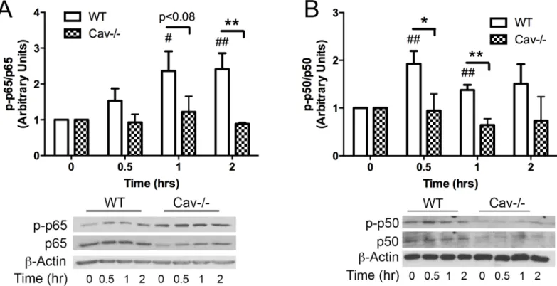
To test the involvement of NF-
κ
B in MP induced signaling, cells were treated with MPs for
up to 2hrs and processed for Western blot analysis to detect expression and phosphorylation
status of the NF-
κ
B subunits p65 and p50. WT MLECs showed increased phosphorylation of
both p65 and p50 over the time course of study in response to MPs (
Fig 3A
). In
Cav-1-/-MLECs, basal levels of both p65 and p50 were somewhat reduced compared to WT. While
enhanced baseline phosphorylation of p65 was also noted in Cav-1-/- cells, MPs failed to
enhance p65 or p50 levels beyond baseline at all observed time points (
Fig 3B
).
Next, we examined whether PARP-1 was activated (as determined by increased PARP-1
expression) in response to MPs. WT MLECs showed clear activation of PARP-1 in response to
MPs over the course of 2 hrs (
Fig 4A
). Cav-1-/- MLECs had decreased basal levels of PARP-1
and expression did not change in response to MPs (
Fig 4A
). In addition to PARP-1 activity, we
detected changes in the presence of cleaved PARP-1. WT MLECs showed ~ 2 fold increase in
PARP-1 cleaved fragments in response to MPs while Cav-1-/- MLECs had no detectable
PARP-1 fragments at basal levels or in response to MPs (
Fig 4B
).
MP-induced increase in ICAM-1 expression is dependent on EGFR
activation
As a potential upstream mediator of an NF-
κ
B /PARP-1 pathway, we examined the effect of
MPs on EGFR activation. The data showed that MPs caused a rise in EGFR phosphorylation,
which peaked at 1hr after treatment (
Fig 5
). Cav-1-/- MLECs, while displaying an enhanced
baseline level of the receptor, showed no change in receptor phosphorylation status in response
to MPs (
Fig 5
). Although the EGFR had been implicated in MP induced activation of the
endo-thelium, the consequences of receptor activation have not been determined. Here, we examined
whether inhibiting EGFR activation using AG1478 would block downstream NF-
κ
B
phosphor-ylation and the induction of ICAM-1 expression. We observed a decrease in phosphorphosphor-ylation
of the NF-
κ
B p65 subunit at 30 min after stimulation with MPs (
Fig 6A
) as well as no change
in the expression of ICAM-1 at 24 hrs in cells pretreated with AG1478 (
Fig 6B
).
PARP-1 inhibition blocks MP-induced PARP-1 activation and
upregulation of ICAM-1
which leads to upregulation of ICAM-1. We utilized the PARP-1 inhibitor PJ34, which has
pre-viously been shown to block PARP-1 activation in endothelial cells [
25
]. PJ34 pretreatment
effectively blocked MP-induced PARP-1 activation and cleavage (
S2 Fig
). In addition, PARP-1
inhibition did not affect NF-
κ
B phosphorylation but was able to attenuate ICAM-1
upregula-tion (
Fig 7
).
Fig 2. MP-induced ICAM-1 expression requires caveolin-1/caveolae.WT and Cav1-/- MLEC’s were treated with MPs (Approx 40,000 MPs/mL) for 24 hrs. Cells were collected, homogenized, lysed and total cellular protein separated by 5–15% SDS-PAGE followed by Western blotting to detect ICAM-1 and
β-Actin. Densitometric quantification showed ~2 fold increase in ICAM-1 expression in WT cells in response to MPs. In Cav-1-/- cells, basal expression of ICAM-1 was lower than that detected in WT cells and it remained unchanged in response to MPs. (Avg±SEM two-way ANOVA and bonferroni’s post hoc
analysis, n = 8,*p<0.05,**p<0.01).
doi:10.1371/journal.pone.0149272.g002
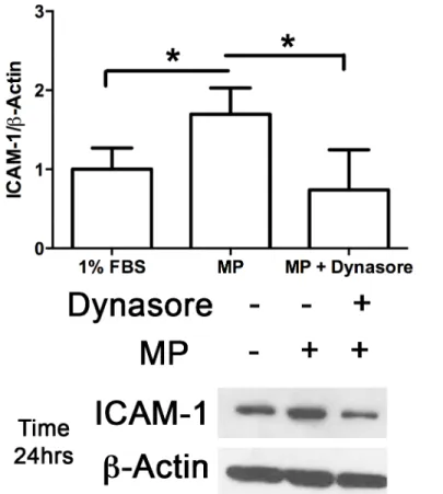
Pharmacological inhibition of caveolae endocytosis attenuates
MP-induced increase in ICAM-1 expression
To determine whether the MP-induced increase in ICAM-1 expression requires
caveolae-mediated endocytosis, we pretreated WT endothelial cells with dynasore, a dynamin-2
inhibi-tor. Inhibition of caveolae endocytosis blocked the MP-induced increase in ICAM-1 expression
(
Fig 8
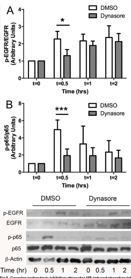
). To determine the involvement of caveolae endocytosis in MP-induced signaling we
also examined the effect of dynasore on p65 and EGFR phosphorylation. Inhibition of caveolae
endocytosis attenuated MP-induced p65 phosphorylation through all observation periods and
EGFR phosphorylation at 30 min but not at later time points (
Fig 9A and 9B
).
Discussion
The major finding of this study is that MPs induce activation of the endothelium through a
mechanism requiring caveolin-1/caveolae endocytosis.
A number of studies have shown that in response to MPs, the endothelium increases
expres-sion of adheexpres-sion molecules
in vitro
[
6
,
7
,
26
,
27
] and
in vivo
[
7
], which causes increased rolling,
and adhesion of leukocytes [
26
]. Increased adhesion molecule expression is a fundamental step
in the progression of a number of cardiovascular diseases such as atherosclerosis [
28
]. MPs
have been shown to interact with the endothelium surface and in some cases are internalized
[
14
]. However, a
“
MP-receptor
”
has yet to be determined and the mechanisms by which MPs
activate the endothelium remain unclear.
Fig 3. MPs induce phosphorylation of NF-κB is absent in Cav-1-/- MLECs.WT and Cav-1-/- cell cultures were treated with MPs (Approx 40,000 MPs/mL)
for indicated times (0, 0.5, 1, 2 hrs) and processed for Western blot analysis. WT cells showed increased phosphorylation of both p65 (A) and p50 (B) in response to MPs. However, MP’s did not induce phosphorylation of either subunit in Cav-1-/- cells. All values are normalized to t = 0 for each cell type respectively (Avg±SEM two-way ANOVA and bonferoni post-hoc analysis A. p-p65/p65: WT and Cav-1-/- n = 4 each, B. p-p50/p50: WT n = 6 and Cav1-/-n = 4 for all time poiCav1-/-nts except oCav1-/-ne which was excluded from each group at t = 0.5 hr based oCav1-/-n Grubb’s test for outliers p<0.05. # denotes comparison with WT t = 0, #p<0.05, ##p<0.01,*denotes comparisons between WT and Cav-1-/- cells*p<0.05,**p<0.01.)
Fig 4. Loss of caveolin-1/caveolae abolish MP-induced production of cleaved and uncleaved PARP-1.
WT and Cav-1-/- MLECs were treated with MPs for indicated times (0, 0.5, 1, 2 hrs). Proteins from whole cell lysates were separated using SDS-page (5–15%, transferred to nitrocellulose membranes, which were probed for PARP-1. MPs induced a ~ 2 fold increase in cleaved and uncleaved PARP-1 in WT cells. Basal expression of PARP-1 was reduced in Cav-1-/- cells. PARP-1 expression and cleavage pattern remained unchanged following treatment with MPs. All values are normalized to t = 0 for each cell type respectively (Avg±SEM Two-way ANOVA and bonferroni post hoc analysis*p<0.05 A. PARP-1: WT and Cav1-/- n = 5, B. Cleaved PARP-1: Cav1-/- n = 5 except t = 2 hr n = 4, cleaved PARP-1: WT n = 4 for all time points except one was excluded at t = 2hrs using Grubbs test for outliers p<0.05; # denotes comparisons to WT t = 0,
#p<0.05, ##p<0.01, ###p<0.001,*denotes comparisons between WT and Cav-1-/- cells,*p<0.05).
doi:10.1371/journal.pone.0149272.g004
By virtue of their signaling properties, lipid rafts and more specifically caveolae, play
impor-tant roles in many aspects of endothelial cell function [
16
]. To date, there are very few studies,
which have investigated the relationship between MPs and these membrane microdomains.
Related to MP production, one study showed that Ang II-induced MP release from the
endo-thelium could be blocked by pretreating cells with methy
–
β
-cyclodextrin, a compound which
disrupts raft integrity. In the present study, we asked whether caveolin-1/caveolae could
influ-ence MP-induced responses in the endothelium.
Upregulation of adhesion molecules by MPs has been reported in several systems and
involve upstream activation of the NF-
κ
B transcription factor [
27
,
29
–
33
]. Here we report that
MP-induced activation of NF-
κ
B in MLECs (
Fig 3A
) followed the same timeline for activation
as reported in other cell types [
27
] where phosphorylation of p65 occurs by 30 min and
remains elevated through 2hr of exposure to MPs. In our studies we found that the loss of
caveolin-1/caveolae prevented MP-induced phosphorylation of both NF-
κ
B subunits (p65 and
p50) (
Fig 3A and 3B
). This finding is the first to show an association between Cav-1/caveolae
and MP signaling and is consistent with a role for caveolae in NF-
κ
B activation by
proinflam-matory agents [
34
]. Although the loss of caveolin-1 prevented activation of NF-
κ
B, we
Fig 5. Caveolin-1/Caveolae mediates MP-induced activation of EGFR.WT and Cav-1-/- MLECs were treated with MPs for 0, 30, 1hr, and 2 hrs and processed for Western blotting. WT cells showed an increase in EGFR phosphorylation that peaked at ~3 fold after 1 hr of MP treatment. Cav-1-/- cells had increased expression of pEGFR and EGFR, however, EGFR phosphorylation was unchanged in response to MPs. All values are normalized to t = 0 for each cell type respectively (Avg±SEM two-way ANOVA and bonferroni
post hoc analysis, n = 5 each, # denotes comparisons to WT t = 0, #p<0.05, ##p<0.01, ###p<0.001,* denotes comparisons between WT and Cav-1-/- cells,*p<0.05,***p<0.001).
Fig 6. Inhibition of EGFR activation attenuates MP-induced phosphorylation of p65 and blocks upregulation of ICAM-1.WT cells were incubated with DMSO or EGFR inhibitor AG1478 (1–100μm) for 30 min prior to treatment with MPs (Approx 40,000 MPs/mL) for time points indicated (0, 0.5, 1, 2, 24 hrs). Cells were then harvested, separated by SDS-PAGE and blotted for the indicated proteins. Inhibition of EGFR with AG1478 blocked phosphorylation of p65 (A) and inhibited upregulation of ICAM-1 in a concentration dependent manner (B). (Avg±SEM Two-way ANOVA and bonferroni post-hoc analysis for p65 and dunnetts post-hoc test for ICAM-1 A. DMSO: n = 6 AG1478: n = 5 # denotes comparison with DMSO t = 0, #p<0.05, ####p<0.0001,*denotes
comparison between DMSO and AG1478 treated cells,*p<0.05, $ denotes comparison with AG1478 at t = 0, $p<0.05. B. ICAM-1 expression: 1% FBS and
MP n = 9, AG1478 n = 4 for each concentration*p<0.05,**p<0.01,***p<0.001)
doi:10.1371/journal.pone.0149272.g006
Fig 7. PARP-1 inhibition blocks upregulation of ICAM-1.MLECs were pretreated with the PARP-1 inhibitor PJ34 2μM for 1 hr prior to incubation with MPs for the indicated time points (0, 0.5, 1, 2, 24 hrs). Cells were then harvested, separated by SDS-Page and blotted for the proteins indicated. The PARP-1 inhibitor PJ34 clearly blocked MP induced upregulation of ICAM-1 (A). In addition, PARP-1 inhibition did not block
NF-κB phosphorylation indicating that NF-κB activation is upstream of PARP-1 (B) (Avg±SEM two-way ANOVA
and bonferroni post hoc test n = 3 each condition).
doi:10.1371/journal.pone.0149272.g007
observed that the basal expression of both p-p65 and p-65 were upregulated (
Fig 2B
) while
basal expression of ICAM-1 was downregulated in caveolin-1 null cells (
Fig 2
). These findings
indicate that Cav-1 expression is required for the conversion of activated NF-
κ
B to the
tran-scription of ICAM-1. In addition, increased basal p65 expression suggests that caveolin-1 or
another caveolae-associated protein may be involved in regulating p65 expression. The
mecha-nism for this is unclear, however, Tiruppathi et al. (2008) have reported that the Cav-1 gene
sequence contains 2 NF-
κ
B binding domains that transcriptionally regulate Cav-1 [
35
] making
it feasible that Cav-1 is in turn involved in p65 regulation.
PARP-1 is a very versatile metabolite of nicotinamide adenine dinucleotide (NAD) and is
known for its involvement in transcription, DNA damage and repair and apoptosis [
36
]. Loss
of PARP-1 has been shown to prevent NF-
κ
B activation in response to H2O2 [
37
] and TNF-
α
[
38
] as well as the upregulation of ICAM-1 [
38
]. PARP-1 is thought to mediate nucleus
translo-cation of NF
κ
B [
38
,
39
], however, PARP-1 is not required for upregulation of all adhesion
mol-ecules such as VCAM-1 [
38
]. Here we evaluated the role of PARP-1 as a signaling mediator in
relationship to MP and caveolae mediated pathways. We found increased PARP-1 expression
in WT cells in response to MPs (
Fig 4A
). Interestingly, Cav-1-/- cells showed reduced basal
lev-els of PARP-1, which did not change in response to MPs (
Fig 4A
). In addition to increased
PARP-1 expression, we also found an increase in PARP-1 fragments (
Fig 4C
). To evaluate the
Fig 8. Inhibition of Caveolae endocytosis blocks upregulation of ICAM-1 expression.MLECs were pretreated with either 60μM Dynasore or 0.1% DMSO for 30 min prior to incubation with MPs (Approx 40,000 MPs/mL) for the time points indicated (0, 0.5, 1, 2, 24 hrs). Cells were then harvested, separated with SDS-PAGE and blotted for ICAM-1 andβ-Actin. MP-induced upregulation of ICAM-1 protein expression was significantly inhibited in cell treated with the dynamin-2 inhibitor dynasore. (Avg±SEM two-tailed t-test, n = 4 each condition,*p<0.05).
Fig 9. Caveolae endocytosis inhibition attenuates MP-induced phosphorylation of p65 and EGFR.
MLECs were pretreated with 60μM Dynasore or 0.1% DMSO for 30 min prior to incubation with MPs for indicated time points (0, 0.5, 1, 2 hrs). Cells were then harvested, separated by SDS-Page and blotted for the proteins indicated. Inhibition of caveolae endocytosis attenuated MP-induced phosphorylation of p65 (A) and EGFR (B) at 30 min. (Avg±SEM two-way ANOVA and bonferroni post hoc analysis for p65 and
role of PARP-1 in MP-induced activation of the endothelium we utilized a PARP-1 inhibitor.
The PARP-1 inhibitor PJ34 effectively blocked the MP-induced PARP-1 activation and
cleav-age (
S2 Fig
) and attenuated the MP-induced upregulation of ICAM-1. However, it did not
affect NF-
κ
B phosphorylation (
Fig 7
), which is supported by recent studies on the involvement
of PARP-1 in NF
–
κ
B signaling and transcriptional activation [
40
]. PARP-1 fragments are
widely accepted to be biomarkers of protease activity involved in cellular apoptosis pathways
[
41
]. The induction of PARP fragments suggests the initiation of pro-apoptotic pathways. MPs
have been shown to induce cleavage of caspase 3, another indicator of activation of apoptosis
pathways [
42
]. Our data indicates that Cav-1-/- cells did not produce PARP-1 fragments (
Fig
4
) suggesting a protective effect against MP-induced apoptosis. This is in contrast to findings
by Xu et al. (2014) who found that Cav-1 knock-down in gastric cancer cells had higher levels
of cleaved PARP-1 in response to TNF-
α
-related apoptosis-inducing ligand (TRAIL)
com-pared to cells containing Cav-1 [
43
]. Previous studies have found Cav-1 linked to cleavage of
PARP-1 through the estrogen receptor ER
α
. ER
α
becomes dissociated from caveolin-1 upon
de-palmitoylation and in turn activates p38 and PARP cleavage [
44
]. Other hormone receptors
also rely heavily on palmitoylation and could provide the mechanism for the involvement of
caveolin-1 in the generation of PARP fragments. One possible candidate is EGFR, which has
been reported to be involved in MP-induced activation of the endothelium [
7
].
EGFR is expressed in the endothelium and involved in regulating endothelial cell functional
phenotype [
45
]. In addition, recent reports have shown that inhibiting EGFR activation with
gefitinib prevented MP-induced upregulation of VCAM-1 in the endothelium [
7
].
Examina-tion of EGFR phosphorylaExamina-tion showed increased activity in WT MLECs in response to MPs
within 30 min that was sustained over the 2 hr observation period (
Fig 5
). EGFR activation has
been linked to both PI3K/Akt and p38 activation which results in downstream activation of
NF-
κ
B [
46
]. We found that EGFR inhibition with AG1478 blocked NF-
κ
B activation (
Fig 6A
)
and upregulation of ICAM-1 (
Fig 6B
) supporting previous reports [
7
].
A number of reports demonstrate a link between caveolin-1 and EGFR transactivation [
47
–
49
] and that loss of caveolin-1 prevents EGFR phosphorylation in response to TGF-
β
[
49
] and
tumor necrosis factor-related apoptosis-inducing ligand (TRAIL) [
43
]. In addition, EGFR has
been reported to co-localize with caveolin-1 [
50
–
53
]. However, whether Cav-1 is a positive or
negative regulator of EGFR seems to be cell type dependent [
54
]. In the present study, we
found that Cav-1-/- cells had increased baseline levels of pEGFR and EGFR expression, which
remained unchanged in response to MPs (
Fig 5
) supporting previous reports of caveolin-1
involvement in EGFR activation. Additionally, some reports indicate an inverse relationship
between caveolin expression and either EGFR [
55
] or phosphorylation of EGFR [
54
,
56
],
which could explain the difference in EGFR expression between WT and Cav-1-/- cells.
EGFR is internalized through both clathrin dependent [
57
] and independent pathways [
58
],
which can depend on the ligand concentration and have consequences regarding the intended
receptor fate (recycled vs. degradation) [
59
]. Both phosphorylation of EGFR and Cav-1 have
been shown to occur simultaneously in response to TRAIL [
43
] and caveolin-1
phosphoryla-tion regulates caveolae endocytosis [
60
]. Dynamin-2 is required for caveolae
“
pinching
”
off
from the membrane [
61
] and dynamin-2 inhibitors are commonly used to block caveolae
endocytosis [
61
,
62
]. Here, we found that inhibition of caveolae endocytosis prevented
MP-induced upregulation of ICAM-1 (
Fig 8
) and attenuated NF-
κ
B activation (
Fig 9B
) as well as
EGFR phosphorylation (
Fig 9A
) and identifies caveolae endocytosis as an important process in
Wallis test**p<0.01 for EGFR A. p65: n = 6 dmso n = 4 dynasore # denotes comparison with DMSO t = 0,###p<0.001 B. n = 4 for each condition*p<0.05).
MP-induced signaling. We recognize that clathrin-mediated endocytosis is also
dynamin-dependent [
63
]. However, caveolae constitute 50
–
70% of the surface of endothelium [
64
] and
the lung endothelium specifically contain very few clathrin-coated pits [
65
] which makes the
use of dynasore specific for blocking caveolae endocytosis in our experiments.
Our studies have demonstrated a role for caveolin/caveolae endocytosis in the mechanism
of MP-induced activation of the endothelium. In our model, we have utilized MPs derived
from microvascular endothelial cells. However, it is important to note that the composition of
MPs depend on the type of endothelial cell (renal and brain microvascular (MiVEC) and
coro-nary macrovascular) from which they were derived [
21
]. We are not aware of any studies that
have examined whether MPs derived from ECs of different vascular linages induce unique
effects in EC signaling and activation. However, ECs of the vasculature can have different
phe-notypes, protein and gene expression profiles. For instance, capillaries in the lung, heart and
skeletal muscle are highly enriched in caveolae [
65
,
66
], while the brain endothelium has a
reduced number of caveolae [
66
]. Therefore, the response of the endothelium to MPs is likely
dependent on the vascular linage. However, this is beyond the scope of this work but warrants
future study in order to fully understand the effects of MPs on the endothelium.
Based on our results, we propose the following mechanism for MP-induced activation of
the endothelium (
Fig 10
). Following MP interaction with the endothelial surface, EGFR
becomes phosphorylated and is endocytosed via a caveolae-dependent mechanism. This leads
Fig 10. Proposed Mechanism of MP-induced activation of the endothelium.Based on the studies presented here we propose the following mechanism for MP-induced activation of the endothelium. 1. MPs interact with the cell surface and induce the 2. EGFR phosphorylation and endocytosis of caveolae 3. EGFR is further phosphorylated followed by 4. phosphorylation of NF-κB subunits p65 and p50. After NF-κB translocation to the nucleus, ICAM-1 transcription is initiated which is dependent on PARP-1 activation and PARP-1 generation.
doi:10.1371/journal.pone.0149272.g010
to phosphorylation of NF-
κ
B and increased PARP-1 activity and cleavage. Activation of
NF-κ
B ultimately leads to transcriptional upregulation of ICAM-1, which is PARP-1 dependent.
Collectively, our results identify a role for caveolin-1/caveolae in the propagation of signaling
events involved in MP-induced activation of the endothelium. These findings may further
explain the cardiovascular protective effects seen in the Cav-1 -/- mouse.
Supporting Information
S1 Fig. Cav-1 expression in WT and Cav-1-/- MLECs.
Cell lysates were harvested from 3
sam-ples for each WT and Cav-1-/- MLECs. Lysates were separated by SDS-Page and blotted for
the proteins indicated. Western blots demonstrate the lack of Cav-1 expression in the
Cav-1-/-MLECs.
(TIF)
S2 Fig. Representative Western blot of PARP-1 inhibition by PJ34.
WT MLECs were
pre-treated with the 2
μ
M of PJ34 for 1 hr prior to treatment with MPs. Cells lysates were harvested,
separated SDS-Page and blotted for the proteins indicated. Western blots demonstrate
inhibi-tion by PJ34 of the MP-induced PARP-1 activainhibi-tion and cleavage.
(TIF)
Acknowledgments
We would like to Thank Dr. Rosario Scalia (Temple Univ.) for his guidance in the flow
cytome-try experiments.
Author Contributions
Conceived and designed the experiments: AMA VR. Performed the experiments: AMA VR.
Analyzed the data: AMA. Contributed reagents/materials/analysis tools: VR. Wrote the paper:
AMA VR.
References
1. Werner N, Wassmann S, Ahlers P, Kosiol S, Nickenig G. (2006). Circulating CD31(+) annexin V+ apo-ptotic microparticles correlate with coronary endothelial function in patients with coronary artery dis-ease. Arteriosclerosis Thrombosis and Vascular Biology; 26(1):112–6.
2. Boulanger CM, Amabile N, Tedgui A. (2006). Circulating microparticles—A potential prognostic marker for atherosclerotic vascular disease. Hypertension; 48(2):180–6. PMID:16801490
3. Mause SF, Weber C. (2010). Microparticles Protagonists of a Novel Communication Network for Inter-cellular Information Exchange. Circulation Research; 107(9):1047–57. doi:10.1161/CIRCRESAHA. 110.226456PMID:21030722
4. Terrisse AD, Puech N, Allart S, Gourdy P, Xuereb JM, Payrastre B, et al. (2010). Internalization of microparticles by endothelial cells promotes platelet/endothelial cell interaction under flow. Journal of Thrombosis and Haemostasis; 8(12):2810–9. doi:10.1111/j.1538-7836.2010.04088.xPMID:
21029362
5. Jansen F, Yang X, Hoyer FF, Paul K, Heiermann N, Becher MU, et al. (2012). Endothelial Microparticle Uptake in Target Cells Is Annexin I/Phosphatidylserine Receptor Dependent and Prevents Apoptosis. Arteriosclerosis Thrombosis and Vascular Biology; 32(8):1925–U475.
6. Jansen F, Yang X, Franklin BS, Hoelscher M, Schmitz T, Bedorf J, et al. (2013). High glucose condition increases NADPH oxidase activity in endothelial microparticles that promote vascular inflammation. Cardiovascular research; 98(1):94–106. doi:10.1093/cvr/cvt013PMID:23341580
8. Abid Hussein MN, Boing AN, Biro E, Hoek FJ, Vogel GM, Meuleman DG, et al. (2008). Phospholipid composition of in vitro endothelial microparticles and their in vivo thrombogenic properties. Thrombosis research; 121(6):865–71. PMID:17889282
9. Sabatier F, Roux V, Anfosso F, Camoin L, Sampol J, Dignat-George F. (2002). Interaction of endothe-lial microparticles with monocytic cells in vitro induces tissue factor-dependent procoagulant activity. Blood; 99(11):3962–70. PMID:12010795
10. Martin S, Tesse A, Hugel B, Martinez MC, Morel O, Freyssinet JM, et al. (2004). Shed membrane parti-cles from T lymphocytes impair endothelial function and regulate endothelial protein expression. Circu-lation; 109(13):1653–9. PMID:15023873
11. Mostefai HA, Agouni A, Carusio N, Mastronardi ML, Heymes C, Henrion D, et al. (2008). Phosphatidyli-nositol 3-kinase and xanthine oxidase regulate nitric oxide and reactive oxygen species productions by apoptotic lymphocyte microparticles in endothelial cells. Journal of immunology; 180(7):5028–35.
12. Raposo G, Stoorvogel W. (2013). Extracellular vesicles: exosomes, microvesicles, and friends. The Journal of cell biology; 200(4):373–83. doi:10.1083/jcb.201211138PMID:23420871
13. Chironi GN, Boulanger CM, Simon A, Dignat-George F, Freyssinet JM, Tedgui A. (2009). Endothelial microparticles in diseases. Cell and tissue research; 335(1):143–51. doi:10.1007/s00441-008-0710-9
PMID:18989704
14. Terrisse AD, Puech N, Allart S, Gourdy P, Xuereb JM, Payrastre B, et al. (2010). Internalization of microparticles by endothelial cells promotes platelet/endothelial cell interaction under flow. Journal of thrombosis and haemostasis: JTH; 8(12):2810–9. doi:10.1111/j.1538-7836.2010.04088.xPMID:
21029362
15. Fleming I, Bauersachs J, Busse R. (1996). Paracrine functions of the coronary vascular endothelium. Mol Cell Biochem; 157(1–2):137–45. PMID:8739240
16. Parton RG, del Pozo MA. (2013). Caveolae as plasma membrane sensors, protectors and organizers. Nature reviews Molecular cell biology; 14(2):98–112. doi:10.1038/nrm3512PMID:23340574 17. Parton RG, Simons K. (2007). The multiple faces of caveolae. Nature reviews Molecular cell biology; 8
(3):185–94. PMID:17318224
18. Minshall RD, Sessa WC, Stan RV, Anderson RG, Malik AB. (2003). Caveolin regulation of endothelial function. American journal of physiology Lung cellular and molecular physiology; 285(6):L1179–83. PMID:14604847
19. Engel D, Beckers L, Wijnands E, Seijkens T, Lievens D, Drechsler M, et al. (2011). Caveolin-1 defi-ciency decreases atherosclerosis by hampering leukocyte influx into the arterial wall and generating a regulatory T-cell response. FASEB journal: official publication of the Federation of American Societies for Experimental Biology; 25(11):3838–48.
20. Takayanagi T, Crawford KJ, Kobayashi T, Obama T, Tsuji T, Elliott KJ, et al. (2014). Caveolin 1 is criti-cal for abdominal aortic aneurysm formation induced by angiotensin II and inhibition of lysyl oxidase. Clinical science; 126(11):785–94. doi:10.1042/CS20130660PMID:24329494
21. Jimenez JJ, Jy W, Mauro LM, Soderland C, Horstman LL, Ahn YS. (2003). Endothelial cells release phenotypically and quantitatively distinct microparticles in activation and apoptosis. Thrombosis research; 109(4):175–80. PMID:12757771
22. Ferreira AC, Peter AA, Mendez AJ, Jimenez JJ, Mauro LM, Chirinos JA, et al. (2004). Postprandial hypertriglyceridemia increases circulating levels of endothelial cell microparticles. Circulation; 110 (23):3599–603. PMID:15569844
23. Dey-Hazra E, Hertel B, Kirsch T, Woywodt A, Lovric S, Haller H, et al. (2010). Detection of circulating microparticles by flow cytometry: influence of centrifugation, filtration of buffer, and freezing. Vascular health and risk management; 6:1125–33. doi:10.2147/VHRM.S13236PMID:21191433
24. van Ierssel SH, Van Craenenbroeck EM, Conraads VM, Van Tendeloo VF, Vrints CJ, Jorens PG, et al. (2010). Flow cytometric detection of endothelial microparticles (EMP): effects of centrifugation and stor-age alter with the phenotype studied. Thrombosis research; 125(4):332–9. doi:10.1016/j.thromres. 2009.12.019PMID:20117824
25. Rom S, Zuluaga-Ramirez V, Dykstra H, Reichenbach NL, Ramirez SH, Persidsky Y. (2015). Poly(ADP-ribose) polymerase-1 inhibition in brain endothelium protects the blood-brain barrier under physiologic and neuroinflammatory conditions. J Cereb Blood Flow Metab; 35(1):28–36. doi:10.1038/jcbfm.2014. 167PMID:25248836
26. Liu ML, Scalia R, Mehta JL, Williams KJ. (2012). Cholesterol-induced membrane microvesicles as novel carriers of damage-associated molecular patterns: mechanisms of formation, action, and detoxifi-cation. Arteriosclerosis, thrombosis, and vascular biology; 32(9):2113–21. doi:10.1161/ATVBAHA. 112.255471PMID:22814745
27. Wang JG, Williams JC, Davis BK, Jacobson K, Doerschuk CM, Ting JP, et al. (2011). Monocytic micro-particles activate endothelial cells in an IL-1beta-dependent manner. Blood; 118(8):2366–74. doi:10. 1182/blood-2011-01-330878PMID:21700772
28. Sprague AH, Khalil RA. (2009). Inflammatory cytokines in vascular dysfunction and vascular disease. Biochemical pharmacology; 78(6):539–52. doi:10.1016/j.bcp.2009.04.029PMID:19413999 29. Bardelli C, Amoruso A, Federici Canova D, Fresu L, Balbo P, Neri T, et al. (2012). Autocrine activation
of human monocyte/macrophages by monocyte-derived microparticles and modulation by PPAR-gamma ligands. British journal of pharmacology; 165(3):716–28. doi:10.1111/j.1476-5381.2011. 01593.xPMID:21745193
30. Neri T, Armani C, Pegoli A, Cordazzo C, Carmazzi Y, Brunelleschi S, et al. (2011). Role of NF-kappaB and PPAR-gamma in lung inflammation induced by monocyte-derived microparticles. The European respiratory journal; 37(6):1494–502. doi:10.1183/09031936.00023310PMID:21148223
31. Distler JH, Jungel A, Huber LC, Seemayer CA, Reich CF 3rd, Gay RE, et al. (2005). The induction of matrix metalloproteinase and cytokine expression in synovial fibroblasts stimulated with immune cell microparticles. Proceedings of the National Academy of Sciences of the United States of America; 102 (8):2892–7. PMID:15701693
32. Tesse A, Martinez MC, Hugel B, Chalupsky K, Muller CD, Meziani F, et al. (2005). Upregulation of proinflammatory proteins through NF-kappaB pathway by shed membrane microparticles results in vascular hyporeactivity. Arteriosclerosis, thrombosis, and vascular biology; 25(12):2522–7. PMID:
16210570
33. Mastronardi ML, Mostefai HA, Meziani F, Martinez MC, Asfar P, Andriantsitohaina R. (2011). Circulat-ing microparticles from septic shock patients exert differential tissue expression of enzymes related to inflammation and oxidative stress. Critical care medicine; 39(7):1739–48. PMID:21494101
34. Mirza MK, Yuan J, Gao XP, Garrean S, Brovkovych V, Malik AB, et al. (2010). Caveolin-1 deficiency dampens Toll-like receptor 4 signaling through eNOS activation. The American journal of pathology; 176(5):2344–51. doi:10.2353/ajpath.2010.091088PMID:20304961
35. Tiruppathi C, Shimizu J, Miyawaki-Shimizu K, Vogel SM, Bair AM, Minshall RD, et al. (2008). Role of NF-kappaB-dependent caveolin-1 expression in the mechanism of increased endothelial permeability induced by lipopolysaccharide. The Journal of biological chemistry; 283(7):4210–8. PMID:18077459 36. Burkle A. (2005). Poly(ADP-ribose). The most elaborate metabolite of NAD+. The FEBS journal; 272
(18):4576–89. PMID:16156780
37. Boulares AH, Zoltoski AJ, Sherif ZA, Jolly P, Massaro D, Smulson ME. (2003). Gene knockout or phar-macological inhibition of poly(ADP-ribose) polymerase-1 prevents lung inflammation in a murine model of asthma. American journal of respiratory cell and molecular biology; 28(3):322–9. PMID:12594058 38. Zerfaoui M, Suzuki Y, Naura AS, Hans CP, Nichols C, Boulares AH. (2008). Nuclear translocation of
p65 NF-kappaB is sufficient for VCAM-1, but not ICAM-1, expression in TNF-stimulated smooth muscle cells: Differential requirement for PARP-1 expression and interaction. Cellular signalling; 20(1):186–94. PMID:17993261
39. Oumouna-Benachour K, Hans CP, Suzuki Y, Naura A, Datta R, Belmadani S, et al. (2007). Poly(ADP-ribose) polymerase inhibition reduces atherosclerotic plaque size and promotes factors of plaque stabil-ity in apolipoprotein E-deficient mice: effects on macrophage recruitment, nuclear factor-kappaB nuclear translocation, and foam cell death. Circulation; 115(18):2442–50. PMID:17438151
40. Kauppinen TM, Gan L, Swanson RA. (2013). Poly(ADP-ribose) polymerase-1-induced NAD(+) deple-tion promotes nuclear factor-kappaB transcripdeple-tional activity by preventing p65 de-acetyladeple-tion. Biochim Biophys Acta; 1833(8):1985–91. PMID:23597856
41. Tewari M, Quan LT, O'Rourke K, Desnoyers S, Zeng Z, Beidler DR, et al. (1995). Yama/CPP32 beta, a mammalian homolog of CED-3, is a CrmA-inhibitable protease that cleaves the death substrate poly (ADP-ribose) polymerase. Cell; 81(5):801–9. PMID:7774019
42. Schock SC, Edrissi H, Burger D, Cadonic R, Hakim A, Thompson C. (2014). Microparticles generated during chronic cerebral ischemia deliver proapoptotic signals to cultured endothelial cells. Biochemical and biophysical research communications; 450(1):912–7. doi:10.1016/j.bbrc.2014.06.096PMID:
24976400
43. Xu L, Qu X, Li H, Li C, Liu J, Zheng H, et al. (2014). Src/caveolin-1-regulated EGFR activation antago-nizes TRAIL-induced apoptosis in gastric cancer cells. Oncology reports; 32(1):318–24. doi:10.3892/ or.2014.3183PMID:24840271
44. Marino M, Ascenzi P. (2008). Membrane association of estrogen receptor alpha and beta influences 17beta-estradiol-mediated cancer cell proliferation. Steroids; 73(9–10):853–8. PMID:18206197 45. Makki N, Thiel KW, Miller FJ Jr. (2013). The epidermal growth factor receptor and its ligands in
46. Xu X, Steere RR, Fedorchuk CA, Pang J, Lee JY, Lim JH, et al. (2011). Activation of epidermal growth factor receptor is required for NTHi-induced NF-kappaB-dependent inflammation. PloS one; 6(11): e28216. doi:10.1371/journal.pone.0028216PMID:22132240
47. Zhang B, Peng F, Wu D, Ingram AJ, Gao B, Krepinsky JC. (2007). Caveolin-1 phosphorylation is required for stretch-induced EGFR and Akt activation in mesangial cells. Cellular signalling; 19 (8):1690–700. PMID:17446044
48. Zhang Y, Peng F, Gao B, Ingram AJ, Krepinsky JC. (2010). Mechanical strain-induced RhoA activation requires NADPH oxidase-mediated ROS generation in caveolae. Antioxidants & redox signaling; 13 (7):959–73.
49. Moreno-Caceres J, Caja L, Mainez J, Mayoral R, Martin-Sanz P, Moreno-Vicente R, et al. (2014). Caveolin-1 is required for TGF-beta-induced transactivation of the EGF receptor pathway in hepato-cytes through the activation of the metalloprotease TACE/ADAM17. Cell death & disease; 5:e1326.
50. Masuelli L, Budillon A, Marzocchella L, Mrozek MA, Vitolo D, Di Gennaro E, et al. (2012). Caveolin-1 overexpression is associated with simultaneous abnormal expression of the E-cadherin/alpha-beta catenins complex and multiple ErbB receptors and with lymph nodes metastasis in head and neck squamous cell carcinomas. Journal of cellular physiology; 227(9):3344–53. doi:10.1002/jcp.24034
PMID:22213373
51. Takaguri A, Shirai H, Kimura K, Hinoki A, Eguchi K, Carlile-Klusacek M, et al. (2011). Caveolin-1 nega-tively regulates a metalloprotease-dependent epidermal growth factor receptor transactivation by angiotensin II. Journal of molecular and cellular cardiology; 50(3):545–51. doi:10.1016/j.yjmcc.2010. 12.009PMID:21172357
52. Yoshida J, Ishibashi T, Yang M, Nishio M. (2010). Amlodipine, a Ca2+ channel blocker, suppresses phosphorylation of epidermal growth factor receptor in human epidermoid carcinoma A431 cells. Life sciences; 86(3–4):124–32. PMID:19951711
53. Abulrob A, Lu Z, Baumann E, Vobornik D, Taylor R, Stanimirovic D, et al. (2010). Nanoscale imaging of epidermal growth factor receptor clustering: effects of inhibitors. The Journal of biological chemistry; 285(5):3145–56. doi:10.1074/jbc.M109.073338PMID:19959837
54. Chen X, Whiting C, Borza C, Hu W, Mont S, Bulus N, et al. (2010). Integrin alpha1beta1 regulates epi-dermal growth factor receptor activation by controlling peroxisome proliferator-activated receptor gamma-dependent caveolin-1 expression. Molecular and cellular biology; 30(12):3048–58. doi:10. 1128/MCB.00892-09PMID:20368353
55. Park SS, Kim JE, Kim YA, Kim YC, Kim SW. (2005). Caveolin-1 is down-regulated and inversely corre-lated with HER2 and EGFR expression status in invasive ductal carcinoma of the breast. Histopathol-ogy; 47(6):625–30. PMID:16324201
56. Agelaki S, Spiliotaki M, Markomanolaki H, Kallergi G, Mavroudis D, Georgoulias V, et al. (2009). Caveo-lin-1 regulates EGFR signaling in MCF-7 breast cancer cells and enhances gefitinib-induced tumor cell inhibition. Cancer biology & therapy; 8(15):1470–7.
57. Henriksen L, Grandal MV, Knudsen SL, van Deurs B, Grovdal LM. (2013). Internalization mechanisms of the epidermal growth factor receptor after activation with different ligands. PloS one; 8(3):e58148. doi:10.1371/journal.pone.0058148PMID:23472148
58. Sigismund S, Woelk T, Puri C, Maspero E, Tacchetti C, Transidico P, et al. (2005). Clathrin-indepen-dent endocytosis of ubiquitinated cargos. Proceedings of the National Academy of Sciences of the United States of America; 102(8):2760–5. PMID:15701692
59. Sigismund S, Argenzio E, Tosoni D, Cavallaro E, Polo S, Di Fiore PP. (2008). Clathrin-mediated inter-nalization is essential for sustained EGFR signaling but dispensable for degradation. Developmental cell; 15(2):209–19. doi:10.1016/j.devcel.2008.06.012PMID:18694561
60. del Pozo MA, Balasubramanian N, Alderson NB, Kiosses WB, Grande-Garcia A, Anderson RG, et al. (2005). Phospho-caveolin-1 mediates integrin-regulated membrane domain internalization. Nature cell biology; 7(9):901–8. PMID:16113676
61. Oh P, Horner T, Witkiewicz H, Schnitzer JE. (2012). Endothelin induces rapid, dynamin-mediated bud-ding of endothelial caveolae rich in ET-B. The Journal of biological chemistry; 287(21):17353–62. doi:
10.1074/jbc.M111.338897PMID:22457360
62. Sinha B, Koster D, Ruez R, Gonnord P, Bastiani M, Abankwa D, et al. (2011). Cells respond to mechan-ical stress by rapid disassembly of caveolae. Cell; 144(3):402–13. doi:10.1016/j.cell.2010.12.031
PMID:21295700
63. Hinshaw JE, Schmid SL. (1995). Dynamin self-assembles into rings suggesting a mechanism for coated vesicle budding. Nature; 374(6518):190–2. PMID:7877694
64. Massey KA, Schnitzer JE. (2009). Targeting and imaging signature caveolar molecules in lungs. Pro-ceedings of the American Thoracic Society; 6(5):419–30. doi:10.1513/pats.200903-011AWPMID:
19687214
65. Oh P, McIntosh DP, Schnitzer JE. (1998). Dynamin at the neck of caveolae mediates their budding to form transport vesicles by GTP-driven fission from the plasma membrane of endothelium. The Journal of cell biology; 141(1):101–14. PMID:9531551