Inhibition of SK4 Potassium Channels
Suppresses Cell Proliferation, Migration and
the Epithelial-Mesenchymal Transition in
Triple-Negative Breast Cancer Cells
Panshi Zhang1, Xiaowei Yang2, Qian Yin1, Jilin Yi1, Wenzhuang Shen1, Lu Zhao1, Zhi Zhu1, Jinwen Liu1*
1Department of Thyroid and Breast Surgery, Tongji Hospital, Tongji Medical College, Huazhong University of Science and Technology, Wuhan, China,2Department of General Surgery, the First Affiliated Hospital, Anhui Medical University, Hefei, China
*liujinwen@tjh.tjmu.edu.cn
Abstract
Treatments for triple-negative breast cancer (TNBC) are limited; intermediate-conductance calcium-activated potassium (SK4) channels are closely involved in tumor progression, but little is known about these channels in TNBC. We aimed to investigate whether SK4 chan-nels affect TNBC. First, by immunohistochemistry (IHC) and western blotting (WB), increased SK4 protein expression in breast tumor tissues was detected relative to that in non-tumor breast tissues, but there was no apparent expression difference between various subtypes of breast cancer (p>0.05). Next, functional SK4 channels were detected in the TNBC cell line MDA-MB-231 using WB, real-time PCR, immunofluorescence and patch-clamp recording. By employing SK4 specific siRNAs and blockers, including TRAM-34 and clotrimazole, in combination with an MTT assay, a colony-formation assay, flow cytometry and a cell motility assay, we found that the suppression of SK4 channels significantly inhib-ited cell proliferation and migration and promoted apoptosis in MDA-MB-231 cells (p<0.05). Further investigation revealed that treatment with epidermal growth factor (EGF)/basic fibro-blast growth factor (bFGF) caused MDA-MB-231 cells to undergo the epithelial-mesenchy-mal transition (EMT) and to show increased SK4 mRNA expression. In addition, the down-regulation of SK4 expression inhibited the EMT markers Vimentin and Snail1. Collectively, our findings suggest that SK4 channels are expressed in TNBC and are involved in the pro-liferation, apoptosis, migration and EMT processes of TNBC cells.
Introduction
Breast cancer is the most frequently diagnosed cancer and the leading cause of cancer death among females worldwide, with an estimated 1.7 million new cases and 521,900 deaths in 2012
[1]. TNBC is one major subtype of breast cancer, defined as tumors that lack expression of the
a11111
OPEN ACCESS
Citation:Zhang P, Yang X, Yin Q, Yi J, Shen W, Zhao L, et al. (2016) Inhibition of SK4 Potassium Channels Suppresses Cell Proliferation, Migration and the Epithelial-Mesenchymal Transition in Triple-Negative Breast Cancer Cells. PLoS ONE 11(4): e0154471. doi:10.1371/journal.pone.0154471
Editor:Aamir Ahmad, University of South Alabama Mitchell Cancer Institute, UNITED STATES
Received:December 4, 2015
Accepted:April 14, 2016
Published:April 28, 2016
Copyright:© 2016 Zhang et al. This is an open access article distributed under the terms of the Creative Commons Attribution License, which permits unrestricted use, distribution, and reproduction in any medium, provided the original author and source are credited.
Data Availability Statement:All relevant data are within the paper and its Supporting Information files.
estrogen receptor (ER), the progesterone receptor (PR), and HER2 [2]. TNBC accounts for approximately 15% of breast cancers and is associated with a higher risk of relapse and a worse overall survival compared with other breast cancer subtypes, including ER-positive (luminal)
and HER2-positive (HER2 over-expression) subtypes [3]. Although sensitive to chemotherapy,
TNBC has relatively limited targeted drug treatment options at present. Thus, it is crucial to further explore the biological features and mechanism of TNBC progression to develop potent
clinical targets [4].
Intermediate-conductance calcium-activated potassium channels, abbreviated as SK4,
KCa3.1 or hIKCa1, have been recently studied in various types of cancer [5]. Blockage of SK4
inhibits the progression of human endometrial cancer [6]. Our previous research demonstrated
that SK4 plays an important role in the proliferation of hepatocellular carcinoma cells [7].
Another study indicated that SK4 can mediate apoptosis in D54-MG glioma cells through the
activation of the intrinsic pathway [8]. Moreover, inhibition of SK4 activity reduces cell
motil-ity in glioblastoma-derived cancer stem cells [9]. The above studies provide evidence that
SK4 channels participate in cancer progression, including cell proliferation, apoptosis and metastasis.
In addition, recent research indicates that SK4 channels participate in the regulation of the EMT. In idiopathic pulmonary fibrosis, SK4 channels promote the differentiation of fibroblasts
to myofibroblasts [10]. In colorectal cancer, SK4 participates in EMT induced by regenerating
liver-3 [11]. Another study proves that inhibition of SK4 reduces the TGF-β1-induced
myofi-broblast phenotype transition of mesangial cells [12].
These findings support the increased interest in investigating SK4 as a tumor marker and therapeutic target for cancers. Although SK4 in breast cancer has been studied in detail, little is known regarding SK4 in TNBC. Thus, in this study, we investigated the relative downstream effects of SK4 on TNBC, with a particular focus on changes in cell proliferation, apoptosis and migration. We also investigated whether SK4 channels are involved in the EMT process of TNBC. We found significant expression of SK4 channels in TNBC tissues and cell lines and identified the electrophysiological function of SK4 channels in MDA-MB-231 cells. We also showed that the inhibition of SK4 channels suppressed cell proliferation and migration and the EMT process.
Materials and Methods
Ethics Statement
The study considered Declaration of Helsinki to be a statement of ethical principles. Written informed consent was obtained from all subjects, and all protocols were approved by the Ethi-cal Committee of Tongji Hospital, Tongji MediEthi-cal College, Huazhong University of Science and Technology (IRB ID: TJ-C20120212).
Cell culture and treatments
Four classic human breast cancer cell lines, MCF-7, T47D, MDA-MB-231 and MDA-MB-468, were obtained from the China Center for Type Culture Collection (Wuhan University, China; Catalogue number: GDC055, GDC045, GDC0297) and Type Culture Collection of the Chinese Academy of Sciences (Shanghai, China; Catalogue number: TCHu136) separately in 2012.
MCF-7 cells were cultured in high-glucose Dulbecco’s Modified Eagle’s Medium (DMEM;
Gibco, USA) supplemented with 10% heat-inactivated fetal bovine serum (FBS; Gibco, USA). T47D, MDA-MB-231 and MDA-MB-468 cells were cultured in Roswell Park Memorial Insti-tute (RPMI) 1640 medium (Gibco, USA) supplemented with 10% heat-inactivated FBS. All cell
lines were maintained at 37°C in a 5% CO2humidified atmosphere. The media were changed
every second or third day.
For some experiments, cells were incubated in medium containing either 1-(2-chlorophe-nyl)diphenylmethyl-1H-pyrazole (TRAM-34; Sigma-Aldrich, USA), clotrimazole
(Sigma-Aldrich, USA), transforming growth factor-β1 (TGF-β1; PeproTech, USA), EGF (PeproTech,
USA) and bFGF (PeproTech, USA).
Tissue specimens
Breast cancer tissues were obtained from 50 patients who were admitted to the Department of Thyroid and Breast Surgery, Tongji Hospital, Tongji Medical College, Huazhong University of Science and Technology between 2012 and 2015. All patients received surgery treatment with no chemotherapy, radiotherapy or any other adjuvant therapy. All samples were subsequently diagnosed as primary breast cancers (including a molecular classification) by the Department of Pathology, Tongji Hospital.
Immunohistochemistry and scoring
Tissues were fixed, paraffin embedded, and serially sectioned at a 4-μm thickness. Then, the
sections were stained using the 2-step plus detection system (Golden Bridge International,
USA) according to the manufacturer’s instructions. Briefly, the sections were deparaffinized,
rehydrated, blocked with H2O2, washed and then incubated with rabbit polyclonal anti-SK4
antibody (1:200) (Bioss, Beijing, China) overnight at 4°C. The next day, the slides were washed, incubated with Reagent Polymer Helper, washed, incubated with Reagent Polyperoxidase-anti-rabbit IgG and developed with diaminobenzidine (DAB) solution. Then, the slides were exam-ined under an optical microscope (Motic, Xiamen, China).
A specialist histopathologist scored the breast cancer tissues. Briefly, scoring standards included the intensity of staining, scored as 0 (negative), 1 (weak), 2 (moderate), and 3 (strong),
and the percentage of positive cells, scored as 0 (no staining), 1 (<25%), 2 (25–50%), 3 (50–
75%), and 4 (>75%). The product of the score of the staining intensity and the score of staining
extent constituted the total score. Each slide was scored at 5 different fields under high
magnifi-cation, and the average was obtained. Negative cases were defined as a score of<2; weak
posi-tive for a score of 3–6; positive for a score of 6–8; and strong positive for a score of>8. For
quantitative analysis, low SK4 expression was defined as a score of6, and high expression
was defined as a score of>6.
Real-time quantitative PCR
Cellular total RNA was extracted using TRIzol Reagent (Invitrogen, USA) according to the
manufacturer’s instructions. A ReverTra Ace qPCR RT Kit (TOYOBO, Japan) was used to
syn-thesize cDNA. Real-time PCR was performed on a StepOne Plus Real-Time PCR System (Applied Biosystems, USA) using SYBR Green Real-time PCR Master Mix (TOYOBO, Japan). The housekeeping gene GAPDH was used as an internal control. The primer sequences are
shown inS1 Table. All reactions were run in triplicate. The relative amount of target gene was
calculated using the formula 2-ΔΔCt.
siRNA knockdown of SK4 expression
A negative control siRNA and 3 siRNA molecules targeting SK4 were designed and synthesized
(RiboBio, Guangzhou, China;S2 Table). Transfection was performed according to
medium and cultured overnight. Then, each well was transfected with 50 nM siRNA using riboFECT CP Reagent (RiboBio, Guangzhou, China). Western blotting and real-time PCR were used to determine the siRNA efficiency.
Western blotting
According to the manufacturer’s instructions, proteins were extracted from cells or tissues
using Western-IP Lysis Buffer (Beyotime, Shanghai, China). The protein concentration was determined using a bicinchoninic acid (BCA) protein concentration determination assay kit (Beyotime, Shanghai, China). Prepared samples were electrophoresed in a 10% SDS-PAGE gel and blotted onto a polyvinylidene fluoride (PVDF) membrane (Millipore, USA) using the Tetra Handcast system (Bio-Rad, USA). The membranes were blocked for 3 h at room temper-ature and incubated overnight at 4°C with an appropriate primary antibody in Tris-buffered saline with 0.05% Tween (TBST) containing 5% non-fat milk. After washing in TBST, the membranes were incubated with secondary antibody, washed and visualized using a supersen-sitive enhanced chemiluminescence (ECL) kit (Beyotime Institute of Biotechnology, China)
according to the manufacturer’s protocol. The protein bands were detected and quantified
using the Gene Gnome Syngene Bio Imaging System (SYNGENE, UK).
Primary antibodies used for Western blotting included a mouse monoclonal SK4 anti-body (1:100; Alomone Labs, Israel), a rabbit polyclonal ER antianti-body (1:1,000; a gift from Dr. Yibing Hu), a rabbit monoclonal anti-EMT antibody sampler kit (1:1,000; Cell Signaling Tech-nology, USA) and a mouse monoclonal anti-tubulin antibody (1:500; abcam, USA).
Immunofluorescence analysis
Cells were plated on Lab-Tek Chamber Slides (Thermo Fisher Scientific, USA) in complete
media at 37°C with 5% CO2. Cells were fixed with 1% paraformaldehyde for 20 min at room
temperature, permeabilized with 0.1% Triton X-100, blocked in 10% serum at 37°C for 30 min, and finally incubated with the SK4 antibody (1:20) (Alomone Labs, Israel) at 4°C overnight. Then, the cells were incubated with a matched Alexa Fluor 594-conjugated secondary antibody (1:300; Thermo Fisher Scientific, USA) and DAPI for 30 min in dark at room temperature and visualized and recorded using fluorescence microscopy (Nikon, Japan).
Electrophysiological recordings
Patch-clamp recordings were performed in the whole-cell configuration using a MultiClamp 700A amplifier. Electrodes were fabricated using a Narishige two-stage puller. Electrodes had a
resistance of 3–5 MOwhen filled with recording solution and the seal resistance was greater
than 2 GO. Before whole-cell recordings, the cells were cultured appropriately at a density of
1×10⁴/ml on 35-mm dishes. The bath solution contained (in mM) 140 NaCl, 5 KCl, 3 CaCl2,
1.2 MgSO4, 10 glucose and 10 HEPES, pH 7.4. The pipette solution contained (in mM) 135
KCl, 2 MgSO4, 2.5 ATP, 0.1 EGTA and 10 HEPES, pH 7.2. Free calcium ions applied in the
pipette solution were buffered with 10 mM EGTA and calculated using Winmaxc32 software (Chris Patton, Hopkins Marine Station, Stanford University). For example, to produce 350 nM
of free calcium, 7 mM CaCl2and 10 mM EGTA were used (pH 7.2). The currents were evoked
MTT proliferation assay
Cells were plated onto 96-well plates at 5×10³(MDA-MB-231) or 1×10⁴(T47D) cells per well
and divided into control and treatment groups. After 24 h, for the treatment group, TRAM-34
or clotrimazole was added, with concentrations ranging from 4μM to 20μM. After an
addi-tional 48 h, 20μl of MTT solution (5 mg/ml in PBS; Biosharp, Hefei, China) was added to each
well and incubated (4 h, 37°C). The medium was then removed, and 150μl of dimethyl
sulfox-ide (DMSO) was added and incubated (37°C, 10 min). Afterwards, the optical density at 490 nm of each well was measured using a Synergy 2 Muti-mode microplate reader (BioTek, USA). The experiments were performed in quintuplicate and repeated 3 times.
Apoptosis assay
Cell apoptosis was analyzed using the Annexin V-FITC Apoptosis with propidium iodide (PI)
Detection Kit (PeproTech, USA) according to the manufacturer’s instructions. Cells were
har-vested, quantified, washed in binding buffer and centrifuged. Then, the supernatant was
aspi-rated, and the cells were resuspended in 100μl of binding buffer per 105cells. After that, 5μl of
Annexin V-FITC and 10μl of PI solution per 106cells was added, mixed well and incubated
for 15 min in the dark at room temperature. Then, 400μl of binding buffer was added, mixed
and analyzed using flow cytometry (BD, USA) and FlowJo software. The experiments were repeated 3 times.
Colony-formation assay
About 200–500 cells per well were seeded in 6-well plates, and 24 h later, 10μM or 20μM
TRAM-34 was administered. Media were changed twice a week. After approximately 2–3
weeks, colonies were fixed in 1% paraformaldehyde and stained with 0.1% crystal violet. Colo-nies with no less than 50 cells per colony were counted. The experiments were repeated 3 times.
Cell motility assay
Four groups were used in the cell motility assay, namely, the parental MDA-MB-231 cells as a
control, the cells transfected with SK4 or control siRNA, and the cells treated with 10μM
TRAM-34. Up to 1×10⁴cells per well were plated in medium containing 5% FBS in the upper
chamber of a 24-well Boyden chamber with an 8-μm pore (Corning, USA). For the SK4 siRNA
and control siRNA groups, the cells were transfected properly and then plated in the upper
chamber at the appropriate time. For the TRAM-34-treatment group, 10μM TRAM-34 was
added to the upper chamber. The lower chamber that served as a chemoattractant was filled
with medium containing 15% FBS. Cells were incubated for 24 h at 37°C and 5% CO2.Then,
non-invading cells were removed with a cotton swab, and cells that had moved through the pores to the lower surface of the membranes were fixed with methanol and stained with 0.1% crystal violet. Five visual fields were randomly selected from each membrane, and the cell num-bers were counted via a light microscope. The experiments were performed in triplicate.
Wound-healing assay
Up to 1×10⁶MDA-MB-231 cells per well were plated in 6-well plates. Cells were incubated
divided by the wound distance at 0 h. Experiments were performed in duplicate and repeated at 3 times.
Statistical analyses
Data are expressed as the mean ± standard error (SD). The comparison of SK4 expression in
breast cancer tissues was analyzed using Fisher’s exact test. For the other comparisons,
Stu-dent’st-test or an ANOVA test was used. All statistical evaluation was performed using
Graph-Pad Prism1
5.0. The results were considered significant atp<0.05,p<0.01 andp<0.001.
Results
SK4 proteins are expressed in human primary breast cancer tissue
SK4 proteins were first detected by performing IHC on 50 breast cancer samples of various subtypes. In total, there were 12 cases of TNBC, 9 cases of HER2 (ER/PR-negative, HER2-posi-tive) and 29 cases of luminal (ER-posiHER2-posi-tive) breast cancer. The SK4 protein was detectable in all
of the studied cases (Fig 1A–1D). Next, we investigated whether SK4 expression correlated
with the molecular subtype of breast cancer. We assigned the subtypes into two groups, 38
cases of non-TNBC (luminal and HER2) and 12 cases of TNBC, and used Fisher’s exact test to
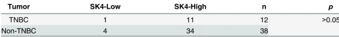
make a quantitative analysis as reported inTable 1. We found a high SK4 expression in 91.7%
(n = 11) and 89.5% (n = 34) of TNBC and non-TNBC cells, respectively, with no significant
difference between the two groups (p>0.05). We also compared SK4 and E-cadherin protein
expression in breast cancer tissues and non-tumor breast tissues using WB (Fig 1E). We
observed increased SK4 expression and decreased E-cadherin expression in the tumor tissues.
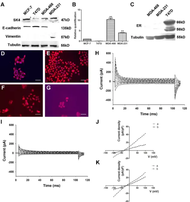
SK4 channels are expressed functionally in breast cancer cell lines
Four well-characterized breast cancer cell lines, MCF-7, T47D, MDA-MB-231 (MDA-231) and MDA-MB-468 (MDA-468), were included. We first found that SK4 proteins were expressed in
MCF-7, MDA-468 and MDA-231 cells but showed little expression in T47D cells (Fig 2A). At
the same time, we detected the EMT state of the cell lines and found that MCF-7, T47D and MDA-468 cells expressed the epithelial cell marker E-cadherin but did not express the mesen-chymal cell marker Vimentin, whereas MDA-231 cells exhibited the opposite expression
pat-tern (Fig 2A). Next, we measured the expression levels of SK4 mRNA in breast cancer cells.
SK4 mRNA was detected in all cell lines (Fig 2B). With MCF-7 cells as the control, the
relative SK4 mRNA levels were 1.00 ± 0.06 times in MCF-7 cells, 0.06 ± 0.01 in T47D cells, 10.38 ± 1.09 in MDA-468 cells, and 3.97 ± 0.27 in MDA-231 cells. Again, we investigated the
expression of ER in the T47D, MDA-231 and MDA-468 cell lines.Fig 2Cshows a clear and
specific band in T47D cells but not in MDA-468 or MDA-231 cells, indicating that ER protein was only expressed in T47D cells. We also located the SK4 proteins in these four cell lines. In
Fig 2D–2F, SK4 proteins (red color) were clearly detected in these 3 cell lines with an enriched
signal on the cell membrane, while the signal was relatively negative in T47D cells (Fig 2G).
These data suggested that SK4 protein was expressed in MCF-7, MDA-468 and MDA-231 cells but showed little expression in T47D cells.
Next, we determined whether SK4 channels were functional in MDA-231 cells. First, with
350 nM free Ca2+in the pipette solution and a step voltage stimulation, apparent outward
cur-rents were detected (Fig 2H). Three minutes after MDA-231 cells were treated with 5μM of
the SK4-specific blocker TRAM-34, the outward currents decreased apparently (Fig 2I). The
voltage-current density curve shown inFig 2Jindicates that TRAM-34 could block the outward
was added in the pipette solution, the reversal potential turned right to nearly -20 mV, and the
block effect of TRAM-34 weakened (Fig 2K).
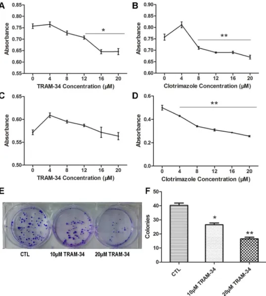
The inhibitory effect of the SK4-specific blocker on MDA-MB-231 cell
proliferation
To determine whether SK4 channels have an effect on the proliferation of breast cancer cells, we applied the SK4-specific inhibitors TRAM-34 and clotrimazole to MDA-231 and T47D
cells. For the MDA-231 cells, the MTT assay indicated that the absorbance (OD) of the 20-μM
Fig 1. SK4 proteins expressed in breast cancer tissue.(A-D) SK4 IHC in fours subtypes of breast cancer tissues including Luminal A (A), Luminal B (B), HER2 (C), and TNBC (D). Scale bars, 50μm. (E)
Immunoblotting of SK4 and E-cadherin in breast cancer tissues (BC1 and BC2) and non-tumor breast tissues (Non-Tumor1 and Non-Tumor2).
doi:10.1371/journal.pone.0154471.g001
Table 1. Comparison of SK4 expression in tumor subtypes for 50 patients using Fisher’s exact test.
Tumor SK4-Low SK4-High n p
TNBC 1 11 12 >0.05
Non-TNBC 4 34 38
For the 12 cases of TNBC and 38 cases of luminal and HER2 breast cancer (Non-TNBC), low SK4 expression was defined as an IHC score of6, and high,>6.
TRAM-34- and clotrimazole-treated groups was 0.65 ± 0.02 and 0.67 ± 0.01, respectively,
which was significantly lower compared with that of the control group (0.76 ± 0.02;
p<0.05;
Fig 3A and 3B). However, for T47D cells, the OD values of the 20-μM TRAM-34 and control
group were not apparently different, with values of 0.57 ± 0.01 and 0.56 ± 0.01, respectively
(p>0.05;Fig 3C). However, the data suggested that clotrimazole (4–20μM) could inhibit T47D
cell proliferation significantly in a dose-dependent manner (
p<0.01;Fig 3D). As a result,
Fig 2. Functional expression of SK4 channels in breast cancer cells.(A) Immunoblotting of SK4 and EMT-related proteins (E-cadherin and Vimentin) in breast cancer cell lines. (B) Comparison of SK4 mRNA expression in 4 breast cancer cell lines as determined by real-time PCR; n = 3. (C) Immunoblotting of ER protein in MDA-MB-468, MDA-MB-231 and T47D cells. (D-G) Immunostaining of SK4 (red) and nuclear marker DAPI (blue) in MDA-MB-231 (D), MDA-MB-468 (E), MCF-7 (F) and T47D (G) cells. Scale bars, 50μm. (H, I) Whole-cell recording of MDA-MB-231 cells before (H) and after (I) 5-μM TRAM-34
treatment. (J, K) With (J) or without (K) 350 nM free Ca2+in the pipette solution, the voltage-current density curves show the currents changes before (a) and after (b) TRAM-34 treatment. The currents were evoked by step voltage ranging from -100 mV to +100 mV in steps of 10 mV every 100 ms. Dunnett’s Multiple Comparison Test was applied in comparison,**p<0.01.
TRAM-34 and clotrimazole could inhibit MDA-231 cell proliferation, while T47D cell prolifer-ation could only be inhibited by clotrimazole.
TRAM-34 suppresses the colony formation of MDA-MB-231 cells
A colony-formation assay was applied to evaluate the effect of TRAM-34 on the colony-forma-tion ability of MDA-MB-231 cells. There were 40 ± 3.4 colonies in the control group, 27 ± 2.6
in the 10-μM TRAM-34-treated group (p<0.05) and 17 ± 2.4 in the 20-μM TRAM-34-treated
group (
p<0.01;Fig 3E and 3F). The colony numbers in the intervention groups were notably
reduced compared with the control group, indicating that TRAM-34 could inhibit the colony-formation ability of MDA-231 cells.
Fig 3. Blockage of SK4 channels inhibits MDA-MB-231 cell proliferation and colony formation ability, but not that of T47D cells.(A-D) Cell growth of MDA-MB-231 (A, B) and T47D cells (C, D) was analyzed using an MTT assay. The two cell lines were treated with 0–20μM TRAM-34 (A, C) or clotrimazole (B, D) for
48 h, and the absorbance was measured; n = 5. (E, F) Images of the formed MDA-MB-231 colonies in the control group (CTL) and treatment groups (10μM TRAM-34 and 20μM TRAM-34); the bar represents
separate counts of the colonies; n = 4. The data are presented as the mean±SD, and Dunnett’s Multiple
Comparison Test was applied in comparison.*p<0.05,**p<0.01.
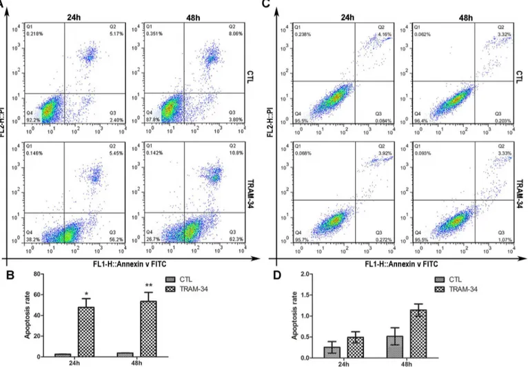
Blockage of SK4 channels promotes MDA-MB-231 cell apoptosis
We then examined whether TRAM-34 could affect the apoptosis of MDA-MB-231 and T47D
cells. For MDA-231, 24 h after treatment with 20μM TRAM-34, the cell early apoptosis rate
(Q3) was 56.2%, and the rate was elevated to 62.3% 48 h later, and both of these rates were
apparently increased compared with those of the controls (Fig 4A and 4B), while T47D cells
showed no significantly different apoptosis rate between the TRAM-34 group and the control
(Fig 4C and 4D). This result indicated that TRAM-34 could facilitate the apoptosis of
MDA-MB-231 cells but not T47D cells.
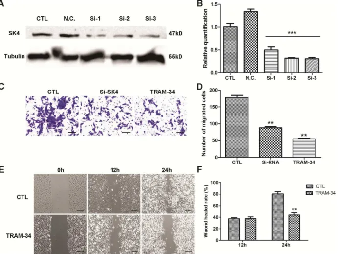
Down-regulation of SK4 channels inhibits MDA-MB-231 cell migration
To suppress the expression of SK4 proteins, a SK4-specific siRNA was used. MDA-MB-231 cells were transfected with SK4 siRNA, and the consequent silencing was confirmed using WB
and real-time PCR (Fig 5A and 5B). Next we evaluated the migration ability of MDA-231 cells.
During a transwell migration assay, the number of migrated cells in the SK4 siRNA- and
Fig 4. Blockage of SK4 channels promotes apoptosis in MDA-MB-231 cells but not T47D cells.MDA-MB-231 (A, B) and T47D (C, D) cells were treated with 20μM TRAM-34 for 24 or 48 h, and cell apoptosis was analyzed by Annexin V-FITC/ PI-PE staining and flow cytometry. The bar of MDA-MB-231
indicates that the apoptosis rate of the TRAM-34-treated group increased apparently compared with that of the control (CTL). For T47D, the difference was not significant. The data are presented as the mean±SD, and unpairedttest was applied in comparison. n = 3;*p<0.05,**p<0.01.
TRAM-34-treated groups was 88 ± 7.0 and 55 ± 4.9, respectively, while in the control group,
the number increased to 178 ± 11.7 (Fig 5C and 5D). In addition, TRAM-34 was applied in a
wound-healing assay, and 24 h after treatment, the wound-healing rate of the 10-μM
TRAM-34-treated group was 43.9% ± 5.4, which was substantially lower than that of the control group
(80.3% ± 5.9;Fig 5E and 5F). These data suggested that down-regulation of SK4 channels
sig-nificantly inhibited the migration ability of MDA-MB-231 cells (p<0.01).
The EGF/bFGF-induced EMT of MDA-MB-231 cells correlates with SK4
channels
Next, we investigated the effect of SK4 channels on the EMT of MDA-MB-231 cells. Firstly, to
construct a breast cancer EMT cell model, we applied TGF-β1 and EGF/bFGF to induce EMT
and found that none of the breast cancer cell lines could undergo a TGF-β1 (5–10
ng/ml)-induced EMT. However, EGF/bFGF could mediate the EMT of 231 and MDA-MB-468 cells. After treatment with EGF (20 ng/ml) and bFGF (10 ng/ml), MDA-231 cells became Fig 5. Down-regulation of SK4 channels inhibits the migration of MDA-MB-231 cells.A negative control siRNA (N.C.) and 3 SK4-specific siRNAs (Si-1, Si-2 and Si-3) were transfected into MDA-MB-231 cells, and 20μM TRAM-34 was added to the TRAM-34-treated group to inhibit SK4 channels. (A, B)
Knockdown of SK4 by siRNA was confirmed using immunoblotting and real-time PCR; n = 3. (C, D) The images and bar of the transwell migration assay indicate that the counts of migrated cells in SK4 siRNA (Si-SK4)- and TRAM-34-treated group were significantly less than those of the control (CTL). Scale bars, 50μm; n = 4. (E, F) The images and bar of the wound-healing assay. The wound-healing rate represents the distance migrated by cells at certain time
divided by the wound distance at 0 h. Scale bars, 100μm; n = 3. The data are presented as the mean±SD, Dunnett’s Multiple Comparison Test was applied
in (B) and (D), and unpairedttest in (F).**p<0.01,***p<0.001.
elongated with enhanced Vimentin protein and mRNA expression, and up-regulation of the
transcription factors (TFs) Slug and Snail1 was observed (Fig 6A–6C). However, with the same
treatment, T47D cells did not show a change in their cell morphology (Fig 6A), and neither did
MCF-7 cells (S1 Fig). When MDA-231 cells underwent EMT, SK4 mRNA expression was
increased (Fig 6C). In addition, down-regulation of SK4 expression suppressed Vimentin and
Snail1 expression (Fig 6D). These results revealed that the EGF/bFGF-induced EMT of
MDA-231 cells correlated with the expression of SK4 channels.
Discussion
SK4, also known as KCa3.1 or hIKCa1, encoded by the gene KCNN4, is one member of the
cal-cium-activated K+channels that open in response to cytosolic calcium. This channel mediates
a link between cytosolic calcium and membrane potential [13–17]. SK4 channels are widely
expressed in epithelial tissues, fibroblasts and hematopoietic-derived cells [18–22]. SK4
chan-nels have physiological effects on fluid secretion, cell volume regulation, cell proliferation and migration.
Fig 6. The EGF/bFGF-induced EMT of MDA-MB-231 cells correlates with SK4 channels.(A) Phase contrast images of MDA-231 and T47D cells treated with (E+b) or without (CTL) EGF/bFGF for 1 day, 3 days and 5 days. Scale bars, 100μm. (B, C) The EGF/bFGF-induced EMT of MDA-231 cells was
confirmed using immunoblotting and real-time PCR of EMT markers (Vimentin, Snail1 and Slug), and the SK4 mRNA level increased after EMT. (D) Immunoblotting of EMT-related proteins (Vimentin and Snail1) was performed 72 h after MDA-231 cells were transfected with negative control siRNA (N.C.) or SK4-specific siRNA (Si-SK4); cells that did not undergo transfection served as a control (CTL). The data are presented as the mean±SD, and pairedttest was applied in comparison. n = 3;*p<0.05.
Moreover, increasing evidence, including our previous research, indicates that SK4 channels participate in the tumor progression. SK4 mRNA is overexpressed in several human cancer cell
lines and tissues, such as glioblastoma cells [23], pancreatic cancer cells [24], prostate cancer
cells [25], melanoma cells [26], endometrial cancer cells [6] and liver cancer tissue [7]. Recent
in vitrostudies indicate that specific blockers of SK4 channels such as TRAM-34 and clotrima-zole can inhibit the proliferation of these cancer cells, further indicating that SK4 channels play
an important role in cancer cell proliferation [24,26]. SK4 channels are also closely related to
cancer cell apoptosis, migration and the EMT process [8–10,27]. However, research on SK4 in
TNBC is limited, prompting us to investigate whether SK4 channels are involved in the biologi-cally malignant behavior of TNBC.
In the present study, we explored the expression of SK4 proteins in various subtypes of breast cancer tissues and cell lines. By employing IHC and WB, we demonstrated that increased levels of SK4 protein were present in breast cancer tissues, as is the case for several other tumor types. However, across different subtypes of breast cancer, including TNBC, lumi-nal and HER2 breast cancer, the SK4 protein was strongly expressed with no apparent differ-ences. Interestingly, though the difference in expression level was not significant, concentrated SK4 proteins were observed at the cytomembrane of the TNBC cells. In consideration of the functions of the SK4 channels in cell proliferation, transition and migration, this phenomenon may explain why TNBC relapses early and easily metastasizes.
Regarding the expression of SK4 mRNA and proteins in 4 different breast cancer cell lines, including two TNBC cell lines (MDA-MB-231 and MDA-MB-468), and two luminal breast
cancer cell lines (MCF-7 and T47D) [28], we found relatively higher expression levels of SK4
mRNA and protein in the TNBC cell lines and reduced expression in T47D cells. SK4 proteins could be detected on the cell membrane using immunofluorescence. Moreover, during the
patch-clamp experiments, we applied 350 nM free Ca2+in the intracellular fluid and found
apparent outward currents. The currents were apparently decreased after the TRAM-34
treat-ment. When no free Ca2+was applied in the pipette solution, the reversal potential turned to
the right with diminished TRAM-34 block effect. This result suggests that calcium-activated channels were functional in MDA-MB-231 cells and they could be inhibited by SK4-specific blocker TRAM-34. All of these data indicate that SK4 channels were functionally expressed in TNBC cells.
EMT, the first step towards metastasis, is a transition process in which epithelial cells acquire mesenchymal characteristics. The hallmarks of the EMT can be summarized as the loss of epithelial cells markers such as E-cadherin, the loss of apico-basal polarity, the acquisition of mesenchymal cells markers such as Vimentin, and the acquisition of migratory and invasive
properties [29,30]. An increasing number of studies have indicated that EMT is involved not
only in metastatic events, but also in other events such as the resistance to cell death,
chemo-therapy and immunochemo-therapy [31]. In the present study, we evaluated ER expression and the
expression of the EMT-related markers E-cadherin and Vimentin in several cancer cell lines and tissues, confirming that T47D cells were epithelial luminal breast cancer cells and MDA-MB-231 cells were mesenchymal-like TNBC cells. This result suggests that epithelial
and mesenchymal cells can be regarded as two extremes of a transition [32]. Besides, the
inter-mediate phenotype of the transition corresponds to a partial EMT that is also observed in
cer-tain pathological conditions such as carcinosarcoma, renal and liver epithelial fibrosis [33–35].
Thus, MCF-7, T47D and MDA-468 cells are in the“epithelial”state with relatively higher
polarity and lower motility, and MDA-231 cells are in the“mesenchymal”state, with less
polar-ity and greater motilpolar-ity.
apoptosis of SK4-positive TNBC cells MDA-MB-231. The results are in agreement with the
work of Ouadid-Ahidouchet al., who reported that the block of SK4 channels in MCF-7
inhibited cell cycle progression [36]. Interestingly, despite expressing little SK4 protein, T47D
cell proliferation was significantly inhibited by clotrimazole and weakly promoted by TRAM-34. This observation can be explained by the fact that clotrimazole inhibits human
cyto-chrome P450 enzymes [37–39], while TRAM-34 selectively inhibits SK4 without blocking
cytochrome P450 enzymes [40]. Moreover, another study demonstrated that TRAM-34
stim-ulated the proliferation of breast cancer cells via the activation of ERs [41]; thus, TRAM-34
may stimulate the growth of ER-positive T47D cells. Therefore, in our study, it is reasonable that TRAM-34 could inhibit cell proliferation and promote the apoptosis of the ER-negative MDA-MB-231 cells.
Given the evidence that calcium-activated potassium channels play an important role in
tumor cell motility [9,42], we investigated the effect of down-regulating SK4 channels on
TNBC cell movement. Using SK4-specific siRNA, SK4 coding gene KCNN4 was successfully knocked down, and the motility of the siRNA-SK4 MDA-MB-231 cells was apparently weak-ened. The blockage of SK4 channels by TRAM-34 had a similar effect. Thus, SK4 may contrib-ute to TNBC cell migration.
Although the half maximal inhibitory concentration (IC50) of TRAM-34 measured by
patch-clamp technique is 20 nM [40], high concentration of TRAM-34 was appliedin vitroor/
andin vivostudies to generate effective effects. For instance, in one study, 30μM TRAM-34
was employed to suppress the growth of human endometrial cancer cells and inhibit the
pro-gression of human endometrial carcinoma in nude mice [6]. Another study proved that
migra-tion-associated secretion of melanoma is diminished by TRAM-34 at a concentration of 40μM
[27]. During the investigation the effects of SK4 channels on breast cancer cell growth and
migration, we found that the effective concentration of TRAM-34 was greater than 8μM.
Finally, based on our founding that SK4 is up-regulated and E-cadherin is down-regulated in breast cancer tissues, we explored whether SK4 channels participated in the EMT progress
of breast cancer cells. As TGF-β1-mediated induction of an EMT is a rare eventin vitro[43],
and we confirmed that the commonly used cell factor TGF-β1 could not induce EMT in the
four breast cancer cell lines. By employing another routine method [31], MDA-MB-231 and
MDA-MB-468 cells, but not MCF-7 and T47D cells, underwent an EMT that was successfully mediated by the cell growth factor EGF combined with bFGF. Approximately 3 days after treat-ment with EGF/bFGF, MDA-MB-231 cells were spindle-like and exhibited up-regulated levels of Vimentin and EMT-related transcription factors. One explanation for these results is that
several breast cancer cell lines are insensitive to TGF-β1 with respect to the EMT; in contrast,
various breast cancer cell lines may in different EMT stages, and TNBC cell lines may more easily adopt a mesenchymal phenotype. In addition, after the EMT of MDA-231, enhanced SK4 mRNA expression was detected. To further confirm the relationship between SK4 chan-nels and EMT, we down-regulated SK4 expression and found that Vimentin and Snail1 expres-sion was also suppressed. These results indicated that the EMT process of MDA-MB-231 cells correlated with the expression of SK4 channels.
In summary, our results provide evidence that SK4 channels are expressed in TNBC and that inhibition of these channels suppresses the proliferation, migration and EMT process of TNBC cells. SK4 may be a potential and novel target for TNBC treatment.
Supporting Information
and 5 days. Scale bars, 100μm.
(TIF)
S1 File. Data statement.Related data in Figs1–6andTable 1were stated separately. (ZIP)
S1 Table. Primer sequences of relevant genes.
(DOCX)
S2 Table. siRNA sequences and target sequences.
(DOCX)
Acknowledgments
We thank Dr. Lu Wang, Dr. Ying Xiang, Dr. Hui Guo, Dr. Yibing Hu, Dr. Yaqi Duan, Dr. Qiang Tang and Ruixue Yue for technical support and helpful suggestions. We apologize to the colleagues whose work was not cited.
Author Contributions
Conceived and designed the experiments: PZ XY JL. Performed the experiments: PZ JY JL QY WS ZZ LZ. Analyzed the data: PZ XY JL. Contributed reagents/materials/analysis tools: PZ JL XY. Wrote the paper: PZ JL.
References
1. Torre LA, Bray F, Siegel RL, Ferlay J, Lortet-Tieulent J, Jemal A. Global cancer statistics, 2012. CA Cancer J Clin. 2015; 65: 87–108. doi:10.3322/caac.21262PMID:25651787
2. Foulkes WD, Smith IE, Reis-Filho JS. Triple-negative breast cancer. N Engl J Med. 2010; 363: 1938– 1948. doi:10.1056/NEJMra1001389PMID:21067385
3. Metzger-Filho O, Tutt A, de Azambuja E, Saini KS, Viale G, Loi S, et al. Dissecting the heterogeneity of triple-negative breast cancer. J Clin Oncol. 2012; 30: 1879–1887. doi:10.1200/JCO.2011.38.2010 PMID:22454417
4. Brewster AM, Chavez-MacGregor M, Brown P. Epidemiology, biology, and treatment of triple-negative breast cancer in women of African ancestry. Lancet Oncol. 2014; 15: e625–e634. doi: 10.1016/S1470-2045(14)70364-XPMID:25456381
5. Pardo LA, Stühmer W. The roles of K(+) channels in cancer. Nat Rev Cancer. 2014; 14: 39–48. doi:10. 1038/nrc3635PMID:24336491
6. Wang ZH, Shen B, Yao HL, Jia YC, Ren J, Feng YJ, et al. Blockage of intermediate-conductance-Ca(2 +) -activated K(+) channels inhibits progression of human endometrial cancer. Oncogene. 2007; 26: 5107–5114. doi:10.1038/sj.onc.1210308PMID:17310992
7. Yang X, Liu J, Zhang R, Yin Q, Shen W, Yi J. Inhibitory effects of blockage of intermediate conductance Ca(2+)-activated K (+) channels on proliferation of hepatocellular carcinoma cells. J Hua Zhong U Sci Technol Med Sci. 2013; 33: 86–89. doi:10.1007/s11596-013-1076-0
8. McFerrin MB, Turner KL, Cuddapah VA, Sontheimer H. Differential role of IK and BK potassium chan-nels as mediators of intrinsic and extrinsic apoptotic cell death. Am J Physiol Cell Physiol. 2012; 303: C1070–C1078. doi:10.1152/ajpcell.00040.2012PMID:22992678
9. Ruggieri P, Mangino G, Fioretti B, Catacuzzeno L, Puca R, Ponti D, et al. The inhibition of KCa3.1 chan-nels activity reduces cell motility in glioblastoma derived cancer stem cells. PLOS ONE. 2012; 7: e47825. doi:10.1371/journal.pone.0047825PMID:23110108
10. Roach KM, Wulff H, Feghali-Bostwick C, Amrani Y, Bradding P. Increased constitutiveαSMA and
Smad2/3 expression in idiopathic pulmonary fibrosis myofibroblasts is KCa3.1-dependent. Respir Res. 2014; 15: 155. doi:10.1186/s12931-014-0155-5PMID:25476248
12. Fu RG, Zhang T, Wang L, Du Y, Jia LN, Hou JJ, et al. Inhibition of the K+ channel K(Ca)3.1 reduces TGF-beta1-induced premature senescence, myofibroblast phenotype transition and proliferation of mesangial cells. PLOS ONE. 2014; 9: e87410. doi:10.1371/journal.pone.0087410PMID:24489911 13. Ghanshani S, Coleman M, Gustavsson P, Wu AC, Gargus JJ, Gutman GA, et al. Human
calcium-acti-vated potassium channel gene KCNN4 maps to chromosome 19q13.2 in the region deleted in dia-mond-blackfan anemia. Genomics. 1998; 51: 160–161. doi:10.1006/geno.1998.5333PMID:9693050 14. Wei AD, Gutman GA, Aldrich R, Chandy KG, Grissmer S, Wulff H, et al. Nomenclature and molecular
relationships of calcium-activated potassium channels. Pharmacol Rev. 2005; 57: 463–472. doi:10. 1124/pr.57.4.9PMID:16382103
15. Joiner WJ, Wang L-, Tang MD, Kaczmarek LK. hSK4, a member of a novel subfamily of calcium-acti-vated potassium channels. Proc Natl Acad Sci USA. 1997; 94: 11013–11018. doi:10.1073/pnas.94.20. 11013PMID:9380751
16. Ishii TM, Silvia C, Hirschberg B, Bond CT, Adelman JP, Maylie J. A human intermediate conductance calcium-activated potassium channel. Proc Natl Acad Sci USA. 1997; 94: 11651–11656. doi:10.1073/ pnas.94.21.11651PMID:9326665
17. Logsdon NJ, Kang J, Togo JA, Christian EP, Aiyar J. A novel gene, hKCa4, encodes the calcium-acti-vated potassium channel in human T lymphocytes. J Biol Chem. 1997; 272: 32723–32726. doi:10. 1074/jbc.272.52.32723PMID:9407042
18. Wulff H, Castle NA. Therapeutic potential of KCa3.1 blockers: recent advances and promising trends. Expert Rev Clin Pharmacol. 2010; 3: 385–396. doi:10.1586/ecp.10.11PMID:22111618
19. Peña TL, Rane SG. The fibroblast intermediate conductance K(Ca) channel, FIK, as a prototype for the cell growth regulatory function of the IK channel family. J Membr Biol. 1999; 172: 249–257. doi:10. 1007/s002329900601PMID:10568794
20. Neylon CB, Lang RJ, Fu Y, Bobik A, Reinhart PH. Molecular cloning and characterization of the inter-mediate-conductance Ca(2+)-activated K(+) channel in vascular smooth muscle: relationship between K(Ca) channel diversity and smooth muscle cell function. Circ Res. 1999; 85: e33–e43.PMID: 10532960
21. Kohler R, Wulff H, Eichler I, Kneifel M, Neumann D, Knorr A. Blockade of the intermediate-conductance calcium-activated potassium channel as a new therapeutic strategy for restenosis. Circulation. 2003; 108: 1119–1125. doi:10.1161/01.CIR.0000086464.04719.DDPMID:12939222
22. Chen MX, Gorman SA, Benson B, Singh K, Hieble JP, Michel MC, et al. Small and intermediate con-ductance Ca(2+)-activated K+ channels confer distinctive patterns of distribution in human tissues and differential cellular localisation in the colon and corpus cavernosum. Naunyn Schmiedebergs Arch Pharmacol. 2004; 369: 602–615. doi:10.1007/s00210-004-0934-5PMID:15127180
23. Fioretti B, Castigli E, Micheli MR, Bova R, Sciaccaluga M, Harper A, et al. Expression and modulation of the intermediate- conductance Ca2+-activated K+ channel in glioblastoma GL-15 cells. Cell Physiol Biochem Int J Exp Cell Physiol Biochem Pharmacol. 2006; 18: 47–56. doi:10.1159/000095135 24. Jager H, Dreker T, Buck A, Giehl K, Gress T, Grissmer S. Blockage of intermediate-conductance Ca2
+-activated K+ channels inhibit human pancreatic cancer cell growth in vitro. Mol Pharmacol. 2004; 65: 630–638. doi:10.1124/mol.65.3.630PMID:14978241
25. Parihar AS, Coghlan MJ, Gopalakrishnan M, Shieh C. Effects of intermediate-conductance Ca2+-acti-vated K+ channel modulators on human prostate cancer cell proliferation. Eur J Pharmacol. 2003; 471: 157–164. doi:10.1016/S0014-2999(03)01825-9PMID:12826234
26. Tajima N, Schönherr K, Niedling S, Kaatz M, Kanno H, Schönherr R, et al. Ca2+-activated K+ channels in human melanoma cells are up-regulated by hypoxia involving hypoxia-inducible factor-1alpha and the von Hippel-Lindau protein. J Physiol. 2006; 571: 349–359. doi:10.1113/jphysiol.2005.096818 PMID:16396931
27. Schmidt J, Friebel K, Schönherr R, Coppolino MG, Bosserhoff A. Migration-associated secretion of melanoma inhibitory activity at the cell rear is supported by KCa3.1 potassium channels. Cell Res. 2010; 20: 1224–1238. doi:10.1038/cr.2010.121PMID:20733613
28. Neve RM, Chin K, Fridlyand J, Yeh J, Baehner FL, Fevr T, et al. A collection of breast cancer cell lines for the study of functionally distinct cancer subtypes. Cancer Cell. 2006; 10: 515–527. doi:10.1016/j. ccr.2006.10.008PMID:17157791
29. Nieto MA. Epithelial plasticity: a common theme in embryonic and cancer cells. Science. 2013; 342: 1234850. doi:10.1126/science.1234850PMID:24202173
30. Sleeman JP, Thiery JP. SnapShot: the epithelial-mesenchymal transition. Cell. 2011; 145: 162–162.e1. doi:10.1016/j.cell.2011.03.029PMID:21458675
32. Nieto MA. The ins and outs of the epithelial to mesenchymal transition in health and disease. Annu Rev Cell Dev Biol. 2011; 27: 347–376. doi:10.1146/annurev-cellbio-092910-154036PMID:21740232 33. Sarrio D, Rodriguez-Pinilla SM, Hardisson D, Cano A, Moreno-Bueno G, Palacios J.
Epithelial-mesen-chymal transition in breast cancer relates to the basal-like phenotype. Cancer Res. 2008; 68: 989–997. doi:10.1158/0008-5472.CAN-07-2017PMID:18281472
34. Iwano M, Plieth D, Danoff TM, Xue C, Okada H, Neilson EG. Evidence that fibroblasts derive from epi-thelium during tissue fibrosis. J Clin Invest. 2002; 110: 341–350. doi:10.1172/JCI15518PMID: 12163453
35. Zeisberg M, Yang C, Martino M, Duncan MB, Rieder F, Tanjore H, et al. Fibroblasts derive from hepato-cytes in liver fibrosis via epithelial to mesenchymal transition. J Biol Chem. 2007; 282: 23337–23347. doi:10.1074/jbc.M700194200PMID:17562716
36. Ouadid-Ahidouch H, Roudbaraki M, Delcourt P, Ahidouch A, Joury N, Prevarskaya N. Functional and melecular identification of intermediate-conductance Ca(2+)-activated K(+) channels in breast cancer cells: assocication with cell cycle progression. American journal of physiology Cell physiology. 2004; 287(1):C125–34. doi:10.1152/ajpcell.00488.2003PMID:14985237
37. Ayub M, Levell MJ. The inhibition of human prostatic aromatase activity by imidazole drugs including ketoconazole and 4-hydroxyandrostenedione. Biochem Pharmacol. 1990; 40: 1569–1575. doi:10. 1016/0006-2952(90)90456-UPMID:2222512
38. Maurice M, Pichard L, Daujat M, Fabre I, Joyeux H, Domergue J, et al. Effects of imidazole derivatives on cytochromes P450 from human hepatocytes in primary culture. FASEB J. 1992; 6: 752–758. PMID: 1371482
39. Fowler SM, Riley RJ, Pritchard MP, Sutcliffe MJ, Friedberg T, Wolf CR. Amino acid 305 determines cat-alytic center accessibility in CYP3A4. Biochem. 2000; 39: 4406–4414. doi:10.1021/bi992372u 40. Wulff H, Miller MJ, Hansel W, Grissmer S, Cahalan MD, Chandy KG. Design of a potent and selective
inhibitor of the intermediate-conductance Ca2+-activated K+ channel, IKCa1: a potential immunosup-pressant. Proc Natl Acad Sci USA. 2000; 97: 8151–8156. doi:10.1073/pnas.97.14.8151PMID: 10884437
41. Roy J, Cowley E, Blay J, Linsdell P. The intermediate conductance Ca2+-activated K+ channel inhibitor TRAM-34 stimulates proliferation of breast cancer cells via activation of oestrogen receptors. Br J Phar-macol. 2010; 159: 650–658. doi:10.1111/j.1476-5381.2009.00557.xPMID:20050851
42. Potier M, Joulin V, Roger S, Besson P, Jourdan ML, Leguennec JY, et al. Identification of SK3 channel as a new mediator of breast cancer cell migration. Molecular cancer therapeutics. 2006; 5(11):2946– 53. doi:10.1158/1535-7163.MCT-06-0194PMID:17121942