Herceptin against HER-2/Neu-Expressing Breast Cancer
Cells
Aamir Ahmad1, Shadan Ali2, Alia Ahmed1, Azfur S. Ali1, Avraham Raz1,2, Wael A. Sakr1, KM Wahidur Rahman1*
1Department of Pathology, Karmanos Cancer Institute, Wayne State University School of Medicine, Detroit, Michigan, United States of America,2Division of Hematology/ Oncology, Karmanos Cancer Institute, Wayne State University School of Medicine, Detroit, Michigan, United States of America
Abstract
Herceptin failure is a major clinical problem in breast cancer. A subset of breast cancer patients with highHER-2/neulevels eventually experience metastatic disease progression when treated with Herceptin as a single agent. Mechanistic details of development of this aggressive disease are not clear. Therefore, there is a dire need to better understand the mechanisms by which drug resistance develops and to design new combined treatments that benefit patients with aggressive breast cancer and have minimal toxicity. We hypothesized that 3, 39-diindolylmethane (DIM), a non-toxic agent can be combined with Herceptin to treat breast cancers with high levels ofHER-2/neu. Here, we evaluated the effects of Herceptin alone and in combination with DIM on cell viability, apoptosis and clonogenic assays in SKBR3 (HER-2/neu-expressing) and MDA-MB-468 (HER-2/neu negative) breast cancer cells. We found that DIM could enhance the effectiveness of Herceptin by significantly reducing cell viability, which was associated with apoptosis-induction and significant inhibition of colony formation, compared with single agent treatment. These results were consistent with the down-regulation ofAktandNF-kB p65. Mechanistic investigations revealed a significant upregulation ofmiR-200and reduction ofFoxM1expression in DIM and Herceptin-treated breast cancer cells. We, therefore, transfected cells with pre-miR-200 or silenced FoxM1 in these cells for understanding the molecular mechanism involved. These results provide experimental evidence, for the first time, that DIM plus Herceptin therapy could be translated to the clinic as a therapeutic modality to improve treatment outcome of patients with breast cancer, particularly for the patients whose tumors express high levels ofHER-2/neu.
Citation:Ahmad A, Ali S, Ahmed A, Ali AS, Raz A, et al. (2013) 3, 39-diindolylmethane Enhances the Effectiveness of Herceptin against HER-2/Neu-Expressing Breast Cancer Cells. PLoS ONE 8(1): e54657. doi:10.1371/journal.pone.0054657
Editor:Ming Tan, University of South Alabama, United States of America
ReceivedJune 4, 2012;AcceptedDecember 17, 2012;PublishedJanuary 22, 2013
Copyright:ß2013 Ahmad et al. This is an open-access article distributed under the terms of the Creative Commons Attribution License, which permits
unrestricted use, distribution, and reproduction in any medium, provided the original author and source are credited.
Funding:This work was supported in part by the Department of Pathology, Wayne State University, Detroit, Michigan, National Institutes of Health-National Cancer Institute grant R01CA51714 (AR) and Department of Defense grant W81XWH-05-1-0505 (KMWR). The funders had no role in study design, data collection and analysis, decision to publish, or preparation of the manuscript.
Competing Interests:The authors have declared that no competing interests exist.
* E-mail: kmrahman@med.wayne.edu
Introduction
Herceptin (Trastuzumab), a humanized monoclonal antibody targetingHER-2/neu, has shown some benefit as a treatment for patients with HER-2/neu-expressing breast cancer, but clinical studies show that patients with high levels ofHER-2/neuwho are treated with a single dose of Herceptin progress to metastatic disease within one year [1–5]. The potential mechanisms underlying Herceptin failure are found in alteredEGFRreceptors, increasedAkt activity and IGF-IRsignaling, reduced p27kip1and
PTENlevel in breast cancer cell [3,6]. Interestingly, these signaling pathways have been reported to be modulated by a natural non-toxic agent, 3, 3-diindolylmethane (DIM) [7–9] which raises the possibility that combination of DIM with Herceptin might help to enhance the antitumor activity of Herceptin againstHER-2/neu -expressing breast cancer. Nevertheless, as not much is known about the mechanism(s) that lead to the development of aggressive disease, the mechanisms by which DIM and Herceptin may affect this process is unclear. Our current study provides new insight into the mechanism(s) of this drug combination and, therefore, has the
potential of advancing our understanding of the biology of aggressive breast cancer.
Dietary and epidemiological studies suggest that consumption of vegetables and fruits are associated with reduced risks for certain cancers, including breast cancer [10–12]. DIM is a plant-derived, natural autolytic compound found in cruciferous vegetables that selectively kills breast cancer cells without affecting normal cells [13–18] and enhances the efficacy of chemotherapeutic agents [13,14,19]. Previously we showed that DIM could reduce the concentration of Taxotere needed to induce an inhibitory effect on breast cancer cells [13,14,17,19], which raises the possibility that combination of DIM with Herceptin might help overcome the shortcomings (both toxicity and molecular cause of Herceptin failure) and increased higher efficacy of Herceptin. We believe that combination of DIM and Herceptin is more potent than individual compounds, which could maximize the effect of Herceptin-based therapies and target multiple signaling pathways.
function as oncogenes or tumor suppressor genes [20]. Among them, changes in miR-200 (miR-200a, miR-200b and miR-200c) levels have been associated with enhanced tumorigenesis and significantly correlated with decreased survival [21–24]. Further-more, down-regulation ofmiR-200is observed in aggressive breast cancer [21,25–27]. FoxM1, an oncogenic transcription factor is known to play important role in the development and progression of many malignancies including breast cancer [28–31]. Interest-ingly, it has been indicated that over-expression of FoxM1could led to decreased expression of miRNAs includingmiR-200[32,33]. With an inverse relationship between FoxM1 and miR-200, the increased expression of FoxM1, promotes oncogenesis and progression of various carcinomas, and contributes to chemother-apeutic resistance. However, the interrelationship betweenFoxM1
andmiR-200that are involved in progression of breast cancer has not yet been clarified. Furthermore, FoxM1 has been shown to confer resistance to Herceptin and microtubule-stabilizing drug Paclitaxel in breast cancer cells [34]. Our recent studies have shown that inactivation of FoxM1, and its target genes, by DIM could enhance the therapeutic efficacy of Taxotere in breast and prostate cancer cells [19] suggesting that DIM in combination with Herceptin could be useful to upregulate miR-200 and down-regulate FoxM1 which should help to develop therapeutic strategies for the prevention and/or treatment of breast cancer.
Here, we report for the first time that DIM upregulates miRNA-200and down-regulatesFoxM1inHER-2/neuexpressing, SKBR3 breast cancer cells. We also report that DIM has moderated effect on miR-200 and FoxM1 in HER-2/neu negative, MDA-MB-468 breast cancer cells. More importantly, combination of DIM and Herceptin is much more effective than either agent alone in HER-2/neuexpressing, breast cancer cells, suggesting that combination-mediated alterations inmiR-200, along with inactivation ofAktand
NF-kB p65, could be a novel approach for the treatment of patients with breast cancer, particularly for the patients whose tumors express high levels ofHER-2/neu.
Materials and Methods
Cell Lines and Reagents
Breast cancer cell lines, SKBR3 (HER-2/neu-expressing) and MDA-MB-468 (HER-2/neu negative) were obtained from ATCC (Manassas, VA). The cell lines have been tested and authenticated in core facility Applied Genomics Technology Center at Wayne State University. Primary antibodies forFoxM1, Akt, pAkt,NF-kB p65 and anti-poly (ADP-ribose) polymerase (PARP) were pur-chased from Santa Cruz Biotechnology and anti-b-actin was purchased from Sigma-Aldrich (St. Louis, MO). All secondary antibodies were obtained from Pierce. FoxM1 siRNA and control siRNA were obtained from Santa Cruz Biotechnology. Lipofec-tAMINE 2000 was purchased from Invitrogen (Carlsbad, CA). Chemiluminescence detection of proteins was done with a kit from Amersham Biosciences (Piscataway, NJ). Protease inhibitor cock-tail, MTT reagent and all other chemicals were obtained from Sigma (St. Louis, MO). DIM (marketed as BR-DIM with enhanced bioavailability), generously provided by Dr. Michael Zeligs (BioResponse, CO), was dissolved in DMSO to make a 50 mmol/L stock solution. Herceptin (Genentech, Inc) was provided by Karmanos Cancer Institute dissolved in Bacterolactic water and BWFI (1.1% benzyl alcohol) to make 21 mg/ml stock solution.
Cell Viability Assay
Cells were seeded in 96-well plates. After 24 hours, they were treated with DIM (10, 15 or 20mM) followed by treatment with
Herceptin (0.25, 0.75 or 1.00mg/ml) for 24, 48 or 72 hours. Cell growth studies were performed by 3-(4,5-Dimethylthiazol-2-yl)-2,5-diphenyltetrazolium Bromide (MTT) as described earlier [13,15].
Clonogenic Assay
Survival of breast cells was tested by clonogenic assay, as described before [17,21]. Briefly, cells were plated in 6-well plates, treated, trypsinized, re-plated in 100-mm Petri dishes and cultured at 37uC in a 5% CO2/5% O2/90% N2incubator. Colonies were stained with 2% crystal violet, counted, and quantitated.
Quantification of Apoptosis by Enzyme-linked Immunosorbent Assay
Cell death detection enzyme-linked immunosorbent assay (ELISA) kit (Roche Applied Science) was used to detect apoptosis. Cells, seeded in six-well plates, were treated with 15mM DIM,
0.75mg/ml Herceptin or their combination for 48 h, trypsinized,
and ,10,000 cells were assayed as described earlier [13,14,16]. TECAN’s microplate fluorometer (TECAN) was used to measure color intensity at 405 nm.
Western Blot Analysis
Cells were lysed in 62.5 mmol/L Tris-HCl and 2% SDS, and protein concentration was measured using BCA protein assay (Pierce). Proteins were subjected to SDS-PAGE and electropho-retically transferred to nitrocellulose membrane. Membranes were incubated with specific primary antibodies, secondary antibody conjugated with peroxidase and the signal was detected using the chemiluminescent detection system (Pierce) as described earlier [16,19,35].
RNA Isolation and Real-time Reverse Transcription-PCR (RT-PCR) Analysis
Total RNA from DIM and Herceptin-treated cells was isolated by TRIzol (Invitrogen) according to the manufacturer’s protocol and subjected to analyses by RT-PCR, as described earlier [36].
miRNA Real-time Reverse Transcription-PCR
To verify the alterations in the expression ofmiR-200in DIM and Herceptin-treated breast cancer cells, we performed RT-PCR analysis using TaqMan MicroRNA Assay Kit (Applied Biosystems) following the manufacturer’s protocol. RT reaction conditions and primers used in this study were, exactly the same as described previously [21]. Briefly, 5 ng of total RNA from each sample were subjected to reverse transcription with a specific miRNA primer (Applied Biosystems). Real-time PCRs were then carried out in a total of 25mL reaction mixture in SmartCycler II (Cepheid). Data were analyzed according to the comparative Ct method and normalized by RNU48 expression in each sample.
Transfection with Pre2/anti-miRNAs
after the last transfection, cells were seeded in 96-well plates for MTT assay and multiple wells of six well plates for real time RT-PCR and western blot analysis.
FoxM1 siRNA Transfection
To determine the effect of FoxM1 siRNA transfection, cells were plated in 100 mm petri dishes overnight and transfected with 100 nmol/l of FoxM1 siRNA or the control siRNA for 48 h by Lipofectamine 2000 following the manufacturer’s protocol.
Statistical Analysis
The data are presented as the mean values6SE. Comparisons between groups were evaluated by Student’s t test. Values of
P,0.05 were considered statistically significant.
Results
Cell Growth Inhibition by DIM and Herceptin Treatment We tested several doses of DIM and Herceptin at different time points (data not shown) and found that treatment of SKBR3 and MDA-MB-468 breast cancer cells with 15mM DIM for 72 h caused 40–60% growth inhibition which is irrespective ofHER-2/ neustatus. However, a combination of these doses resulted in 80% growth inhibition of SKBR3 breast cancer cells (Fig. 1A), suggesting a significant inhibitory effect of combination treatment inHER-2/neu-expressing breast cancer cells. As observed in the MTT assays, treatment with Herceptin alone did not show any decreased cell viability in MDA-MB-468 (HER-2/neu negative) breast cancer cells which confirms the specificity of Herceptin action. Also, DIM and Herceptin combination did not show any significant decrease of cell viability in MDA-MB-468 breast cancer cells, compared to DIM alone. Previous studies have shown the effects of different doses of Herceptin in breast cancer cells [37]. Our results show that the combination of DIM with a lower dose of Herceptin elicited significantly greater inhibition of cancer cell growth compared with either agent alone (Fig. 1A).
Effect of DIM and Herceptin on Clonogenicity of Breast Cancer Cells
The effect of DIM and Herceptin treatment on anchorage-dependent clonogenic growth was assessed by clonogenic assay (Fig. 1B). Treatment with 15mM DIM and 0.75mg/ml Herceptin resulted in a significant inhibition (90%) of colony formation in SKBR3 cells when compared with single-agent treatment (Fig. 1C). On the other hand there was no significant decrease in number of colonies in MDA-MB-468 breast cancer cells (Fig. 1C).
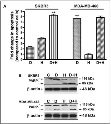
Induction of Apoptosis by DIM and Herceptin
The inhibition of cell viability was further evaluated by determining the apoptotic effects, using the cell death detection ELISA. Here we found that the combination of DIM (15mM) and Herceptin (0.75mg/ml) resulted in a significant induction of apoptosis in SKBR3 breast cancer cell lines tested (DIM, 2.25-fold; Herceptin, 4.0-fold; and DIM plus Herceptin, 8.25-fold) (Fig. 2A). In contrast, we found apoptotic effects of DIM in MDA-MB-468 cells. However, combination treatment did not induce apoptosis in MDA-MB-468 cells. Fold changes in DIM and Herceptin-treated SKBR3 and MDA-MB-468 cells relative to untreated cells are shown in (Fig. 2A). We also observed that the DIM and Herceptin combination treatment resulted in significantly more cleaved PARP compared with mono-treatment (Fig. 2B), suggesting that the combination treatment could induce greater apoptosis in breast cancer cells, particularly in HER-2/neu expressed breast
cancer cells. These results are consistent with the cell viability assay. Since inhibition of cell proliferation observed by MTT and clonogenic assay could be due to altered regulation of several genes such as FoxM1, Akt and NF-kB p65, the underlying mechanism for the inhibition of cell viability was further studied by western blot analysis to determine the effects of DIM and Herceptin on the expression of selective proteins.
Effects of DIM and Herceptin on the Expression of Selective Proteins
Previously we reported that DIM down-regulates FoxM1 in
HER-2/neu-expressing breast cancer cells [19]. Thus, we hypoth-esized that combining DIM with Herceptin may be an effective strategy for treatingHER-2/neu-expressing breast cancer.Aktand
NF-kBplay a critical role in cancer progression and our previous report suggests thatAkt/NF-kBcould be efficiently inactivated by DIM [13,15]. We further hypothesized that down-regulation of
FoxM1signaling by DIM and Herceptin will induce apoptotic cell death, which could be due to inactivation ofFoxM1directly and indirectly via inactivation of Akt and NF-kB. However, a more detailed understanding of the effects of this drug combination and its mechanism of action inHER-2/neu-expressing breast cancer is needed. Therefore, to study the functional relevance of DIM and Herceptin-mediated alteration of FoxM1, Aktand NF-kB expres-sion in breast cancer cells, we treated cells with DIM and Herceptin for 72 hours. Expression ofFoxM1 and pAktproteins was significantly reduced in SKBR3 breast cancer cells treated with DIM and Herceptin when compared with control and single treatment (Fig. 3). Importantly, we found significant down-regulation of FoxM1 in DIM and Herceptin-treated SKBR3 breast cancer cells. Interestingly, we found some inhibitory effect of DIM and Herceptin onFoxM1in MDA-MB-468 breast cancer cells. Moreover, we did not find any alterations in the total protein expression of Akt in DIM and Herceptin-treated SKBR3 and MDA-MB-468 breast cancer cells (Fig. 3). SinceNF-kBwas not found to be a significantly-changed protein after DIM and Herceptin treatment in SKBR3 and MDA-MB-468, we did not present this data in Fig. 3. Further,FoxM1has been found to be elevated in advanced cancers, including breast carcinoma [8,14,19] and our current results further underline the importance ofFoxM1in Herceptin efficacy as well. Next, we questioned the mechanism by which elevated FoxM1 levels might regulate the anti-tumor activity of Herceptin. In particular, we looked at the miRNAs that might be regulated byFoxM1. There is evidence in literature to suggest a regulatory connection betweenFoxM1and
miR-200family of miRNAs [33]. Our earlier publications have independently established a role of FoxM1 and miR-200 in the aggressiveness of breast cancer cells [16,17,19,21] but a relation-ship was never studied. Therefore, we further investigated the effects of DIM and Herceptin on miR-200s.
Upregulation ofmiR-200Expression by DIM and Herceptin
DIM and Herceptin individually, we did not observe any effect on
miR-200cof these compounds individually. However, interestingly, a very significant up-regulation of miR-200cwas observed in the combinational treatment (Fig. 4B). In MDA-MB-468 cells, DIM alone was found to upregulatemiR-200a,miR-200bbut Herceptin alone was found to have no effect and no significant up-regulation of miR-200c was observed in the combinational treatment in MDA-MB-468 cells (Fig. 4C). As expected, we did not observe any effect of Herceptin alone onmiR-200a/bexpression in MBA-MB-468 cells. However, surprisingly, in combination with DIM, Herceptin significantly induced the expression levels ofmiR-200a
and miR-200b. To evaluate an involvement of miR-200s in the cytotoxic action of DIM/Herceptin, we exposed control (trans-fected with non-specific anti-miRNAs) and miR-200s-trans-fected SKBR3 cells (transmiR-200s-trans-fected with a cocktail containing anti-miR-200a+anti-miR-200b+anti-miR-200c) to DIM plus Hercep-tin. The cells transfected with non-specific anti-miRNAs were found to be sensitive to DIM plus Herceptin treatment, however, antagonizing miR-200s significantly (p,0.01) attenuated the effects of DIM/Herceptin (Fig. 4D). These results clearly establish
a mechanistic involvement of miR-200s in DIM plus Herceptin. DIM plus Herceptin work via up-regulation of miR-200s, and antagonizing miR-200s (miR-200a+miR-200b+miR-200c) signifi-cantly diminishes the biological effects of these drugs. To determine that anti-miR-200s transfections reduced the levels of miR-200 family miRNAs, we evaluated the levels of miR-200a, miR-200b as well as miR-200c in SKBR3 cells transfected with anti-miR-200s (anti-miR-200a+anti-miR-200b+anti-miR-200c) vs. SKBR3 cells transfected with non-specific anti-miRNAs, and found that the transfections with anti-miR-200s lowered the levels of all miR-200s by at least half (Fig. 4E, controls in non-specific anti-miRNAs vs. controls in anti-miR-200s). We also looked at the levels of miR-200s after these cells were exposed to DIM plus Herceptin for 72 hours. The results suggested that while DIM plus Herceptin induced the levels of all miR-200s in control SKBR3 cells (transfected with non-specific anti-miRNAs), the induction in anti-miR-200s-transfected SKBR3 cells was considerably reduced (Fig. 4E). Western blot analysis of these samples indicated that DIM plus Herceptin down-regulated FoxM1 and pAkt levels in SKBR3 cells as expected, and this was attenuated by transfections with anti-miR-200s (Fig. 4F). The reduced induction of miR-200s (miR-200a+miR-200b+miR-200c) and inhibition of FoxM1/Akt
activation may help explain the attenuation of DIM plus Herceptin action as observed in Fig. 4D. To further validate whether miR-200 indeed could target the expression levels of
FoxM1,we investigated the effect of transfection of pre-miRNAs and FoxM1 siRNA in breast cancer cells.
Effects of pre-miRNAs and FoxM1 siRNA Transfection on Selective Proteins
Our results, as described above, revealed an effect of DIM plus Herceptin on miR-200 and FoxM1 expression individually. We were now interested in establishing a mechanism for this observation. Since miRNAs modulate the expression of their target genes andFoxM1is a predicted target ofmiR-200, we asked the question whether ectopic expression ofmiR-200can affect the expression ofFoxM1. Such an observation will suggest that DIM/ Herceptin-mediated regulation of miR-200 is important for the eventual regulation of FoxM1, which ultimately defines the aggressiveness, and possibly, the drug resistance of breast cancer cells. SKBR3 and MDA-MB-468 cells were transfected with either pre-miR-200 orFoxM1 siRNA followed by DIM and Herceptin treatment for 48 hours. In fig. 5A, it is clear that in SKBR3 cells, pre-miR-200, DIM plus pre-miR-200, as well as Herceptin plus pre-miR-200, were found to down-regulate FoxM1 and the combination was found to be significantly more effective. On the other hand, the expression ofpAkt and NF-kB p65 was further reduced with both the transfections (Fig. 5). We did not observe any effect onFoxM1, NF-kb p65andpAktin MDA-MB-468 treated with pre-miR-200, DIM plus pre-miR-200, as well as Herceptin plus pre-miR-200 individually. However, combination treatment down-regulates those genes in MDA-MB-468 breast cancer cells although more significant effects were observed in SKBR3. Conversely, transfection with FoxM1 siRNA in both SKBR3 and MDA-MB-468 cell lines showed inhibitory effects onFoxM1, NF-kb p65 and pAkt, although more significant effects were observed in SKBR3 suggesting that DIM enhances Herceptin Figure 1. Effects on cell growth after DIM and Herceptin treatment.(A) SKBR3 and MDA-MB-468 cells were treated with DIM (15mM) and Herceptin (0.75mg/ml), either alone or combination for 72 hours. DIM in combination with Herceptin significantly inhibited cell proliferation, as measured by MTT assay. (B) SKBR3 and MDA-MB-468 cells were treated with 15mM DIM, 0.75mg/ml Herceptin and combination. Assay for anchorage-dependent clonogenicity was done as described under materials and methods. (C) The bar graphs at the bottom represent quantification of results presented on the top in each case. *,P,0.05; **,P,0.01 relative to control. C, Control; D, DIM; H, Herceptin.
doi:10.1371/journal.pone.0054657.g001
Figure 2. Effects of DIM and Herceptin on apoptosis. (A) Induction of apoptosis in SKBR3 and MDA-MB-468 cells treated with 15mM of DIM, 0.75mg/ml of Herceptin, and their combination. Fold changes in DIM and Herceptin-treated SKBR3 and MDA-MB-468 cells relative to untreated cells are shown. (B) PARP cleavage assay showed that combination treatment with DIM and Herceptin induced signifi-cantly greater apoptosis. The p values (**, P,0.01) represent comparisons between cells treated by either of the drugs and their combinations by using the pairedttest. C, Control; D, DIM (15mM/L DIM); H, Herceptin (0.75mg/ml).
activity in breast cancer cells, but significant effect was found in
HER-2/neu-expressing breast cancer cells. Collectively, these findings clearly suggest that DIM and Herceptin are capable of down-regulating the expression ofFoxM1,which is normally high in malignant tumors, and upregulating the expression ofmiR-200.
Discussion
DIM is a majorin vivoacid-catalyzed condensation product of indole-3-carbinol (I3C) and a plant-derived, non-toxic, dietary agent that selectively kills breast cancer cells without affecting normal cells [15,38–40]. Previously we have shown that I3C can inhibit breast cancer cell growth in a dose dependent manner in
HER-2/neuover-expressing and in normalHER-2/neu-expressing cells by up-regulating Bax, down-regulating Bcl-2[38]. We also reported that DIM significantly inhibited the growth of breast tumor without associated toxicity [13–16,19]. Another study showed that DIM in combination with paclitaxel can inhibit cell proliferation growth by decreasing Bcl-2 in HER-2/neu human breast cancer cells [41]. This report also showed that DIM alone can decrease the activation of the HER-2/neu receptor and the combination decreases the activation ofERK1/ERK2 [41]. DIM sensitizes pancreatic tumor cells to targeted therapeutic agents such as EGFR inhibitor Erlotinib, potentiating apoptotic effect through altered regulation of EGFR signaling [42]. It has been shown that DIM or isoflavones could function as miRNA
regulators leading to the reversal of EMT phenotype [43]. Recently, we have shown that DIM could enhance the therapeutic efficacy of Taxotere in breast and prostate cancer cells [13,14,19] suggesting that DIM can sensitize cancer cells to chemotherapeu-tic agents. These findings led us to hypothesize that the combining effect of DIM and Herceptin may result in an enhanced antitumor response, which could be a useful approach for the treatment of human breast cancers, particularlyHER-2/neu-expressing breast cancer patients.
Current Herceptin-based combination therapies involving conventional chemotherapeutic drug increase response rates, time to disease progression, and survival of patients with high level of
HER-2/neu [44]. However, these treatments are associated with several toxicities [44]. A majority of cancers that initially respond to Herceptin, develop metastatic disease within 12 months [3,45– 47]. Neither Herceptin’s mechanisms of action nor those of disease progression are known, but they likely involveHER-2/neuand its downstream signaling pathways [4,47]. Therefore, identifying the mechanisms responsible for aggressiveness that could be sup-pressed by DIM plus Herceptin is important for the development of new therapeutic strategies. Our data demonstrate the efficacy of DIM to increase the effectiveness of Herceptin by significant killing of breast cancer cells inHER-2/neuexpressing breast cancer cells.
FoxM1 belongs to a family of proteins that is evolutionary conserved and which is marked by the presence of DNA-binding Figure 3. Effects of DIM and Herceptin on selective proteins.Expression ofFoxM1, Akt, pAktandNF-kB p65, andb-actinin SKBR3 and MDA-MB-468 cell lines treated with 15mM DIM, 0.75mg/ml Herceptin or the combination for 72 hours. The bar graphs at the bottom in each case are based on quantitative values of bands of interest normalized tob-actin. *,P,0.05 and **,P,0.01 relative to control.
Figure 4. Effects of DIM and Herceptin onmiR-200family.(A) Comparative expression analysis ofmiR-200in SKBR3 and MDA-MB-468 cells by real-time miRNA RT-PCR. (B and C) Relative expression analysis ofmiR-200in SKBR3 and MDA-MB-468 cells treated with 15mM DIM, 0.75mg/ml Herceptin or their combinations for 24 h. (D) Effect of anti-miR-200s (anti-miR-200a+anti-miR-200b+anti-miR-200c) on cell proliferation of SKBR3 cells treated with 15mM DIM and 0.75mg/ml Herceptin for 72 hours, as measured by MTT assay. (E) Efficacy of anti-miR-200s transfections as well the effect of DIM plus Herceptin (15mM DIM and 0.75mg/ml Herceptin for 72 hours) on expression levels of anti-miR-200s (miR-200a/miR-200b/miR-200c) was evaluated by real time RT-PCR. (F) Effect of anti-miR-200s (anti-miR-200a+anti-miR-200b+anti-miR-200c) on expression ofFoxM1andpAktin SKBR3 cells treated with 15mM DIM and 0.75mg/ml Herceptin for 72 hours, as determined by western blotting. The numbers represent percent of corresponding control normalized tob-actin levels. Her, Herceptin; anti-miR-200s, anti-miR-200a+anti-miR-200b+anti-miR-200c. *,P,0.05 and **,
P,0.01 relative to control.
domain called ‘forkhead box’. The various members of Fox family have been implicated in the progression of many cancers, including those of breast, liver, prostate, brain and lung through their ability to drive cell cycle progression and evasion of growth arrest [31].FoxM1is a key regulator of transition from G1 to S
phase as well as for the progression to mitosis. Loss of FoxM1
expression has been connected to mitotic spindle defects and accumulation of cells in mitosis, which leads to a mitotic catastrophe. Using microarray studies, we first identifiedFoxM1
as a target of DIM [16]. The consequence ofFoxM1expression on Figure 5. The expression ofFoxM1, Akt, pAktandNF-kB p65in breast cancer cells lines.Expression ofFoxM1,Akt, pAktandNF-kB p65in
SKBR3 and MDA-MB-468 cells after transfection with pre-miRNAs (pre-miR-200a+pre-miR-200b+pre-miR-200c) and FoxM1 siRNA followed by 15mM of DIM and 0.75mg/ml of Herceptin for 48 h. D, DIM; H, Herceptin. The numbers represent percent of corresponding control normalized tob-actin levels.
the cell growth, clonogenicity migration and invasion was later evaluated [31]. More recently, we showed that DIM can down-regulate FoxM1 in multiple breast cancer cells leading to a potentiation of Taxotere action [19].
The miRNAs have emerged as principal regulators of various biological and pathologic processes. They are small (19–24 nucleotides) non-coding RNA molecules that down-regulate gene expression by interacting with sequences located in the 39
untranslated region (UTR) of multiple target mRNAs, resulting in either translational repression or degradation of mRNAs. The regulation of oncogenes/tumor suppressor genes by miRNAs is increasingly being realized as a key step in the progression of human malignancies. It has been shown that miRNAs play an important role in modulating multiple cellular pathways, including cell proliferation, differentiation, and apoptosis, and thus may function as oncogenes or tumor suppressor genes [20,48,49]. In particular, decreased expression of miRNA-200 (200a, miR-200b and miR-200c) has been reported in breast cancer [50]. Furthermore, miR-200 family members are associated with resistance to several chemotherapy drugs: docetaxel in non-small cell cancer cells cisplatin in breast cancer cells and gemcitabine in cholangiocarcinoma cells [51–53]. In our study we found that DIM in combination with Herceptin can upregulate miR-200
expression, a master regulator of genes expression and intimately involved in breast cancer. As a direct proof of mechanistic involvement of miR-200 in DIM plus Herceptin action, we demonstrated that antagonizing miR-200 significantly attenuated the cytotoxic effect of DIM plus Herceptin. miR-200 family is particularly involved in the invasion and metastasis of breast cancer cells through its involvement in the regulation of epithelial-mesenchymal transition (EMT) [21]. Thus, effective up-regulation of miR-200 family by DIM can potentially help overcome the aggressive phenotype of breast cancer cells, a phenotype that is hallmark of metastatic as well as drug-resistant breast cancers. Our results suggest that DIM and Herceptin could affect miRNAs, which is likely to aid in designing novel therapies for breast cancer. On the other hand, post-transcriptional regulation of gene expression by miRNA has recently attracted major interest among researchers in relation to its involvement in cancer development [48]. Every miRNA potentially regulates the expression of numerous protein coding genes (tens to hundreds), but it has become increasingly clear that not all miRNAs are equally important. FoxM1 is a transcription factor that is important for cellular processes in carcinogenesis. By increasing expression of
FoxM1,these miR-200s could promote oncogenesis and progres-sion of various carcinomas, and contribute to chemotherapeutic resistance. However, whether FoxM1-trageting miR-200s are involved in progression of breast cancer has not yet been clarified. Consistent with our results, it was shown that down-regulation of
FoxM1by treatment of cells with FoxM1 siRNA and pre-miR-200s transfection decreasedFoxM1expression. The implication of our data would be enormous because natural nontoxic agents could be useful for the down-regulation ofFoxM1mediated via upregula-tion ofmiR-200, and our data further suggest that such strategy could in fact be useful for sensitization of drug-resistant breast cancer cells to other agents that are not very effective by themselves, such as Herceptin. The down-regulation of FoxM1
by DIM and Herceptin would decreaseAktphosphorylation and
NF-kB p65, which would likely contribute to the inhibition of cell growth and induction of apoptosis, and such effects could be exploited in a preclinical animal model in future studies.
In this study, we were puzzled by the observation that a combination of DIM and Herceptin was significantly better than DIM alone in MDA-MB-468 cells. As expected, Herceptin alone
had no to minimal activity in MB-468 cells. Since MDA-MB-468 cells do not expressHER-2/neu, and Herceptin is very specific for HER2, MDA-MB-468 cells were rather insensitive to
HER-2/neu alone for most of the parameters studied. In such a scenario we were expecting a combination of DIM and Herceptin to be no better than DIM alone. However, to our surprise, we observed the combination to be significantly better than either agent alone, in many assays. Herceptin, being a monoclonal antibody, is highly specific forHER-2/neu and though there are reports on its sensitization but such studies have only looked at
HER-2/neu as its target. A careful survey of literature led us to couple of interesting reports. In one of these reports [54], which also used MDA-MB-468 cells, it was shown that knockdown of protein synthesis regulator eEF-2 leads to sensitization of MCF-7 as well as MDA-MB-468 cells to Herceptin. With little to no HER-2/neuexpression, none of these cell lines is expected to respond to Herceptin. Deregulation of a single gene made these cells sensitive to Herceptin, which clearly demonstrates that individual cellular events can make even HER-2/neu negative cells sensitive to Herceptin. In present study, inhibition ofFoxM1by DIM might be the cellular event leading to sensitization to Herceptin treatment (as observed by significantly higher activity in combinational treatment). Also, although Herceptin is believed to be very specific for HER-2/neu, there is evidence to indicate non-specific side effects of this drug inHER-2/neu negative cells [55]. This report looked at non-specific effects of Herceptin on miRNAs in MCF-7 and MDA-MB-231 cells and reported significant upregulation as well as down-regulation of several miRNAs. While miR-200s were not specifically recognized in this particular study, several other miRNAs such as let-7 family miRNAs andmiR-34awere found to be down-regulated by Herceptin in HER-2/neu negative cells. These miRNAs play very similar role asmiR-200in the regulation of EMT. Few other miRNAs mentioned in the study, such as miR-148a and miR-16, have also been linked to similar biological functions as miR-200. Therefore, althoughlet-7s,miR-16,miR-34a,
miR-148aand some other miRNAs were identified as non-specific targets of Herceptin in MCF-7 and MDA-MB-231 cells, it is possible that miR-200s are the non-specific targets in MDA-MB-468 cells. These reports lend credibility to our novel observations but clearly more mechanistic studies need to be performed in future to fully understand this action of Herceptin, which is beyond the scope of our current study.
In conclusion, our results suggest that DIM enhances the efficacy of Herceptin through upregulation of miR-200s in breast cancer cells. This is accompanied by down-regulation ofFoxM1,
AktandNF-kB p65which needs further mechanistic evaluations. Our results could be exploited for the development of a novel therapeutic strategy to treat breast cancer, particularlyHER-2/ neu-expressing breast cancer using the combination of DIM plus Herceptin in the immediate future. These findings also highlight the potential usefulness of DIM, a non-toxic chemoprotective agent for treatingHER-2/neu-expressing breast cancer. We have also provided a direct correlation between miR-200 and DIM plus Herceptin action, however, further in-depth molecular investiga-tions are needed to fully address the mechanisms by which DIM potentiates Herceptin effects.
Author Contributions
References
1. Browne BC, O’Brien N, Duffy MJ, Crown J, O’Donovan N (2009) HER-2 signaling and inhibition in breast cancer. Curr Cancer Drug Targets 9: 419–438. 2. Dean-Colomb W, Esteva FJ (2008) Her2-positive breast cancer: herceptin and
beyond. Eur J Cancer 44: 2806–2812.
3. Nahta R, Esteva FJ (2006) Herceptin: mechanisms of action and resistance. Cancer Lett 232: 123–138.
4. Nahta R (2012) Pharmacological strategies to overcome HER2 cross-talk and Trastuzumab resistance. Curr Med Chem 19: 1065–1075.
5. Tsang RY, Finn RS (2012) Beyond trastuzumab: novel therapeutic strategies in HER2-positive metastatic breast cancer. Br J Cancer 106: 6–13.
6. Bedard PL, de Azambuja E, Cardoso F (2009) Beyond trastuzumab: overcoming resistance to targeted HER-2 therapy in breast cancer. Curr Cancer Drug Targets 9: 148–162.
7. Li Y, Chinni SR, Sarkar FH (2005) Selective growth regulatory and pro-apoptotic effects of DIM is mediated by AKT and NF-kappaB pathways in prostate cancer cells. Front Biosci 10: 236–243.
8. Wang Z, Yu BW, Rahman KM, Ahmad F, Sarkar FH (2008) Induction of growth arrest and apoptosis in human breast cancer cells by 3,3-diindolyl-methane is associated with induction and nuclear localization of p27kip. Mol Cancer Ther 7: 341–349.
9. Wang TT, Schoene NW, Milner JA, Kim YS (2012) Broccoli-derived phytochemicals indole-3-carbinol and 3,39-diindolylmethane exerts concentra-tion-dependent pleiotropic effects on prostate cancer cells: comparison with other cancer preventive phytochemicals. Mol Carcinog 51: 244–256. 10. Block G (1991) Dietary guidelines and the results of food consumption surveys.
Am J Clin Nutr 53: 356S–357S.
11. Block G, Patterson B, Subar A (1992) Fruit, vegetables, and cancer prevention: a review of the epidemiological evidence. Nutr Cancer 18: 1–29.
12. Verhoeven DT, Goldbohm RA, van Poppel G, Verhagen H, van den Brandt PA (1996) Epidemiological studies on brassica vegetables and cancer risk. Cancer Epidemiol Biomarkers Prev 5: 733–748.
13. Rahman KM, Ali S, Aboukameel A, Sarkar SH, Wang Z, et al. (2007) Inactivation of NF-kB by 3, 39-diindolylmethane (DIM) contributes to increased apoptosis induced by chemotherapeutic agent in breast cancer cells. Molecular Cancer Therapeutics 6: 1–9.
14. Rahman KM, Banerjee S, Ali S, Ahmad A, Wang Z, et al. (2009) 3,39 -Diindolylmethane enhances taxotere-induced apoptosis in hormone-refractory prostate cancer cells through survivin down-regulation. Cancer Res 69: 4468– 4475.
15. Rahman KM, Sarkar FH (2005) Inhibition of nuclear translocation of nuclear factor-{kappa}B contributes to 3,39-diindolylmethane-induced apoptosis in breast cancer cells. Cancer Res 65: 364–371.
16. Rahman KW, Li Y, Wang Z, Sarkar SH, Sarkar FH (2006) Gene expression profiling revealed survivin as a target of 3,39-diindolylmethane-induced cell growth inhibition and apoptosis in breast cancer cells. Cancer Res 66: 4952– 4960.
17. Ahmad A, Wang Z, Kong D, Ali S, Li Y, et al. (2010) FoxM1 down-regulation leads to inhibition of proliferation, migration and invasion of breast cancer cells through the modulation of extra-cellular matrix degrading factors. Breast Cancer Res Treat 122: 337–346.
18. Banerjee S, Wang Z, Kong D, Sarkar FH (2009) 3,39-Diindolylmethane enhances chemosensitivity of multiple chemotherapeutic agents in pancreatic cancer. Cancer Res 69: 5592–5600.
19. Ahmad A, Ali S, Wang Z, Ali AS, Sethi S, et al. (2011) 3,39-diindolylmethane enhances taxotere-induced growth inhibition of breast cancer cells through downregulation of FoxM1. Int J Cancer 129: 1781–1791.
20. Esquela-Kerscher A, Slack FJ (2006) Oncomirs - microRNAs with a role in cancer. Nat Rev Cancer 6: 259–269.
21. Ahmad A, Aboukameel A, Kong D, Wang Z, Sethi S, et al. (2011) Phosphoglucose isomerase/autocrine motility factor mediates epithelial-mesen-chymal transition regulated by miR-200 in breast cancer cells. Cancer Res 71: 3400–3409.
22. Gregory PA, Bert AG, Paterson EL, Barry SC, Tsykin A, et al. (2008) The miR-200 family and miR-205 regulate epithelial to mesenchymal transition by targeting ZEB1 and SIP1. Nat Cell Biol 10: 593–601.
23. Korpal M, Kang Y (2008) The emerging role of miR-200 family of microRNAs in epithelial-mesenchymal transition and cancer metastasis. RNA Biol 5: 115– 119.
24. Park SM, Gaur AB, Lengyel E, Peter ME (2008) The miR-200 family determines the epithelial phenotype of cancer cells by targeting the E-cadherin repressors ZEB1 and ZEB2. Genes Dev 22: 894–907.
25. Cochrane DR, Howe EN, Spoelstra NS, Richer JK (2010) Loss of miR-200c: A Marker of Aggressiveness and Chemoresistance in Female Reproductive Cancers. J Oncol 2010: 821717.
26. O’Day E, Lal A (2010) MicroRNAs and their target gene networks in breast cancer. Breast Cancer Res 12: 201.
27. Radisky DC (2011) miR-200c at the nexus of epithelial-mesenchymal transition, resistance to apoptosis, and the breast cancer stem cell phenotype. Breast Cancer Res 13: 110.
28. Millour J, Constantinidou D, Stavropoulou AV, Wilson MS, Myatt SS, et al. (2010) FOXM1 is a transcriptional target of ERalpha and has a critical role in breast cancer endocrine sensitivity and resistance. Oncogene 29: 2983–2995. 29. Kwok JM, Peck B, Monteiro LJ, Schwenen HD, Millour J, et al. (2010) FOXM1
confers acquired cisplatin resistance in breast cancer cells. Mol Cancer Res 8: 24–34.
30. Madureira PA, Varshochi R, Constantinidou D, Francis RE, Coombes RC, et al. (2006) The Forkhead box M1 protein regulates the transcription of the estrogen receptor alpha in breast cancer cells. J Biol Chem 281: 25167–25176. 31. Wang Z, Ahmad A, Li Y, Banerjee S, Kong D, et al. (2010) Forkhead box M1 transcription factor: a novel target for cancer therapy. Cancer Treat Rev 36: 151–156.
32. Bao B, Ali S, Kong D, Sarkar SH, Wang Z, et al. (2011) Anti-tumor activity of a novel compound-CDF is mediated by regulating miR-21, miR-200, and PTEN in pancreatic cancer. PLoS ONE 6: e17850.
33. Bao B, Wang Z, Ali S, Kong D, Banerjee S, et al. (2011) Over-expression of FoxM1 leads to epithelial-mesenchymal transition and cancer stem cell phenotype in pancreatic cancer cells. J Cell Biochem 112: 2296–2306. 34. Carr JR, Park HJ, Wang Z, Kiefer MM, Raychaudhuri P (2010) FoxM1
mediates resistance to herceptin and paclitaxel. Cancer Res 70: 5054–5063. 35. Rahman KM, Sarkar FH, Banerjee S, Wang Z, Liao DJ, et al. (2006)
Therapeutic intervention of experimental breast cancer bone metastasis by indole-3-carbinol in SCID-human mouse model. Mol Cancer Ther 5: 2747– 2756.
36. Wang Z, Banerjee S, Kong D, Li Y, Sarkar FH (2007) Down-regulation of Forkhead Box M1 transcription factor leads to the inhibition of invasion and angiogenesis of pancreatic cancer cells. Cancer Res 67: 8293–8300. 37. Khalili P, Arakelian A, Chen G, Singh G, Rabbani SA (2005) Effect of
Herceptin on the development and progression of skeletal metastases in a xenograft model of human breast cancer. Oncogene 24: 6657–6666. 38. Rahman KM, Aranha O, Glazyrin A, Chinni SR, Sarkar FH (2000)
Translocation of Bax to mitochondria induces apoptotic cell death in indole-3-carbinol (I3C) treated breast cancer cells. Oncogene 19: 5764–5771. 39. Rahman KM, Aranha O, Sarkar FH (2003) Indole-3-carbinol (I3C) induces
apoptosis in tumorigenic but not in nontumorigenic breast epithelial cells. Nutr Cancer 45: 101–112.
40. Rahman KM, Li Y, Sarkar FH (2004) Inactivation of akt and NF-kappaB play important roles during indole-3-carbinol-induced apoptosis in breast cancer cells. Nutr Cancer 48: 84–94.
41. McGuire KP, Ngoubilly N, Neavyn M, Lanza-Jacoby S (2006) 3,39 -diindolylmethane and paclitaxel act synergistically to promote apoptosis in HER2/Neu human breast cancer cells. J Surg Res 132: 208–213.
42. Ali S, Banerjee S, Ahmad A, El-Rayes BF, Philip PA, et al. (2008) Apoptosis-inducing effect of erlotinib is potentiated by 3,39-diindolylmethane in vitro and in vivo using an orthotopic model of pancreatic cancer. Mol Cancer Ther 7: 1708–1719.
43. Li Y, VandenBoom TG, 2nd, Kong D, Wang Z, Ali S, et al. (2009) Up-regulation of miR-200 and let-7 by natural agents leads to the reversal of epithelial-to-mesenchymal transition in gemcitabine-resistant pancreatic cancer cells. Cancer Res 69: 6704–6712.
44. Hahn T, Bradley-Dunlop DJ, Hurley LH, Von-Hoff D, Gately S, et al. (2011) The vitamin E analog, alpha-tocopheryloxyacetic acid enhances the anti-tumor activity of trastuzumab against HER2/neu-expressing breast cancer. BMC Cancer 11: 471.
45. Hubalek M, Brunner C, Mattha K, Marth C (2010) Resistance to HER2-targeted therapy: mechanisms of trastuzumab resistance and possible strategies to overcome unresponsiveness to treatment. Wien Med Wochenschr 160: 506– 512.
46. Nahta R, Takahashi T, Ueno NT, Hung MC, Esteva FJ (2004) P27(kip1) down-regulation is associated with trastuzumab resistance in breast cancer cells. Cancer Res 64: 3981–3986.
47. Wilken JA, Maihle NJ (2010) Primary trastuzumab resistance: new tricks for an old drug. Ann N Y Acad Sci 1210: 53–65.
48. Spahn M, Kneitz S, Scholz CJ, Stenger N, Rudiger T, et al. (2010) Expression of microRNA-221 is progressively reduced in aggressive prostate cancer and metastasis and predicts clinical recurrence. Int J Cancer 127: 394–403. 49. Ambs S, Prueitt RL, Yi M, Hudson RS, Howe TM, et al. (2008) Genomic
profiling of microRNA and messenger RNA reveals deregulated microRNA expression in prostate cancer. Cancer Res 68: 6162–6170.
50. Avril-Sassen S, Goldstein LD, Stingl J, Blenkiron C, Le Quesne J, et al. (2009) Characterisation of microRNA expression in post-natal mouse mammary gland development. BMC Genomics 10: 548.
51. Rui W, Bing F, Hai-Zhu S, Wei D, Long-Bang C (2010) Identification of microRNA profiles in docetaxel-resistant human non-small cell lung carcinoma cells (SPC-A1). J Cell Mol Med 14: 206–214.
53. Sato J, Kimura T, Saito T, Anazawa T, Kenjo A, et al. (2011) Gene expression analysis for predicting gemcitabine resistance in human cholangiocarcinoma. J Hepatobiliary Pancreat Sci 18: 700–711.
54. Cheng Y, Li H, Ren X, Niu T, Hait WN, et al. (2010) Cytoprotective effect of the elongation factor-2 kinase-mediated autophagy in breast cancer cells subjected to growth factor inhibition. PLoS ONE 5: e9715.