Assessment of central venous
catheter-associated infections using semi-quantitative or
quantitative culture methods
Storti, A.1; Manzato, A.J.2; Pizzolitto, A.C.1; Pizzolitto, E.L.1*
1Departamento de Análises Clínicas, Faculdade de Ciências Farmacêuticas, Universidade Estadual Paulista, UNESP, Araraquara, SP, Brasil.
2Departamento de Ciências de Computação e Estatística, Instituo de Biociências, Letras e Ciências Exatas, Universidade Estadual Paulista, UNESP, São Jose do Rio Preto, SP, Brasil.
Recebido 29/11/06 / Aceito 05/04/07
*Autor correspondente: Elisabeth Loshchagin Pizzolitto - Coordenadoria
de Análises Clínicas e Hemoterapia - Unidade Auxiliar de Estrutura Complexa do Núcleo de Atendimento à Comunidade (CACHNAC)
-ABSTRACT
Semiquantitative (Maki) and quantitative (Brun-Buisson) culture techniques were employed in the diagnosis of catheter-related bloodstream infections (CRBSI) in patients who have a short-term central venous catheter (inserted for 30 days). The diagnosis of CRBSI was based on the results of semiquantitative and quantitative culture of material from the removed catheters. Catheter tips (118) from 100 patients were evaluated by both methods. Semiquantitative analysis revealed 34 catheters (28.8%) colonized by 15 colony-forming units (cfu), while quantitative cultures (34 catheters, 28.8%) showed the growth of 103 cfu/mL. Bacteremia was confirmed in four patients by isolating microorganisms of identical species from both catheters and blood samples. Using the semiquantitative culture technique on short-term central venous catheter tips, we have shown that with a cut-off level of 15 cfu, the technique had 100.0% sensitivity, specificity of 68.4%, 25.0% positive predictive value (PPV) and 100.0% negative predictive value (NPV), efficiency of 71.4% and a prevalence of 9.5%. The quantitative method, with a cut-off limit of 103 cfu/mL, gave identical values: the sensitivity was 100.0%, specificity 68.4%, positive predictive value (PPV) 25.0%, negative predictive value (NPV) 100.0%, efficiency 71.4% and prevalence 9.5%. We concluded that the semiquantitative and quantitative culture methods, evaluated in parallel, for the first time in Brazil, have similar sensitivity and specificity. Keywords: central venous catheter; semi-quantitative culture;
quantitative culture; catheter-related bacteremia.
INTRODUCTION
Central venous catheters are frequently used for the assessment and monitoring of critically ill patients (Kamal et al., 1991). Catheters may be inserted for the administration
of fluids, blood products, medication, nutritional solutions, and for hemodynamic monitoring (Polderman & Girbes, 2002). Central venous catheters pose a greater risk of device-related infections than any other types of medical device (Maki, 1994) and are major causes of morbidity and mortality (Elliot, 1988). They are also the main source of bacteremia and septicemia in hospitalized patients (Elliot, 1993). It has been proved that bacteria have the capacity to adhere to and multiply on the surfaces of catheters (Marrie et al., 1983). Microorganisms attach to surfaces and develop biofilms (Donlan, 2002), which have been shown to be present on central venous catheters on either the outside or the inner lumen (Raad et al., 1993; Donlan & Costerton, 2002). The presence of biofilms is critical; usually such infections can be cured only by removing the catheter (Horvath & Collignon, 2003).
The diagnosis of catheter-related bloodstream infection (CRBSI) requires a positive culture of blood from a peripheral vein and clear evidence that the catheter is the source. The culture of 15 or more colonies forming units (cfu) of a pathogen from a catheter tip is diagnostic of CRBSI (Horvath & Collignon, 2003).
Biofilms on central venous catheters have been detected routinely by a semiquantitative procedure, also termed the roll-plate technique or the "Maki roll", first described in 1977 (Hodge & Puntis, 2002), in which the distal tip of the catheter is removed aseptically and rolled over the surface of a nonselective solid medium, which is thus inoculated with those microorganisms adhering to the outer surface. Catheter colonization is indicated by 15 cfu (Hodge & Puntis, 2002). Estimation of the biofilm population on the catheter tip depends on the number of organisms recovered by contact with the agar surface.
A number of investigators have used the semiquantitative culture of the catheter tip to quantitate biofilms and determine the relationship between bloodstream infection and biofilm formation (Corona et al., 1990; Aufwerber et al., 1991; Anaissie et al., 1995; Maki et al., 1997). The semiquantitative method plays an important role Revista de Ciências
Farmacêuticas Básica e Aplicada
Journal of Basic and Applied Pharmaceutical Sciences
Rev. Ciênc. Farm. Básica Apl., v. 27, n.3, p.213-220, 2006
in the diagnosis of CRBSI. However, since this method only recovers microorganisms attached to the outer surface of the catheter, bacteria growing on the luminal surface of the catheter will not be detected (Hodge & Puntis, 2002), and the method has a positive predictive value of 16-31% because most catheter tip cultures are negative (Maki et al., 1977).
For this reason, other diagnostic approaches have been investigated. In several studies it has been shown that central venous catheter-related infections may also be confirmed after removal of the catheter by the Brun-Buisson et al. (1987) technique, in which the catheter tips are subjected to quantitative culture. This is a sensitive and specific method to detect biofilm and diagnose catheter-related infections. Several methods (semiquantitative and quantitative) have been applied to catheter tips after removal of the device (Maki et al., 1977; Cleri et al., 1980; Linares et al., 1985; Brun-Buisson et al. 1987; Cercenado et al., 1990; Raad et al., 1992; Raad et al., 1994; Bouza et al., 2004; Catton et al, 2005). Culture of the catheter tip by semiquantitative or quantitative methods is the standard microbiological test for the diagnosis of catheter colonization and CRBSI (Rijnders et al., 2002; Bouza et al., 2004).
In Brazil, our group has assessed catheter tip colonization by semiquantitative (Aoki et al., 2004; Storti et al., 2005) or quantitative culture methods (Aoki et al., 2005). The objective of the present study was to examine the effectiveness and practicality of the semiquantitative and the quantitative techniques of central venous catheter tip culture, when employed in parallel in the diagnosis of catheter-related bloodstream infections in patients with short-term central venous catheters.
MATERIAL AND METHODS
Sampling procedures
This study was carried out on 100 catheterized Intensive Care Unit (ICU) patients in hospitals in the State of Sao Paulo (Brazil), from January 2004 to January 2005. One hundred and eighteen catheters were removed and blood samples were taken from peripheral veins before removal of the device.
The patients were divided into III groups. From group I, comprising 30 live patients, 36 catheter tips and 36 blood samples were taken and studied. Group II consisted of 5 patients who subsequently died, and 12 catheter tip and 6 blood samples were examined from patients in this group. Group III comprised 65 deceased patients and 70 catheter tips were taken from this group and studied. All catheters removed were studied by culture.
The data on catheter tips and patients were recorded in accordance with pre-established protocols, as follows: age, sex, date of ICU admission, type of disease on arrival, location of insertion, cause of catheter removal, length of time in ICU, duration of catheter insertion, reason for catheter insertion, suspicion of infection at the site of insertion and
fever after removal. The hospital ethics committee velted and agreed to this study. Table 1 shows the clinical and demographic characteristics of the patients and the catheter tips.
Samples were collected as follows. A 10-mL sample of blood was collected from a peripheral vein from groups I and II at the time of catheter removal. The intravascular catheters were aseptically removed by the ICU medical team. The tip (distal 5.0 to 6.0 cm of the catheter) was cut off with sterile scissors and sent to the laboratory in a dry sterile Falcon tube (15.0 mL), where it was subsequently sectioned into two 2.0 cm fragments. The two segments were cultured, one by means of the Maki et al. (1977) technique and other by means of the Brun-Buisson et al. (1987) method.
Microbiological studies
Blood cultures
Qualitative blood cultures were performed using the Oxoid Signal Blood culture system (OXOID®, Hamsphire, England).
Semiquantitative technique
Catheter tips were cultured by the semiquantitative method proposed by Maki et al. (1977). A 2.0 cm catheter segment was placed on a plate of agar containing sheep blood and rolled back and forth at least four times. The culture was incubated at 37°C and examined daily for five days, and resulting colonies were counted. This technique permitted differentiation between colonized (infected) catheters ( 15 cfu per catheter tip) and contaminated catheters ( 15 cfu per catheter tip). The semiquantitative technique distinguishes infection ( 15 cfu) from contamination and is specific in diagnosis of catheter-related infections.
Quantitative technique
Catheter tips were also cultured by the quantitative method, as described by Brun-Buisson et al. (1987). Sterile water (1mL) was dripped into a 2.0 cm segment that was then placed in a 15.0 mL Falcon tube, to introduce a flow of liquid through the internal lumen of the segment. The tube was vortexed for one minute, and a 0.1 mL fraction of the suspension was removed and spread evenly over the entire surface of a 90-mm diameter 5% sheep blood agar plate. The culture was incubated at 37°C and examined daily for five days in order to observe any bacterial growth. In the quantitative technique, bacterial growth of 103 cfu indicates catheter tip colonization.
Identification of microorganisms
The microorganisms were identified by standard methods (Murray et al., 2003). Staphylococci were determined with the ID API 20 Staph system (bioMérieux®,
Marcy l'Etoile, France) and Gram-negative bacteria with the BBL CRYSTALTM Identification Systems Enteric/ Nonfermenter ID Kit (BD Biosciences, Becton Dickinson and Company, Sparks, MD United States).
Antibiotic susceptibility assay (Disc diffusion)
Disc diffusion tests were performed with Ciprofloxacin (5 g), Clindamycin (2 g), Chloramphenicol (30 g), Erythromycin (15 g), Gentamicin (10 g), Levofloxacin (5 g), Oxacillin (1 g), Penicillin (10 U), Rifampicin (30 g), Tetracycline (15 g), Teicoplanin (30 g), Trimethoprim/sulfamethaxole (1.25/23.75 g) and Vancomycin (30 g), the using Kirby-Bauer method (NCCLS, 2003).
Definitions (O'Grady et al., 2002; Fraenkel et al., 2000)
Colonized catheter: Colonization was revealed by
the growth of 15 bacterial colonies in the semiquantitative culture prepared from the catheter tip (Maki et al., 1977) or 103 cfu/mL detected in the quantitative culture of the tip
(Brun-Buisson et al., 1987).
Catheter-related bacteremia: Blood infection was defined by isolation of the same organism (identical g en u s , s p ec ie s an d an ti b i o t ic t yp e) f r o m a semiquantitative or quantitative culture of the catheter tip and from blood taken from the peripheral vein of the patient, together with clinical symptoms of bloodstream infection and no other apparent sources of infection (Raad & Bodey, 1992).
Statistical analysis
The Fletcher et al. (2003) method was used to calculate sensivity, specificity, positive predictive value (PPV), negative predictive value (NPV), efficiency and Table 1 - Clinical and demographic characteristics of patients and catheter tips
Catheters culture methods
died.) Group I
Live patients
Group II
Live patients who
died later
Group III
Patients who died before
removal
Patient Characteristics N % N % N % Total %
Number in each group 30 30.0 5 5.0 65 65.0 100 100.0
Age (57.8±3.0) (67.7±3.9) (67.6±1.9)
Sex
Male 18 60.0 4 80.0 34 52.3 56 56.0
Female 12 40.0 1 20.0 31 47.7 44 44.0
Length of time in ICU (days) (19.9±2.0) (24.1±2.3) (6.6±0.8)
Catheter tips 36 30.5 12* 10.2 70 59.3 118 100.0
Number of catheterizations
Three 2 6.5 2 40.0 0 0 4 4.0
Two 2 6.5 3 60.0 5 7.7 10 10.0
One 26 87.0 0 0 60 92.3 86 86.0
Site of catheter insertion
Subclavian vein 29 80.5 12 100.0 65 92.8 106 89.9
Internal jugular vein 5 13.9 0 0 2 2.9 7 5.9
External jugular vein 2 5.6 0 0 2 2.9 4 3.4
Right femoral vein 0 0 0 0 1 1.4 1 0.8
Duration of catheterization (days) (16.1±1.2) (16.6±2.1) (4.2±0.5)
Motive for catheter removal
Local inflammation 25 69.4 5 41.7 0 0 30 25.4
Suspension of venous access 4 11.1 1 8.3 0 0 5 4.2
Pain 2 5.6 0 0 0 0 2 1.7
Bad function 5 13.9 0 0 0 0 5 4.2
Death 0 0 6 50.0 70 100.0 76 64.5
Bacteremia
Positive 3 8.3 1 16.7 0 0 4 9.5
Negative 33 91.7 5 83.3 0 0 38 90.5
prevalence. Semiquantitative and quantitative tests were compared by means of the coefficient of linear correlation (r) (Zar, 1999), the Kappa correlation, and McNemar tests (Soares & Siqueira, 2002).
RESULTS
Catheter tips (n=118) were all tested by the semiquantitative and quantitative culture techniques. All the catheters assessed were central venous catheters made of Vialon and inserted in the subclavian, femoral, on internal jugular veins, the majority being located in the subclavian vein. The average duration of catheterization, in days, was 16.1 1.2 (group I), 16.2 2.1 (group II) and 4.2 0.5 (group III).
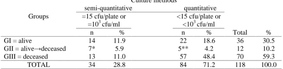
The semi-quantitative and quantitative methods gave similar results. Out of 118 catheters analyzed, 34 (28.8%) showed growth of 15 cfu/plate in the former test and the same number showed 103 cfu/mL in the latter; thus, 84
(71.2%) had <15 cfu/plate or <103 cfu/mL. Distribution of
the data obtained from the three groups of patients is shown in Table 2.
From the semi-quantitative plates prepared from the 34 colonized catheters, 55 microorganisms were isolated, 32 (58.2%) being classified as Gram-positive bacteria
Culture methods
semi-quantitative quantitative Groups =15 cfu/plate or
=103 cfu/ml
<15 cfu/plate or <103 cfu/ml
n % n % Total %
GI = alive 14 11.9 22 18.6 36 30.5
GII = alive deceased 7* 5.9 5** 4.2 12 10.2
GIII = deceased 13 11.0 57 48.4 70 59.3
TOTAL 34 28.8 84 71.2 118 100.0
cfu=colony-forming units; *(2 alive and 5 deceased); **(4 alive and 1 deceased ); G=groups Table 2 - Frequency of culture results using the semi-quantitative and quantitative methods.
(mainly Staphylococcus aureus), 19 (34.5%) as Gram-negative and 4 (7.3%) as Candida species.
By means of the quantitative catheter culture method, 52 microorganisms were isolated. Of these, 32 (61.6%) were classified as Gram-positive (mainly
Staphylococcus aureus), 19 (36.5%) as Gram-negative and
one (9.0%) as Candida sp.
The five microorganisms isolated most commonly from catheter tips were, in decreasing order: Staphylococcus
aureus, Staphylococcus epidermidis, Acinetobacter baumannii, Pseudomonas aeruginosa, and Stenotrophomonas maltophilia.
Results regarding the resistance patterns of the
Staphylococcus aureus isolates from catheter tips and blood
cultures are shown in Table 3.
The Staphylococcus aureus isolates were oxacillin-resistant, but vancomycin and teicoplanin were active against all S. aureus isolates from catheter tips and blood culture.
Of the 34 cultures identified by the semi-quantitative method, 14 (41.2%) were polymicrobial and 20 (58.8%) were monomicrobial. Of the 34 cultures identified by the quantitative method, 13 (38.2%) were polymicrobial and 21 (61.8%) were monomicrobial.
All catheter tips giving positive cultures by the quantitative method were also positive by the semi-quantitative method. A comparison of the two methods
Antibiotics Abbreviation Concentration n and % of the strains
S I R
Gentamicin GEN 10 g 0 0 37(100.0)
Rifampicin RIF 30 g 5(13.5) 14(37.8) 18(48.7)
Chloramphenicol CLO 30 g 0 0 37(100.0)
Ciprofloxacin CIP 5 g 0 0 37(100.0)
Levofloxacin LVX 5 g 0 0 37(100.0)
Teicoplanin TEC 30 g 37(100.0) 0 0
Vancomycin VAN 30 g 37(100.0) 0 0
Clindamycin CLI 2 g 0 0 37(100.0)
Erytromycin ERI 15 g 0 0 37(100.0)
Oxacillin OXA 1 g 0 0 37(100.0)
Penicillin PEN 10 UI 0 0 37(100.0)
Trimethoprim/sulfamethaxole SUT 25 g 0 0 37(100.0)
Tetracycline TET 30 g 0 0 37(100.0)
(S)=sensitive (I)=intermediate (R)=resistante
Table 3 - Resistance pattern of 37 Staphylococcus aureus isolates to commonly tested antimicrobial agents as determined by disc diffusion method.
showed a Kappa coefficient of 89% and the McNemar concordance test showed 2=0.107, p=0.75.
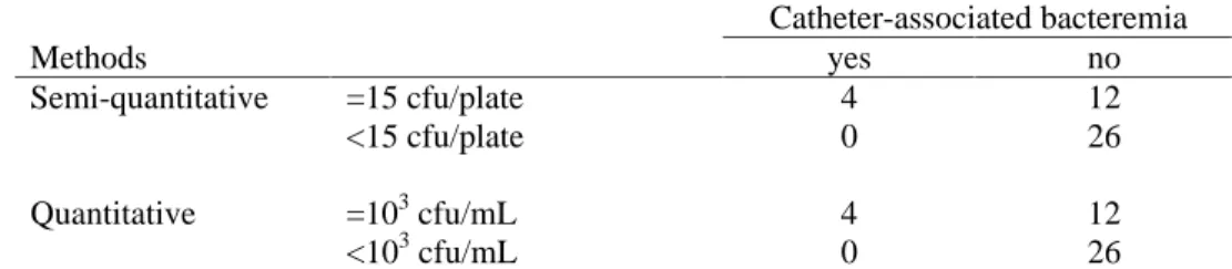
Of the 42 catheters paired with blood cultures, in 16 (38.1%) there was a significant development of colony-forming units in both the semi-quantitative and quantitative methods (Table 4).
The data in Table 4 show that the two methods had the same results, with maximal correlation (r=1), and that there was neither a significant number of colony-forming units isolated, nor did catheter-associated bacteremia exist, in 26 (61.9%) catheter tips examined by the semi-quantitative or quantitative methods. In 12 (28.5%) cases, (a significant number colony formation of colony-forming units) was found, which did not cause bacteremia. In the four catheter-associated bacteremia cases, the microorganisms isolated from the catheter tips were also isolated from the blood. Two cases showed Staphylococcus aureus, one case showed
Stenotrophomonas maltophilia and remaining case, Citrobater freundii. The number of colony-forming units
were uncountable by either the semi-quantitative or the quantitative methods. The CRBSI diagnosis for 4 (9.5%) patients with short-term catheter insertion was confirmed by both semi-quantitative and quantitative cultures of their removed catheters. Thus, for short-term central venous catheter insertion, a cut-off point of 15 cfu or 103 cfu/
mL, togheter with peripheral blood cultures, has a sensitivity of 100.0%, a specificity of 68.4%, a positive predictive value (PPV) of 25.0%, a negative predictive value (NPV) of 100.0%, efficiency of 71.4% and prevalence of 9.5% for the diagnosis of CRBSI.
DISCUSSION
Data presented here demonstrated that either the semi-quantitative or quantitative culture method for the assessment of central venous catheter-associated infections can be used as an indicator of bacteremia. These data on both technique tested concomitantly are described for the first time in Brazil.
The semi-quantitative culture of central venous catheter tips shows that catheter-associated bacteremia was only observed in catheters in which semi-quantitative cultures indicated the presence of over 15 cfu/plate. In a study using mostly peripheral venous catheters, Maki et al. (1977) showed that the accepted indicator for the risk of bacteremia
was the presence of at least 15 colonies/plate. Bouza et al. (2004) chose this positivity criterion ( 15 cfu), because the majority of patients with lower numbers ( 15 cfu) did not present symptoms of an infection, while all the bacteremia cases had counts above 15 cfu.
Positivity of at least one blood culture was used in the present study as a factor for the interpretation and to establish the diagnosis of catheter-associated infection (Raad & Bodey 1992). The criteria for establishing catheter-associated infection used here were based on studies carried out by Raad & Bodey (1992) and confirmed by Bouza et al. (2003), according to whom a certain microorganism has to be isolated from the catheter tip by the semi-quantitative or quantitative method and also from the peripheral blood. As pointed out by Stratton (1998), the isolated microorganism must be of the same genus and species, and have the same antibiotic sensitivity test profile. In the present study, the isolates of the three species (S. aureus,
S. maltophylia and C. freundii) obtained from catheter tips
and from blood each had the same profile of antibiotic sensitivity. However, this does not necessarily means that these bacteria are from the same clone. For this to be verified, molecular genotyping would have to be done. Futher studies are planned, in which molecular methods will be applied to these isolates.
According to data presented here the same microorganisms were isolated from the catheter tips by both culture methods and peripheral vein blood. The semiquantitative test showed 100% sensitivity and a specificity of 68.4%. These results differ from those of Collignon et al. (1986), who obtained values of 92% sensitivity and 83% specificity. However, Bouza et al. (2004) observed that the specificity of the semi-quantitative technique was 76%. The current results are also in good agreement with those obtained by Gutiérrez et al. (1992), who showed the method described by Maki et al. (1977) has 100% sensitivity and 76%-79% specificity for the diagnosis of catheter-associated bacteremia. Whereas, Cercenado et al. (1990), noted that the semi-quantitative method, used as a standard, is unable to show the degree of colonization inside the catheter, Fätkenheuer et al. (2003) suggested that the semi-quantitative culture method be considered the standard, even if it only captures bacteria that adhere to the external surface of the catheter. An increase of 15 or more colony-forming units on a plate is considered proof of catheter-associated infection.
Catheter-associated bacteremia
Methods yes no
Semi-quantitative =15 cfu/plate <15 cfu/plate
4 0
12 26
Quantitative =103 cfu/mL <103 cfu/mL
4 0
12 26 cfu = colony forming units
Table 4 - Relationship between catheter-related bacteremia and the different methods of catheter culture.
The present studies demonstrate that the quantitative culture from the catheter tips is able to identify catheter-associated bacteremia. This method is considered a sensitive and reliable method (Blot et al., 1998). The comparison of the semi-quantitative and quantitative methods showed them to give similar results, as described by Brun-Buisson et al. (1987) and, in addition, the quantitative technique proved to be more sensitive, with highly positive predictive values (Blot et al., 1998). The present results showed that the quantitative technique was 100% sensitive in detecting catheter-related bacteremia. Siegman-Ingra et al. (1997) compared six methods, and proved that the most accurate one, with sensitivity and specificity above 90%, was a quantitative method, similar to that of Maki et al. (1977).
The present results show a similar correlation with both methods. The semi-quantitative and quantitative methods are highly sensitive, with 100% negative predictive value, meaning that both methods are appropriate for detecting the absence of catheter-associated bacteremia. In the present study, the positive predictive value was low (25.0%), similar to that reported by Sitges-Serra and Linares (1988), who found that the Maki method showed a low positive predictive value, varying from 10% to 75% depending on the duration of the catheterization. In the present study, the duration of catheterization was 12.4 1.3 days and the positive predictive values of this method were low (16-31%). The majority (71.2%) of the catheter tips showed negative results.
Regarding the specificity of the methods, the values obtained in the present study were lower (65.0%) than expected, compared with those of other authors: (Raad et al. (1992) 86%, Gutierrez et al. (1992) 83%, and Widmer et al. (1992) 96%. The results reported here showed no significant difference between the two techniques in sensitivity, specificity, PPV, NPV, efficiency, or prevalence. Nevertheless, the semi-quantitative method is the more recommended, as it is rapid and easy to perform, routinely, in the clinical microbiology laboratory, as pointed out by Fätkenheur et al. (2003).
Concluding, it can be reported that, in the semi-quantitative and semi-quantitative methods employed in parallel tests here, all cultures from catheters with significant numbers of colony-forming units ( 15cfu/plate for semiquantitative culture or 103cfu/mL for quantitative
culture) have a 9.5% probability of indicating catheter-associated infections and high sensitivity. The confidence in both methods is similar, and both techniques can be recommended, although the semi-quantitative method is more widely used in clinical microbiology laboratories, because of its simplicity and low cost.
ACKNOWLEDGEMENTS
We thank the NAC (Unidade Auxiliar de Estrutura Complexa) of the School of Pharmaceutical Sciences, UNESP, Araraquara (SP, Brazil) for financial support.
RESUMO
Avaliação do cateter venoso central associado à infecção por métodos de cultura semi-quantitativa e quantitativa
As técnicas de cultura semiquantitativa (Maki) e quantitativa (Brun-Buisson) foram usadas para o diagnóstico da infecção relacionada ao cateter em pacientes com cateter venoso central de curta duração ( 30 dias). O diagnóstico da infecção relacionada ao cateter foi baseado nos resultados das culturas semiquantitativa e quantitativa do cateter removido. As pontas de cateter (118) de 100 pacientes foram avaliadas usando ambos os métodos. A análise semi-quantitativa revelou que 34 cateteres (28,8%) tiveram crescimento 15 unidades formadoras de colônias (ufc) e, as culturas quantitativas (34 cateteres, 28,8%) crescimento 103 ufc/ mL. A bacteriemia foi confirmada em quatro pacientes por isolamento de microrganismos de espécie idêntica, em ambos, cateteres e amostras de sangue. Usando a técnica semi-quantitativa de cultura em cateteres de curta duração, mostra-se que com ponto de corte 15 ufc, a técnica apresentou 100% de sensibilidade, especificidade 68,4%, valor preditivo positivo (VPP) 25,0% e valor preditivo negativo (VPN) 100%, eficiência 71,4 % e prevalência 9,5%. Para o método quantitativo usando limite de cut-off 103 ufc/ml, a sensibilidade foi 100%, especificidade 68,4%, valor preditivo positivo (VPP) 25,0%, valor preditivo negativo (VPN) 100%, eficiência 71,4% e prevalência 9,5%. Conclui-se que as técnicas de cultura semi-quantitativa e quantitativa avaliadas concomitantemente neste estudo, pela primeira vez no Brasil, apresentam especificidade e sensibilidade similares.
Palavras-chave: cateter venoso central; cultura
semi-quantitativa; cultura semi-quantitativa; bacteriemia relacionada ao cateter.
REFERENCES
Anaissie E, Samonis G, Kontoyiannis D, Costerton JW, Sabharwal U, Bodey G, Raad II. Role of catheter colonization and infrequent hematogenous seeding in catheter-related infections. Eur J Clin Microbiol Infect Dis 1995; 14:135-7. Aoki EE, Garcia LB, Pizzolitto AC, Pizzolitto EL Uso do método de cultura semi-quantitativa para estudo de bacteriemia relacionada ao cateter venoso central utilizado por pacientes em hemodiálise. Rev Bras Anal Clin 2004; 36:159-62.
Aoki EE, Pizzolitto AC, Garcia LB, Pizzolitto EL Staphylococcus aureus biofilms on central venous haemodialysis catheters. Braz J Microbiol 2005; 36:342-6. Aufwerber E, Ringertz S, Ransjo U. Routine semiquantitative cultures ands central venous catheter-related bacteremia.
Blot F, Schmidt E, Nitenberg G, Tancrede C, Leclercq B, Laplanche A, Andremont A. Earlier positivity of central venous versus peripheral blood cultures is highly predictive of catheter-related sepsis. J Clin Microbiol 1998; 36:105-9. Bouza E, Liñares J, Pascual A. Diagnóstico microbiológico de las infecciones asociadas a cateteres intravasculares 2004. Disponível em URL: http://www.seimc.org/protocolos/ microbiologia/cap15.htm. [17 jun 2005].
Bouza E, Muñoz P, López-Rodrígues J, Jesús Pérez M, Rincón C, Martín Rabadán P, Sánchez C, Bastida E. A needleless closed system device (CLAVE) protects from intravascular catheter tip and hub. J Hosp Infect 2003; 54:279-87.
Bouza E, San Juan R, Muñoz P, Pascau J, Voss A, Desco M, Cooperative Group of the European Study on Nosocomial Infections (ESGNI). A European perspective on intravascular catheter-related infections: report on the microbiology workload, aetiology and antimicrobial susceptibility (ESGNI-005 Study). Clin Microbiol Infect 2004; 10:838-42. Brun-Buisson C, Abrouk F, Legrand P, Huet Y, Larabi S, Rapin M. Diagnosis of central venous catheter-related sepsis - critical level of quantitative tip cultures. Arch Intern Med 1987; 147:873-7.
Catton JA, Dobbins BM, Kite O, Wood JM, Eastwood K, Sugden S, Sandoe JA, Burke D, McMahon MJ, Wilcox MH In situ diagnosis of intravascular catheter-related bloodstream infection: a comparison of quantitative culture, differential time to positivity, and endoluminal brushing. Crit Care Med 2005; 33:787-91.
Cercenado E, Ena J, Rodrìgues-Créixens M, Romero I, Bouza E. A conservative procedure for the diagnosis of catheter-related infections. Arch Intern Med 1990; 150:1417-20. Cleri DJ, Corrado ML, Seligman SJ Quantitative culture of intravenous catheters and other intravascular inserts. J Infect
Dis 1980; 141:781-6.
Collignon PJ, Soni N, Pearson IY, Woods WP, Munro R, Sorrell TC,. Is semi-quantitative culture of central vein catheter tips useful in the diagnosis of catheter-associated bacteremia? J Clin Microbiol 1986; 24:532-5.
Corona ML, Peters SG, Narr BJ, Thompson RL. Subspecialty clinics critical care medicine. Infections related to central venous catheters. Mayo Clin Proc 1990; 65:979-86. Donlan RM. Biofilms: microbial life on surfaces. Emerg
Infect Dis 2002; 8:1-19.
Donlan RM, Costerton JW. Biofilms: survival mechanisms of clinically relevant microorganisms. Clin Microbiol Rev 2002; 15:167-93.
Elliot TSJ. Intra-vascular-device infections. J Med Microbiol 1988; 27:161-7.
Elliot TSJ. Line associated bacteraemias. Comm Dis Rep 1993; 3:91-5.
Fätkenheur G, Buchleidt D, Cornely OA, Fuhr HG, Karthaus M, Kisro J, Leithäuser M, Salwender H, Südhoff T, Szelényi H, Weissinger F. Central venous catheter (CVC)-related infections in neutropenic patients. Guidelines of the infectious diseases working party (AGIHO) of the German Society of Hematology and Oncology (DGHO). Ann
Hematol 2003; 82:149-57.
Fletcher RH, Fletcher SW, Wagner EH. Epidemiologia
clínica: elementos essenciais. Porto Alegre: ArtMed; 2003. 281p.
Fraenkel DJ, Rickard C, Lipman J. Can we ackieve consensus on central venous catheter-related infections? Anaesth
Intensive Care 2000; 28:475-90.
Gutiérrez J, Leon C, Matamoros R, Nogales C, Martín E. Catheter-related bacteremia and fungemia. Reliability of two methods for catheter culture. Diagn Microbiol Infect Dis 1992; 15:575-8.
Hodge D, Puntis JWL Diagnosis, prevention, and management of catheter related bloodstream infection during long term parenteral nutrition. Arch Dis Child Fetal Neonatal
Ed 2002; 87:F21-4.
Horvath R, Collignon P Controlling intravascular catheter infections. Aust Prescr 2003; 26:41-3.
Kamal GD, Pfaller MA, Rempe LE, Jebson PJ. Reduced intravascular catheter infection by antibiotic bonding. JAMA 1991; 265: 2364-8.
Linares JA, Sitges-Serra A; Garau J, Pérez JL, Martin R. Pathogenesis of catheter sepsis: a prospective study with quantitative and semiquantitative cultures of catheter hub and segments. J Clin Microbiol 1985; 21:357-60.
Maki DG. Infections caused by intravascular devices used for infusion therapy: pathogenesis, prevention, and management. In: Bisno AL and Waldovogel FA (ed.),
Infections associated with indwelling medical devices. 2nd.ed.
Washington, D.C.: American Society for Microbiology, 1994. p.155-212.
Maki DG, Stolz SM, Wheeler S, Mermel LA. Prevention of central venous catheter-related bloodstream infection by use of an antiseptic-impregnated catheter. A randomized, controlled trial. Ann Intern Med 1997; 127:257-66. Maki DG, Weise CE, Sarafin HW. A semiquantitative culture method for identifying intravenous-caheter-related infection.
N Engl J Med 1977; 296:1305-9.
Marrie TJ, Noble MA, Costerton JW. Examination of the morphology of bacteria adhering to peritoneal dialysis catheters by scanning and transmission electron microscopy.
J Clin Microbiol 1983; 18:1388-98.
Murray PR, Baron EJ, Jorgensen JH, Pfaller MA, Yolken RH. Manual of clinical microbiology. Washington, DC: American Society for Microbiology; 2003. 2v
NCCLS. National Committee for Clinical Laboratory Standards. Performance standards for antimicrobial disk
susceptibility testing; twelfth informational supplement,
M100-S13. Pennsylvania: National Committee for Clinical Laboratory Standards; 2003.
O'Grady NP, Alexander M, Dellinger EP, Gerberding JL, Heard SO, Maki DG, Masur H, McCormick RD, Mermel LA, Pearson ML, Raad II, Randolph A, Weinstein RA. Guidelines for the prevention of intravascular catheter-related infections. Pediatrics 2002; 110:1-40.
Polderman KH, Girbes ARJ. Central venous catheter use.
Intensive Care Med 2002; 28:18-28.
Raad II, Bodey GP. Infectious complications of indwelling vascular catheters. Clin Infect Dis 1992; 15:197-210. Raad II, Costerton JW, Sabharwal U, Sacilowski M, Anaissie W, Bodey GP. Ultrastructural analysis of indwelling vascular catheters: a quantitative relationship between luminal colonization and duration of placement. J Infect Dis 1993; 168:400-7.
Raad II, Luna M, Khalil SAM, Costerton JW, Lam C, Bodey GP The relationship between the thrombotic and infectious complications of central venous catheters. JAMA 1994; 271:1014-6.
Raad II, Sabbagh MF, Rand KH, Sherertz RJ. Quantitative tip culture methods and the diagnosis of central venous catheter-related infections. Diagn Microbiol Infect Dis 1992; 15:13-20.
Rijnders BJ, Van Wijngaerden E, Peetermans WE Catheter-tip colonization as a surrogate end point in clinical studies on catheter-related bloodstream infection: how strong is the evidence? Clin Infect Dis 2002; 35:1053-8.
Siegman-Ingra Y, Anglim AM, Shapiro DE, Adal KA, Strain BA, Farr BM. Diagnosis of vascular catheter-related bloodstream infection: a meta-analysis. J Clin Microbiol 1997; 35:928-36.
Sitges-Serra A, Liñares J. Limitations of semiquantitative methods for catheter culture. J Clin Microbiol 1988; 26:1074-8. Soares JF, Siqueira AL. Introdução à estatística médica. Belo Horizonte: Coopmed; 2002. 300p.
Storti A, Pizzolitto AC, Pizzolitto EL Detection of mixed microbial biofilms on central venous catheters removed from intensive care unit patients. Braz J Microbiol 2005; 36:275-80. Stratton CW. Catheter-associated infections: a necessary evil.
Antimicrob Infect Dis Newsl 1998; 17:49-56.
Widmer AF, Nettleman M, Flint K, Wenzel RP. The clinical impact of culturing central venous catheters. Arch Intern Med 1992; 152:1299-302.
Zar JH. Biostatistical analysis. New Jersey: Pretice Hall; 1999. 663p.