Article
ISSN 0102-695X
http://dx.doi.org/10.1590/S0102-695X2011005000136 Received 14 Sep 2010 Accepted 25 Feb 2011 Available online 5 Aug 2011
of the naphthoquinone
5-methoxy-3,4-dehydroxanthomegnin
Rodrigo Rezende Kitagawa,
*,1Wagner Vilegas,
2Iracilda Zeppone
Carlos,
3Maria Stella Gonçalves Raddi
31Departamento de Ciências Farmacêuticas,Universidade Federal do Espírito Santo,
Brazil,
2Instituto de Química de Araraquara, Universidade Estadual Paulista “Júlio de Mesquita
Filho”, Brazil,
3Faculdade de Ciências Farmacêuticas de Araraquara, Universidade Estadual Paulista
“Júlio de Mesquita Filho”, Brazil.
Abstract: Large number of quinones has been associated with antitumor, antibacterial, antimalarial and antifungal activities. In this work we describe the effect of the naphthoquinone, 5-methoxy-3,4-dehydroxanthomegnin, on murine tumor cells (LP07 and LM2) and its immunomodulatory effect on nitric oxide (NO) production on LPS-stimulated macrophages. The results have shown that 5-methoxy-3,4-dehydroxanthomegnin was a significant inhibitor of LPS-stimulated NO generation from macrophage (inhibition percentage ranged from 97.4 to 98.9%) and a strong cytotoxic agent against both tumor cells LP07 and LM2 (CI50 6.2±0.36 µM and 74.6±1.9 µM, respectively). These results indicate that the 5-methoxy-3,4-dehydroxanthomegnin may show promising activity in the treatment of murine breast and lung cancer by immunomodulatory and antiproliferative activities.
Keywords:
5-methoxy-3,4-dehydroxanthomegnin cytotoxic activity immunomodulatory naphthoquinone
Introduction
Natural products have had major impact as template or in direct treatments of cancers and infective diseases. In the cancer area, from the 92 drugs commercially available prior to 1983 in the United States, or approved worldwide between 1983 and 1994, approximately 62% can be related to a natural product origin (Ravelo et al., 2004). In this context, phytochemicals offer promising new options for the development of more effective chemopreventive and chemotherapeutic strategies (Punathil & Katiyar, 2009). Mass screening programs of natural products by National Cancer Institute have identified quinone moiety as an important pharmacophoric element for cytotoxic activity (Pérez-Sacau et al, 2007).
Quinone derivatives are ubiquitous in nature, have been found in plants, fungi and bacteria and are also present in many drugs such as anthracyclines, daunorubicin, doxorubicin and mitomycin, which are used clinically in the therapy of solid cancers (Verma, 2006). Large number of quinone derivatives has been associated with antitumor, antibacterial, antimalarial and antifungal activities (Huang et al., 2002).
The antitumor activity of naturally occurring quinones is exhibited predominantly by three main
groups such as benzoquinone, naphthoquinone and anthraquinone. Mitomycin and streptonigrin possess
p-benzoquinone moiety with heterocyclic groups whereas anthracyclines, doxorubicin and daunorubicin consist of anthraquinone moiety. Some naphthaquinone antibiotics such as lapachol and lapinone are also found to be cytotoxic to tumor cells (Inbaraj et al., 1999).
In some tumor types over 70% of the mass of the tumor consists of infiltrating leukocytes. These tumor-associated leukocytes, specially macrophages, release angiogenic factors, mitogens, nitric oxide (NO), proteolytic enzymes and chemotactic factors, recruiting more inflammatory cells (Yan et al., 2006; Le Bitoux & Stamenkovic, 2008). The mediators and cellular effectors of inflammation are important constituents of the local environment of tumors. In some types of cancer, inflammatory conditions are present before a malignant change occurs. Conversely, in other types of cancer, an oncogenic change induces an inflammatory microenvironment that promotes the development of tumors (Mantovani et al., 2008).
exposure of cells to high NO concentrations resulting from iNOS induction during chronic inflammation could have an active role in carcinogenesis. In tumor growth and metastasis, experimental tumor models have provided more convincing evidence for a direct role of NO (Lala & Chakraborty, 2001). NO can, for example, enhanced migration, invasion and metastasis of breast and colon cancer cells by activating the mitogen-activated protein kinase pathway (Le Bitoux & Stamenkovic, 2008). It has been reported that NO mediated promotion of tumor cell migration requires activation of nitric oxide synthase (NOS) and a positive association between endothelial NOS (eNOS) expression in tumor cells and growth and metastasis of tumors (Lala & Orucevic, 1998).
The 5-methoxy-3,4-dehydroxanthomegnin, 1,4-naphthoquinone (1) isolated from the capitula
Paepalanthus latipes Silveira, plant of the Eriocaulaceae family in previous studies, has shown a significant cytotoxic index (CI) for McCoy cells compared to cisplatin, and when associated with ascorbic acid resulted in CI seven times lower due to hydrogen peroxide generated by ascorbate - driven 5-methoxy-3,4-dehydroxanthomegnin redox cycling (Kitagawa et al., 2004; Kitagawa et al., 2008). The purpose of the present study was to investigate the cytotoxic activity of this naphthoquinone in murine lung adenocarcinoma and mammary tumor cells, as well as its ability to act as immunomodulatory agent by determination of NO production in LPS-induced macrophages.
O O
O H3CO
OH
OCH3
CH3 O
1
1
4 11 10
5 6 9
Materials and Methods
Plant material
Paepalanthus latipes Silveira, Eriocaulaceae, was collected at Serra do Cipó, in the Espinhaço Chain, Minas Gerais state, Brazil and authenticated by Prof. Paulo Takeo Sano from Instituto de Biociências, USP, São Paulo. The voucher specimen (CFSC 13846) is on file of the Herbarium of the Departamento de Botânica, Instituto de Biociências, Universidade de São Paulo, Brazil.
Chemicals
Eagle medium was purchase from Adolfo Lutz (São Paulo, Brazil), fetal bovine serum from Cultilab (Campinas, Brazil), dimethylsulfoxide (DMSO), lipopolysaccharide (LPS) and 1,1-diphenyl-2-picryl-hydrazyl (DPPH) from Sigma (St. Louis, MO, USA), neutral red (NR) from Riedel-de-Haën AG (Seelze, Hannover), Trolox from Acros Organics (Geel, Belgium), 5-methoxy-3,4-dehydroxanthomegnin, isolated and characterized as previously described (Kitagawa et al., 2004) was stored as stock solution at 10.0 mg/mL in DMSO. The final concentration of DMSO (2%) used does not interfere cell viability as well as on NO production by macrophages.
Cell culture
The murine mammary tumor cells (LM2) and lung adenocarcinoma tumor cells (LPO7) were provided by Instituto de Oncologia Angel H. Roffo, Buenos Aires, Argentina. The cells were cultured in 96-well plates (Corning, USA) with E-MEM supplemented with 10% (v/v) fetal bovine serum, and maintained at 37 °C in a humidified incubator under 5% CO2.
Cytotoxicity assay
Aliquots (0.2 mL) of medium containing 104
cells/mL were seeded into 96-well tissue-culture plates and incubated at 37 oC under 5% CO
2 for 24 h. After
this period, the medium was removed and the cells were placed into unmodified medium (control) or in medium modified with various concentrations of 5-methoxy-3,4-dehydroxanthomegnin and incubated for 24 h under the same conditions. After, the medium was removed and the plates were prepared for the neutral red (NR) assay (Borenfreund & Puerner, 1985). After brief agitation, the optical density of each well was measured using a 540 nm filter and a 620 nm reference wavelength (Spectra and Rainbow (Shell) Readers - Tecan, Austria). All experiments were performed at least three times, using three wells for each concentration of tested chemical. The cytotoxicity data was standardized by determining absorbance and calculating the correspondent 5-methoxy-3,4-dehydroxanthomegnin concentration. Linear regression analysis with 95% confidence limit was used to define dose-response curve and to compute the concentration needed to reduce absorbance of the NR by 50% (CI50), the so called cytotoxic midpoint (Barile, 1994). Cisplatin was used as reference cytotoxic agent.
Animals
55±5% humidity, 10-18 circulations/h and a 12-h light/ dark cycle), with water and food available ad libitum.
All animal procedures were performed in accordance with the institutional guidelines.
Peritoneal exudates cells
Thioglycollate-elicited peritoneal exsudate cells (PEC) were harvested from Swiss mice using 5.0 mL of sterile PBS, pH 7.4. The cells were washed twice by centrifugation at 200 g for 5 min at 4 ºC and resuspended in appropriate medium for each test.
Inhibition of NO production
PEC (5x10⁶ cells/mL) were incubated in the
medium with LPS (1 µg/mL) plus 5-methoxy-3,4-dehydroxanthomegnin for 24 h at 37º C in a 7.5% CO2 atmosphere. NO was determined by measuring the accumulation of nitrite, a stable end product, in the culture supernatant according to the Griess reaction (Green et al., 1982). Equal volumes of culture supernatant and Griess reagent (100 µL) were mixed and left over for 10 min at room temperature. Absorbance was read at 540 nm (Multiskan, Labsystem) and the nitrite concentration in medium was calculated using sodium nitrite as a standard. Cells incubated with LPS only were used as a positive control.
Antioxidant activity
The antioxidant capacity of the 5-methoxy-3,4-dehydroxanthomegnin was studied through its scavenging activity against 1,1-diphenyl-2-picryl-hydrazyl (DPPH) radical. The bleach of DPPH was monitored at an absorbance of 517 nm. Wherein the bleaching rate of a
stable free radical, DPPH• is monitored at a characteristic
wavelength in the presence of the sample. In its radical
form, DPPH• absorbs at 517 nm, but upon reduction by an
antioxidant or a radical species, its absorption decreases. Lower absorbance of the reaction mixture indicates higher free radical-scavenging activity. The capability to scavenge
the DPPH• radical was calculated using the following equation: DPPH• scavenging effect (%)=(A0-A1/A0)×100 wherein A0is the absorbance of the control reaction and
A1 is the absorbance in the presence of the sample. Trolox was utilized as antioxidant standard (Gülcin et al., 2005).
Statistical analysis
The parameters were expressed as mean+SD. Statistical analysis was performed with analysis of variance (ANOVA). Differences were considered significant at p>0.05.
Results and Discussion
The chemotherapeutic properties of quinones are usually related to their ability to act as prooxidants in the generation of cytotoxic oxygen-centered radicals, their ability to function as cytotoxic alkylating agents, or their ability to form Michael adducts with cellular thiols, including glutathione (Thornton et al., 1995).
The metabolism of quinones in biochemical systems has been attributed to one or two electron
reductions catalyzed by flavoenzymes in the presence
of suitable electron donors. A two electron reduction is catalyzed by NAD(P)H quinone acceptor oxidoreductase (DT-Diaphorase [DTD]). The activity of DTD varies with tissue, and it has been found to be elevated in tumors. Thus, differential levels of the enzyme in tissues, including tumors, can provide a selective tumor cell kill for agents that are good substrates for the enzyme (Gutierrez, 2000).
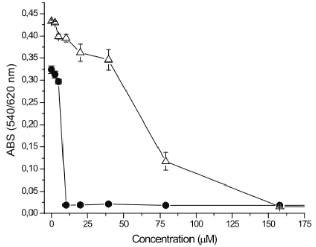
Quinone and quinone derivates redox cycling with participation of ascorbate mimics to some degree the enzymatic redox cycling since both processes have nearly the same elementary stages (Roginsky et al., 1999). In the current study we evaluated the ability of the 5-methoxy-3,4-dehydroxanthomegnin to cause cell death in cell line tumor LP07 and LM2 using the neutral red assay (NR). It was observed that 5-methoxy-3,4-dehydroxanthomegnin demonstrated cytotoxicity for both LP07 and LM2 cells and a noticeable cytotoxicity for LM2 cells when compared to cisplatin (Figure 1). The CI50 employed to evaluate this effect is shown in Table 1.
0 25 50 75 100 125 150 175
0,00 0,05 0,10 0,15 0,20 0,25 0,30 0,35 0,40 0,45
ABS (540/620 nm)
Concentration (µM)
Figure 1. Dose-effect curve of the 5-methoxy-3,4-dehydroxanthomegnin on LM2 (open triangle) and LP07 (solid circle) cells. Cells were plated at a density of 104
Table 1. Cytotoxicity of the
5-methoxy-3,4-dehydroxanthomegnin and cisplatin on LM2 and LP07 cells.
Substances CI50 (µM)
LM2 LP07
5-methoxy-3,4-dehydroxanthomegnin 74.6±1.9 6.2±0.36
cisplatin 168.4±3.9 4.34±0.45
A solid tumor consists of tumor cells and host
derived cells, including tumor-infiltrating leucocytes and
cells of the tumor vasculature, especially endothelial cells (Lala & Chakraborty, 2001).
Clinical and experimental observations of the past two decades increasingly support the notion that immune cells recruited to and activated within the tumor microenvironment play a strong supporting role in tumor progression (Le Bitoux & Stamenkovic, 2008).
Inflammation facilitates the initiation of normal cells
and their growth and progression to malignancy through
production of pro-inflammatory cytokines and oxidants an activation of signaling pathways involved in inflammation
and carcinogenesis. Targets for chemoprevention include signaling pathways (e.g. NF-kB), oxidant-generating enzymes [e.g. inducible NOS (iNOS)], mediators of
inflammation (e.g. cytokines, COX-2), anti-apoptotic factors, ROS/RNS and xenobiotic-metabolizing enzymes (Ohshima et al, 2005).
The NO produced by iNOS, derived from L-arginina, is induced by lipopolysaccharide (LPS) or
proinflammatory cytokines such as TNF-α, IL-1β and IFN-γ. (Li et al., 2007). NO•can react with O2 to form
nitrogen dioxide (NO2-), with O2- to form peroxynitrite (ONOO-), or with transition metal ions to form metal-nitrosyl (M-NO) complexes. Most of these products are
more reactive than NO•. For example, ONOO-, although less stable and shorter-lived than NO•, is much more
cytotoxic - for instance, in causing DNA damage. (Lala & Chakraborty, 2001). This leads to increased mutations and altered functions of important enzymes and proteins (e.g. activation of oncogene products and/or inhibition of
tumor-supressor proteins) in inflamed tissues, thus contributing
to the multistage carcionogenesis process (Oshima et al, 2005).
All of these observations suggest that macrophage
infiltrates potentiate tumor cell growth and dissemination,
at least in part by promoting angiogenesis (Le Bitoux & Stamenkovic, 2008).
As a mimic in vitro model of inflammation,
macrophages were stimulated with LPS, which activates
the macrophage functions and the release of inflammatory
mediators including enhancement of iNOS, generation
of NO, and secretion of pro-inflammatory cytokines.
Since most of NO generated is converted immediately into nitrites, the nitrite level was measured in cell culture supernatants after treatment with
5-methoxy-3,4-dehydroxanthomegnin (79, 158, 316 µM) to investigate
their possible anti-inflammatory activity indicated by the
inhibition of the LPS-stimulated NO. The result revealed that 5-methoxy-3,4-dehydroxanthomegnin exerted immunosuppressive activity on the LPS-stimulated NO generation with inhibition percentage ranged from 97.4 to 98.9% (p>0.05) (Figure 2), implying a potent
anti-inflammatory activity to this 1,4-naphthoquinone. The
inhibition of the generated NO by the tested compound may be due to a direct scavenging capacity of NO, an inhibition of iNOS pathway, and / or a modulation of other factors in the NO cascade such as transcriptional factors.
Figure 2. Effect of the 5-methoxy-3,4-dehydroxanthomegnin on LPS-induce NO production in peritoneal macrophages. Adherent cells (5x10⁶) were incubated for 24 h with
5-methoxy-3,4-dehydroxanthomegnin and LPS (1 µg/mL). Cells incubated just with LPS were used as a positive control and cell in culture medium (RPMI-1640) as a negative control. Nitrite concentrations in the medium were determined using Griess reagent, as described in materials and methods. *p<0.05 vs. LPS control. Each values represents the mean±SD for three different experiments performed in triplicate.
To investigate the radical scavenging activity of 5-methoxy-3,4-dehydroxanthomegnin, we submitted it to DPPH assay, which revealed that the compound had no radical scavenging activity at the tested concentrations (reduction of DPPH absorption of 0.1% at all concentrations
tested) (Figure 3). These findings suggested that the NO
dehydroxanthomegnin. Phytochemistry 69: 2205-2208. Kitagawa RR, Raddi, MSG, Santos LC, Vilegas W 2004. A new
cytotocity naphthoquinone from Paepalanthus latipes. Chem Pharm Bull 52: 1487-1488.
Lala PK 1998. Significance of nitric oxide in carcinogenesis, tumor progression and cancer therapy. Cancer Metast Rev 17: 1-6.
Lala PK, Orucevic, A 1998. Role of nitric oxide in tumor progression: lessons from experimental tumors. Cancer Metast Rev 17: 91-106.
Lala, PK, Chakraborty C 2001. Role of nitric oxide in carcinogenesis and tumor progression. Lancet Oncol 2: 149-156.
Le Bitoux MA, Stamenkovic I 2008. Tumor-host interactions: the role of inflammation. Histochem. Histochem Cell Biol 130: 1079-1090.
Li C, He L, Jin J 2007. Atractylenolide I and atractylenolide III inhibit lipopolysaccharide-induced TNF-α and NO production in macrophages. Phytother. Res. 21: 347-353.
Mantovani A, Allavena P, Sica A, Balkwill F 2008. Cancer-related inflammation. Nature 454: 436-444.
Ohshima H, Tazawa H, Sylla BS, Sawa T 2005. Prevention of human cancer by modulation of chronic inflammatory processes. Mutat Res 591: 110-122.
Pérez-Sacau E, Diaz-Peñate RG, Estévez-Braun A, Ravelo AG, Garcia-Castellano JM, Pardo L, Campillo M 2007. Syntesis and pharmacophore modeling of naphthoquinone derivatives with cytotoxic activity in human promyelocytic leukemia HL-60 cell line. J Med Chem 50: 696-706.
Punathil T, Katiyar SK 2009. Inhibition of non-small cell lung cancer cell migration by grape seed proanthocyanidins is mediated through the inhibition of nitric oxide, guanylate cyclase, and ERK1/2. Mol Carcinogen 48: 232-242. Ravelo AG, Estévez-Braun A, Chávez-Orellana H, Pérez-Sacau
E, Mesa-Siverio D 2004. Recent studies on natural products as anticancer agents. Curr Med Chem 4: 241-265.
Roginsky VA, Barsukova TK, Stegmann HB 1999. Kinetics of redox interaction between substituted quinones and ascorbate under aerobic conditions. Chem-Biol Interact 121: 177-197.
Thornton DE, Jones KH, Jiang Z, Zhang H, Liu G, Cornwell DG 1995. Antioxidant and cytotoxic tocopheryl quinones in normal and cancer cells. Free Radical Bio Med 18: 963-976.
Verma RP 2006. Anti-cancer activities of 1,4-naphthoquinones: a QSAR study. Anti-cancer Agent Me 6: 489-499. Yan L, Anderson GM, Dewitte M, Nakada, MT 2006. Therapeutic
potential of cytokine and chemokine antagonists in cancer therapy. Eur J Cancer 42: 793-802.
*Correspondence
Rodrigo Rezende Kitagawa
Departamento de Ciências Farmacêuticas, Universidade Federal do Espírito Santo
Av. Marechal Campos 1468, 29040-090 Vitória-ES, Brazil kitagawarr@ccs.ufes.br
Fax: +55 27 33357293.
0 50 100 150 200 250
0 20 40 60 80 100
ABS decline (%)
Concentration (µM)
Figure 3. DPPH scavenging activity (%) of the 5-methoxy-3,4-dehydroxanthomegnin (solid triangle) and trolox (solid circle). Each value is expressed as mean±SD of triplicate measurements.
Acknowledgments
This study was supported by a grant of CNPq to
RRK, to FAPESP for financial aid to WV and financial
assistance from the PADC-UNESP.
References
Barile FA 1994. Introduction to in vivo Cytotoxicology: Mechanisms and Methods. Boca Raton: CRC Press. Borenfreund E, Puerner JA 1985. Toxicity determined in vitro
by morphological alterations and neutral red absorption. Toxicol Lett 24: 119-124.
Green LC, Wagner DA, Glogowski J, Skipper PL, Wishnok JS, Tannenbaum SR 1982. Analysis of nitrate, Nitrit and [15N] Nitrit in biological fluids. Anal Biochem 126: 131-138.
Gülcin I, Berashvili D, Gepdiremen A 2005. Antiradical and antioxidant activity of total anthocyanins from Perilla pankinensis Decne. J Ethnopharmacol 101: 287-293. Gutierrez PH 2000. The role of NAD(P)H oxidoreductase
(DT-diaphorase) in the bioactivation of quinine-containing antitumor agents: a review. Free Radical Bio Med 29: 263-275.
Huang S, Kuo H, Hsiao C, Lin Y 2002. Efficient synthesis of redox-switched naphthoquinone thiol-crown ethers and their biological activity evaluation. Bioorg Med Chem Lett 10: 1947-1952.
Inbaraj JJ, Gandhidasan R, Murugesan R 1999. Cytotoxity and superoxide anion generation by some naturally occurring quinones. Free Radical Bio Med 26: 1072-1078. Jadeski LC, Chakraborty C, Lala PK 2003. Nitric oxide-mediated
promotion of mammary tumor cell migration requires sequential activation of nitric oxide synthase, guanylate cyclase and mitogen-activated protein kinase. Int J Cancer 106: 496-504.