molecules
ISSN 1420-3049
www.mdpi.com/journal/molecules Article
Anti-Candida Targets and Cytotoxicity of Casuarinin Isolated
from Plinia cauliflora Leaves in a Bioactivity-Guided Study
Tatiana M. Souza-Moreira 1, Juliana A. Severi 1,†, Keunsook Lee 2, Kanya Preechasuth 2, Emerson Santos 1, Neil A. R. Gow 2, Carol A. Munro 2, Wagner Vilegas 3 and
Rosemeire C. L. R. Pietro 1,*
1
Department of Drugs and Medicines, School of Pharmaceutical Sciences, UNESP-Univ Estadual Paulista, Rodovia Araraquara-Jau, km 1, Araraquara, 14801-902, São Paulo, Brazil; E-Mails: souzatm@gmail.com (T.M.S.M.); juseveri@yahoo.com.br (J.A.S.); emersonsan@gmail.com (E.S.)
2
Department of Microbiology, Institute of Medical Sciences, University of Aberdeen, Foresterhill, Aberdeen, AB25 2ZD, Scotland, UK; E-Mails: k.k.lee@abdn.ac.uk (K.L.);
kanyamt@hotmail.com (K.P.); n.gow@abdn.ac.uk (N.A.R.G.); c.a.munro@abdn.ac.uk (C.A.M.)
3
Department of Organic Chemistry, Institute of Chemistry, UNESP-Univ Estadual Paulista, R. Francisco Degni s/n, Araraquara, 14800-900, São Paulo, Brazil; E-Mail:
vilegasw@gmail.com (W.V.)
† Nowadays this author is at Department of Pharmacy and Nutrition, Centro de Ciências Agrárias,
UFES-Univ Federal do Espírito Santo, Alto Universitário, Guararema, 29500-000, Alegre, ES, Brazil.
* Author to whom correspondence should be addressed; E-Mail: pietrorc@fcfar.unesp.br; Tel.: +55-16-3301-6965; Fax: +55-16-3301-6990.
Received: 27 May 2013; in revised form: 5 July 2013 / Accepted: 5 July 2013 / Published: 9 July 2013
cellular integrity was suggested by the antifungal activity ameliorated when using an osmotic support. The most important target for the tannin fraction studied was suggested by ultrastructural analysis of yeast cell walls revealing a denser mannan outer layer and wall porosity reduced. It is possible to imply that P. cauliflora targeted the C. albicans cell wall inducing some changes in the architecture, notably the outer glycoprotein layer, affecting the cell wall porosity without alteration of the polysaccharide or protein level.
Keywords: Plinia cauliflora (Myrtaceae); ellagitannin; Candida; antifungal activity; cell wall
1. Introduction
formation, an increase in the number of buds and changes in the cell wall ultrastructure of C. albicans treated with that subfraction [13].
Therefore, the objective of this study was to analyze the potential of the compounds from P. cauliflora leaf extract by performing a bioassay-guided chemical study and to investigate the antifungal effect on C. albicans cell, as well as cytotoxic properties against a mammal cell.
2. Results and Discussion
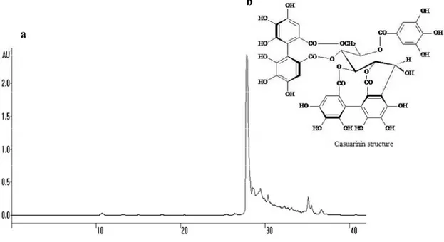
The bio-guided chemical investigation of the hydroalcoholic extract from P. cauliflora leaves was initially carried out by fractionation of the crude extract by liquid-liquid extraction into different polarity fractions. The n-butanol fraction (BF) was more biologically active (see below) against Candida strains and fractionated further, yielding the active subfraction F2. Purification of F2 was achieved by a combination of High Speed Counter-Current Chromatography (HSCCC) and semi-preparative reverse phase High Performance Liquid Chromatography (HPLC) and led to the purification of a major hydrolysable tannin named casuarinin (Figure 1). The structure of the isolated compound was unequivocally assigned based on the ultraviolet (UV) spectral, 1D/2D nuclear magnetic resonance (NMR) and mass (MS) data, in comparison with the literature reference [14]. Casuarinin is the main ellagitannin isolated from Casuarina stricta [14] and was also isolated from the leaves of Melaleuca squarosa, a member of Myrtaceae family [15].
Figure 1. (a) Semi-preparative HPLC-UV chromatogram of F2 from P. cauliflora leaves. Chromatographic conditions: HPLC with UV detector set at 254 nm, the column was RP-18, elution phase acidified with trifluoroacetic acid 0.05% in linear gradient elution of CH3OH/H2O (20:80 in 15 min, 20:80→50:50 in 35 min, 50:50→95:5 in 40 min); flow-rate
of 2.5 mL/min. (b) Structure of casuarinin. This hydrolysable tannin is a C-glycosidic ellagitannin with an open-chain glucose core called casuarinin.
a
2.1. Susceptibility Testing
The in vitro susceptibility tests of the crude extract, fractions and subfractions from the leaves of P. cauliflora against five Candida strains are shown in Table 1. C. krusei was more susceptible to the plant samples than the other species. BF was the most active fraction, with an inhibitory effect on fungal growth with MICs in the range of 19 to 156 µg/mL, depending upon the species. This result guided us to purify and further characterize that fraction. Among the subfractions obtained by HSCCC, just one of them called F2, showed good activity against C. albicans, according to previous reports [16,17]. The major compound isolated from F2 by HPLC, casuarinin, had some antifungal activity and C. krusei was the most susceptible strain. Although casuarinin has some antifungal activity, it did not have as good activity as F2, suggesting that the minor compounds present in F2, as shown in Figure 1, also contributed to the total antifungal activity of F2. All the plant samples showed low fungicidal activity.
Cytotoxicity was assessed indirectly using a mammalian cell line by measuring the fluorescent emission of the molecule of resazurin reduced to resorufin in response to metabolic activity. CC50
indicates the concentration of the plant samples that inhibited 50% of cell viability [18]. Plinia crude extract (PcE), BF, AF and subfraction F2 had low cytotoxic properties with a CC50 concentration
higher than the antifungal MIC (Table 1). Casuarinin was not cytotoxic to mammalian cells at the highest concentration tested (116 µg/mL), but much higher concentrations of casuarinin were required to inhibit the fungal cells with the exception of C. krusei and C. tropicalis, which is a favorable point to study this compound for further application with animals or humans.
Table 1. Antifungal activity and cytotoxicity of P. cauliflora leaf samples, in µg/mL.
C.a. (ATCC) C.a. (SC5314) C.k. C.p. C.t.
Samples MIC MFC MIC MFC MIC MFC MIC MFC MIC MFC CC50
Extract 156 625 156 156 19 39 78 156 312 1250 417
EAF 625 1250 NT NT 19 78 312 625 312 625 221
BF 78 1250 156 156 19 39 19 19 156 1250 767
AF 312 >1250 NT NT 39 78 39 78 312 1250 1500
F2 78 312 156 312 625 >1250 156 1250 312 >1250 >400
casuarinin 580 580 580 >580 26 580 580 580 145 580 >116
FCZ 4 8 2 32 32 32 4 4 32 32 NA
Amph B 0.25 0.25 1 8 0.50 0.50 0.50 0.50 0.50 2 NA
2.2. Ergosterol Levels
In order to examine whether the P. cauliflora active fraction targets the membrane and specifically affects ergosterol levels, the ergosterol content of isolates treated with BF was measured. The sterol quantification methodology is based on the unique spectral absorption pattern produced by ergosterol at 281.5 nm and the spectral absorption pattern produced by 24(28) dihydroergosterol at 281.5 and 230 nm wavelengths. Consequently, the amount of ergosterol can be determined by calculating the total ergosterol and 24(28) dihydroergosterol content [19]. Fluconazole, which inhibits ergosterol synthesis [20] was used as a control and as expected the ergosterol content of fluconazole-treated cells was significantly decreased (Table 2). No evidence was found to implicate P. cauliflora acting on fungal ergosterol biosynthesis.
Table 2. Ergosterol quantification, in percentage (%).
SAMPLES C. albicans C. krusei C. parapsilosis C. tropicalis
Control 1.38 ± 0.32 1.13 ± 0.70 0.34 ± 0.30 1,94 ± 0.10
BF 2.03 ± 0.77(+48) 1.44 ± 0.58(+28) 0.77 ± 0.70(+29) 1,30 ± 0.86(−33) FCZ 0.40 ± 0.23(−71) * 0.68 ± 0.08(−40) 0.05 ± 0.05(−84) * 0(−100) *
Ergosterol content expressed as a percentage (%) of the wet weight of the cell ± standard deviation. In parentheses there is the increased (+) or decreased (−) percentage value relative to the ergosterol content in control. BF = n-butanolic fraction. FCZ = fluconazole. * Statistically significant.
2.3. Reduction of P. Cauliflora Leaf Samples Antifungal Properties 2.3.1. Addition of Exogenous Ergosterol
When a compound binds ergosterol, addition of exogenous ergosterol can reduce the antifungal efficacy of the compound. As a consequence, it is necessary to use a higher concentration of the compound to inhibit yeast growth relative to the MIC without exogenous ergosterol addition [8]. The effect of addition of ergosterol at 200 µg/mL to the drug susceptibility assays was determined. Deoxycholate amphotericin B can be used as positive control in this experiment since it is known to bind ergosterol in the fungal membrane and its antifungal activity is considerably compromised by the addition of 200 µg/mL exogenous ergosterol[20,21]. This finding was re-confirmed and similar results were observed with the crude extract and fractions of the plant in the present study. In our experiments, addition of exogenous ergosterol increased amphotericin B MIC 8 fold against C. albicans and C. tropicalis and 32 fold against C. krusei and C. parapsilosis (Table 3). The MIC of the extract, BF and F2 against C. albicans and C. parapsilosis increased 16 fold in the presence of ergosterol (Table 3), indicating their binding to ergosterol.
2.3.2. Sorbitol Protection Assay
fluconazole and amphotericin B were just 2-fold higher with sorbitol addition in accord with their target being the membrane rather than the wall (Table 3).
Table 3. Antifungal activity of the plant preparations in the presence of ergosterol or sorbitol, in µg/mL.
Ergosterol Sorbitol
Samples C.a. C.k. C.p. C.t. C.a. C.k. C.p. C.t.
Extract 5000 39 312 625 2500 39 2500 156
EAF 5000 39 1250 625 5000 39 2500 312
BF 5000 39 1250 312 5000 39 1250 312
AF 5000 78 2500 625 5000 78 2500 1250
F2 1250 78 1250 312 1250 78 625 312
FCZ NA NA NA NA 8 64 4 64
Amph B 2 16 16 4 0.25 1 1 1
MIC results are expressed as the mode in µg/mL, for 24–48 h. C.a. = C. albicans; C.k. = C. krusei; C.p. = C. parapsilosis; C.t. = C. tropicalis. FCZ = fluconazole (64 to 0.06 µg/mL). Amph B = amphotericin B (16 to 0.03 µg/mL).Test concentrations: 5,000 to 8 µg/mL of extract. EAF = ethyl acetate fraction; BF = n-butanolic fraction; AF = aqueous fraction; F2 = fraction 2 from BF; NA = not applicable.
2.4. Ultrastructural Analysis
We also investigated whether the plant-derived compounds altered the cell wall architecture. C. albicans SC5314 cells were treated with subfraction F2 (156 µg/mL) and processed for transmission electron microscopy (TEM), preserving the ultrastructure of the cell wall. Untreated cells of C. albicans SC5314 had a typical fibrillar outer mannan layer [Figure 2(a)], but cells treated with the F2 had a denser and shorter outer layer [Figure 2(b)] indicating that the plant compounds altered cell wall structure.
Figure 2. Ultrastructural analysis of the C. albicans yeast cell surface. (a) Transmission electron microscopy of cells without treatment (b) And cells treated with 156 µg/mL of subfraction F2 for 16 h. Magnification of 130,000×.
2.5. Assessing C. albicans cell wall treated with F2
To assess whether F2 did indeed target the cell wall a more detailed analysis was performed by biochemically determining cell wall composition and porosity and examining the distribution of chitin in the wall by staining with the fluorescent dye Calcofluor White (CFW). Chitin localization was not affected by treatment with the casuarinin fraction F2 (not shown). No significant changes were observed in the cell wall composition of untreated cells and cells treated with 156 µg/mL of F2 for 16 h as determined by HPAE-PAD (Table 4). As the outer fibrillar layer is rich in glycoproteins the total protein content of isolated cell walls was also measured. F2-treated yeast cell walls had a non-significant decrease in the levels of protein compared to the untreated control (Table 4). In contrast F2-treated hyphal cell walls had a significant increase (10%) in protein levels compared to the untreated samples.
Table 4. Cell wall assay results comparing non-treated and casuarinin fraction treated C. albicans SC5314.
Control cells Treated cells
Yeast Hyphae Yeast Hyphae
Chitin (%) 5.1 ± 1.8 21.8 ± 1.3 4.2 ± 1.4 22.1 ± 4.0
Glucan (%) 66.3 ± 4.9 68.8 ± 0.6 73.4 ± 4.0 67.3 ± 2.0
Mannan (%) 28.6 ± 5.6 9.4 ± 1.6 22.4 ± 5.3 10.6 ± 2.1
Covalently attached cell wall protein (µg/mL) 109.4 ± 5.5 90.2 ± 5.0 114.5 ± 4.2 101.3 ± 2.0 * Non-covalently attached cell wall protein (µg/mL) 226.5 ± 1.5 156.6 ± 0.5 227.6 ± 3.4 173.3 ± 0.1
Results of saccharide content are given as percentage (%) of total dried cell wall, while protein content is given as µg/mL and are the average ± standard deviation of triplicate samples.* Statistically significant.
The cell wall acts as a physical barrier and filter to the external environment. Changes in the cell wall architecture and cross-linking of cell wall polysaccharides to each other is likely to affect the porosity of the wall. The porosity of the cell walls of C. albicans treated with subfraction F2 was compared to untreated cells. Cells were treated with the polycations DEAE-dextran (500 kDa) and poly-L-lysine (50 kDa) which interact with the cell membrane releasing 260 nm UV absorbing compounds [22]. The wild type cell wall is a barrier against the free diffusion of molecules through its depth. Its porosity is a measure of its relative permeability of molecules and is normally reflective of the molecular size and charge of the permeant. Control, untreated C. albicans SC5314, had a relative porosity of 34.8 ± 0.2%. In comparison cells treated with the casuarinin-containing subfraction F2 had significantly reduced porosity (−9.2 ± 5.4%). The F2-treated cell wall were still as porous to poly-L-lysine but were less porous to DEAE-dextran than control cells without treatment.
3. Experimental
3.1. General Procedures
equipped with a RP-18 column (Phenomenex Luna). Thin layer chromatography (TLC) analyses were performed on silica gel plates (Al sheets, F254, 200 m thickness, Sorbent Technologies, Norcross, GA,
USA) and visualized after spraying with anisaldehyde/H2SO4 solution under UV/Vis light. NMR
analyses and 2D experiments were carried out on a Varian INOVA 500 spectrometer, operating at 500 MHz for 1H and 150 MHz for 13C and chemical shifts were given in δ (ppm) using trimethylsilane (TMS) as internal standard. Complementary structural information was achieved by mass spectrometry (MS) analysis using a Thermo Finnigan TSQ Quantum Ultra AM system equipped with a hot electrospray ionization (ESI) source (electrospray voltage 3.0 kV, sheath gas: nitrogen; vaporizer temperature: 50 °C; capillary temperature: 250 °C), operating in positive and negative ionization modes. Reagents were mainly acquired from Sigma-Aldrich (St. Louis, MO, USA). Culture medium reagents were from Acumedia (Lansing, MI, USA), Sigma-Aldrich and Invitrogen (Carlsbad, CA, USA). Microbiological analysis used a VERSAmax tunable microplate reader (Molecular Devices, Sunnyvale, CA, USA), a microplate spectrofluorometer (Tecan, Männedorf, Switzerland) and a spectrophotometer (Shimadzu 1603, Kyoto, Japan). Microscopy visualization utilized a Philips CM10 transmission microscope (FEI UK Ltd, Cambridge, UK) with a Gatan Bioscan 792 (Gatan, UK, Abingdon, UK) for recording the images. Cell wall composition was analyzed by High-Performance Anion Exchange Chromatography from Dionex (Sunnyvale, CA, USA) with pulsed amperometric detection. 3.2. Plant Material
Leaves of P. cauliflora were collected in São Carlos, São Paulo, Brazil in December 2006 and identified by Marcos Sobral from the Department of Natural Sciences of the “Universidade Federal de São João Del-Rei”. A voucher herbarium specimen was deposited under number ESA 96038 at the “Herbário da Escola Superior de Agricultura Luiz de Queiroz” (ESALQ), Piracicaba, São Paulo, Brazil.
3.3. Extraction and Isolation
The leaves were dried at 40 °C and powdered using a knife mill. The crude extract was obtained by percolation of 80 g of the powder with 70% aqueous ethanol (1L, 3 days, ambient temperature). The organic solvent was evaporated at 40 °C under reduced pressure and the aqueous residue lyophilized, yielding a green crude extract (30%). A portion (15 g) of the extract was suspended in water (500 mL) and partitioned successively (v/v, 3 × 500 mL each) with ethyl acetate, followed by n-butanol, yielding the ethyl acetate fraction (EAF, 21.8%), n-butanol fraction (BF, 41.7%) and aqueous fraction (AF, 36.5%). The BF was fractionated by preparative High Speed Counter-Current Chromatography equipped with a multilayer helical column containing two coils (φ = 1.68 mm, internal extreme
β = 0.50, external extreme β = 0.85, revolution ratio n = 10 cm). An amount of 1.25 g of BF was dissolved in 15 mL of the mobile phase composed by a solution of ethyl acetate-n-butanol-water (3.8:1.2:5, v/v), centrifuged and the supernatant injected at a flow-rate of 1.5 mL/min. Rotation was set up at 850 rpm and a total of 130 fractions (4 mL each) were collected and analysed by TLC (CHCl3/MeOH/PrOH/H2O (5:6:1:4, v/v/v/v - organic phase, anisaldehyde/H2SO4 reagent for
linear gradient elution of CH3OH/H2O as follow: 20:80 in 15 min, 20:80→50:50 in 35 min,
50:50→95:5 in 40 min; eluted at a flow-rate of 2.5 mL/min. The effluent was monitored using a ProStar 330 photodiode-array ultraviolet detection system at 254 nm and led to the isolation of the compound casuarinin (1, 13 mg) [14]. Colorless amorphous powder. UV max nm: (MeOH): 213, 232,
257sh. 1H-NMR (DMSO-d6): δ 6.94 (2H, d, J = 2 Hz, galloyl ArH), 6.57, 6.29, 6.25 (1H each, s,
HHDP), 5.39 (1H, dd, J = 5.0, 2.0 Hz, H-1), 5.26 (1H, dd, J = 8.0, 2.0 Hz, H-4), 5.21 (1H, dd, J = 2.0, 8.0, H-3), 5.17 (1H, m, H-5), 4.56 (1H, m, H-2) 4.53 (1H, m, H-6), 4.00 (1H, d, J = 13 Hz, H-6). ESI-MS (positive mode) m/z 937 [M+H]+. ESI-MS (negative mode) m/z 935 [M-H]-. C41H28O26.
Those experiments were performed at the Institute of Chemistry and the School of Pharmaceutical Sciences, UNESP, BRA
3.4. Fungal Strains, Cell Line and Culture Conditions
The fungal strains used were Candida albicans ATCC 64548, Candida krusei ATCC 6258, Candida parapsilosis ATCC 22019 and Candida tropicalis ATCC 750 and one clinical isolate Candida albicans SC5314. Each strain was maintained on YPD agar (1% (w/v) yeast extract, 2% (w/v) mycological peptone, 2% (w/v) glucose, 1.6% (w/v) agar) and cultured in YPD broth at 30 °C with shaking at 200 rpm for 16 h to prepare inocula for experiments. When required, the strains were cultured in Sabouraud broth (1% (w/v) mycological peptone, 2% (w/v) glucose). Minimal inhibitory concentration (MIC) was performed with RPMI 1640 medium. Hyphal growth of strain SC5314 was induced using two different growth media: water with 20% fetal calf serum (FCS) and Lee’s medium [23,24] and incubation at 37 °C. The rabbit corneal cell line (ATCC SIRC CCL-60) was maintained in Eagle’s MEM containing 10% (v/v) fetal bovine serum, 2 mM L-glutamine and 0.2% sodium bicarbonate. The cultures were incubated at 37 °C with 5% CO2 until reaching confluence.
Microbiological and cytotoxic assays were realized at School of Pharmaceutical Sciences in UNESP, BRA and experiments with Candida albicans SC5314 were performed at the Institute of Medical Sciences, University of Aberdeen, UK, including TEM analysis, MIC, hyphal growth and cell wall assays.
3.5. Antifungal Susceptibility Testing
which the number of colony forming units was zero after 2 µL of microplate cultures were incubated at 37 °C for 48 h on YPD agar [26]. Data are presented as the mode values of triplicates.
3.6. Cytotoxicity Testing
The mammalian cytotoxicity of P. cauliflora samples was evaluated as follows: aliquots (300 µL) of a 1×10⁵ cells/mL SIRC cell suspension was added to the wells of 96-well microplate and incubated in Eagle’s MEM containing 10% (v/v) fetal bovine serum, 2 mM L-glutamine and 0.2% sodium bicarbonate at 37 °C, 5% CO2 for 72 h. The medium was removed and replaced with fresh medium
containing extract or fractions (2,000 to 4 µg/mL) and casuarinin (116 to 0.2 µg/mL) and the plate incubated at 37 °C, 5% CO2 for 24 h. A 0.1 mg/mL resazurin solution was added and after 3 h of
incubation the fluorescence of the reduced product resorufin was read using a microplate spectrofluorometer with excitation and emission wavelengths of 530 nm and 590 nm, respectively [18,27]. Then 50% cytotoxic concentration (CC50) was calculated [28]. The test was done
in triplicate.
3.7. Sterol Quantification Method
Total cellular ergosterol of the standard strains treated with BF was quantified [19]. One colony was incubated overnight in Sabouraud broth and used to inoculate (2.5 × 10³ cfu/mL) Sabouraud containing BF at MIC value. After incubation at 35 °C, 18 h, 135 rpm, cells were collected, washed and saponified. Sterols were extracted with hexane. An aliquot of sterol extract was diluted 5-fold in 100% ethanol and scanned in a spectrophotometer between 200 and 300 nm. The ergosterol content was calculated as percentage according to the equations in Arthington-Skaggs et al. [19]. The test was done in triplicate and compared with the results obtained for fluconazole.
3.8. Ultrastructural Analysis by Transmission Electron Microscopy (TEM)
Cells were inoculated (OD600 < 0.2) into YPD broth plus 156 µg/mL subfraction F2 and incubated for 16 h at 30 °C, 200 rpm. The cells were filtered and humidified samples of untreated and treated cells were frozen by high pressure (Leica EM PACT high-pressure freezer, Vienna, Austria). Freeze substitution, sectioning and visualization were done as described previously [29].
3.9. Cell Wall Polymer Quantification
The cell walls were prepared from untreated (control) and treated (156 µg/mL of F2) yeast and hyphal C. albicans SC5314 cells grown as follows. For yeast growth, 1 mL from overnight YPD culture (OD at 600 nm < 0.2) was inoculated in 100 mL YPD and incubated for 16 h at 30 °C, 200 rpm. To induce hyphae formation cells were washed in H2O and inoculated at <2 × 10⁷ cells/mL
into 200 mL of 20% FCS in sterile distilled water and incubated for 16 h at 37 °C, 200 rpm. Cells were harvested by centrifugation and cell walls were isolated as described [30].
and eluted isocratically with 12 mM NaOH [24]. The total g per 1 mg of each cell wall component was obtained by calibration using standard curves of glucosamine, glucose and mannose, and expressed as a percentage of total dry cell wall. All data are presented as the mean values of triplicate experiments. The amount of protein in cell wall fractions was determined by Bradford method [30]. All data are presented as the mean values of triplicate samples.
3.10. Relative Cell Wall Porosity
Cells from an overnight YPD culture of C. albicans SC5314 were inoculated at OD600 < 0.2 into 50 mL YPD broth and incubated for 3 h at 30 °C, 200 rpm. The F2 subfraction was added (final concentration 156 µg/mL) and cells were incubated for 2 h. The cells were harvested and washed with water. Cells at a density of 1 × 10⁸ cells/mL were dispensed in duplicate in 1 mL aliquots, centrifuged and 10 mM of Tris-HCl buffer pH 7.5 was added to each of the cell pellets, which were divided in two sets of reactions: one received 1 mL of 5 µg/mL DEAE-dextran and the other 1 mL of 15 µg/mL poly-L-lysine. The samples were incubated for 30 min at 30 °C, 200 rpm, centrifuged at 16,000 × g for 3 min, the supernatant collected and again centrifuged. The supernatant absorbance was measured at 260 nm. Relative porosity was calculated as [(A260DEAE-dextran—A260buffer)/(A260poly-L-lysine—
A260buffer) × 100]. Each test was performed in triplicate [22].
3.11. Statistical Analysis
Where necessary, statistical analysis was performed using ANOVA. A Dunnett’s T-test was used to compare treated with non-treated cells in the assays of hyphal growth, sterol and cell wall polymers quantification and cell wall porosity. A 5% significance level was adopted.
4. Conclusions
It can be proposed that the specific cell wall changes induced by treatment with subfraction F2 may be due to the formation of a tannin-cell wall protein complex on the outermost layer of C. albicans since tannins are known to complex with macromolecules such as proteins and polysaccharides [31]. To corroborate these potential effects of F2 we observed: (i) a denser outer mannoprotein layer visualized by TEM indicating precipitate formation; (ii) no changes in mannan content and iii) reduced cell wall porosity. Hence, the present study proposes that the tannin-rich fraction F2 of P. cauliflora has the ability to interfere with the outer glycoprotein-rich layer of C. albicans.
Acknowledgments
Conflict of Interest
The authors declare no conflict of interest.
References
1. Sobral, M. Alterações nomenclaturais em Plinia (Myrtaceae). Boletim do Museu Botânico de Curitiba 1985, 63, 1–4.
2. Lorenzi, H. Árvores brasileiras: Manual de identificação e cultivo de plantas arbóreas nativas do Brasil; Instituto Plantarum: Nova Odessa, Brazil, 2000.
3. Agra, M.F.; Silva, K.N.; Basílio, I.J.L.D.; Freitas, P.F.; Barbosa-Filho, J.M. Survey of medicinal plants used in the region northeast of Brazil. Braz. J. Pharmacogn. 2008, 18, 472–508.
4. Reynertson, K.A.; Wallace, A.M.; Adachi, S.; Gil, R.R.; Yang, H.; Basile, M.J.; D’Armiento, J.; Weinstein, I.B.; Kennelly, E.J. Bioactive depsides and anthocyanins from jaboticaba (Myrciaria cauliflora). J. Nat. Prod. 2006, 69, 1228–1230.
5. Souza-Moreira, T.M.; Moreira, R.R.D.; Sacramento, L.V.S.; Pietro, R.C.L.R. Histochemical, phytochemical and biological screening of Plinia cauliflora (DC.) Kausel, Myrtaceae, leaves. Braz. J. Pharmacogn. 2010, 20, 48–53.
6. Calderone, R.A. Candida and Candidiasis; ASM Press: Washington, DC, USA, 2002.
7. Richardson, M.; Lass-Flörl, C. Changing epidemiology of systemic fungal infections. Clin. Microbiol. Infect. 2008, 14, 5–24.
8. Escalante, A.; Gattuso, M.; Pérez, P.; Zacchino, S. Evidence for the mechanism of action of the antifungal phytolaccoside b isolated from Phytolacca tetramera Hauman. J. Nat. Prod. 2008, 71, 1720–1725.
9. Hoehamer, C.F.; Cummings, E.D.; Hilliard, G.M.; Rogers, P.D. Changes in the proteome of Candida albicans in response to azole, polyene, and echinocandin antifungal agents. Antimicrob. Agents Chemother. 2010, 54, 1655–1664.
10. Zhang, J.D.; Xu, Z.; Cao, Y.B.; Chen, H.S.; Yan, L.; An, M.M.; Gao, P.H.; Wang, Y.; Jia, X.M.; Jiang, Y.Y. Antifungal activities and action mechanisms of compounds from Tribulus terrestris L. J. Ethnopharmacol. 2006, 103, 76–84.
11. Newman, D.J.; Cragg, G.M. Natural products as sources of new drugs over the last 25 years. J. Nat. Prod. 2007, 70, 461–477.
12. Gertsch, J.; Tobler, R.T.; Brun, R.; Sticher, O.; Heilmann, J. Antifungal, antiprotozoal, cytotoxic and piscicidal properties of justicidin b and a new arylnaphthalide lignan from Phyllanthus piscatorum. Planta Med. 2003, 69, 420–424.
13. Ishida, K.; de Mello, J.C.P.; Cortez, D.A.G.; Filho, B.P.D.; Ueda-Nakamura, T.; Nakamura, C.V. Influence of tannins from Stryphnodendron adstringens on growth and virulence factors of Candida albicans. J. Antimicrob. Chemother. 2006, 58, 942–949.
15. Yoshimura, M.; Ito, H.; Miyashita, K.; Hatano, T.; Taniguchi, S.; Amakura, Y.; Yoshida, T. Flavonol glucuronides and c-glucosidic ellagitannins from Melaleuca squarrosa. Phytochemistry
2008, 69, 3062–3069.
16. Araujo, M.G.; Hilario, F.; Nogueira, L.G.; Vilegas, W.; Santos, L.C.; Bauab, T.M. Chemical constituents of the methanolic extract of leaves of Leiothrix spiralis Ruhland and their antimicrobial activity. Molecules 2011, 16, 10479–10490.
17. Holetz, F.B.; Pessini, G.L.; Sanches, N.R.; Cortez, D.A.; Nakamura, C.V.; Filho, B.P. Screening of some plants used in the Brazilian folk medicine for the treatment of infectious diseases. Mem. Inst. Oswaldo Cruz. 2002, 97, 1027–1031.
18. Perrot, S.; Dutertre-Catella, H.; Martin, C.; Rat, P.; Warnet, J.-M. Resazurin metabolism assay is a new sensitive alternative test in isolated pig cornea. Toxicol. Sci. 2003, 72, 122–129.
19. Arthington-Skaggs, B.A.; Jradi, H.; Desai, T.; Morrison, C.J. Quantitation of ergosterol content: Novel method for determination of fluconazole susceptibility of Candida albicans. J. Clin. Microbiol. 1999, 37, 3332–3337.
20. Odds, F.C.; Brown, A.J.P.; Gow, N.A.R. Antifungal agents: Mechanisms of action. Trends Microbiol. 2003, 11, 272–279.
21. Lee, S.H.; Lee, J.R.; Lunde, C.S.; Kubo, I. In vitro antifungal susceptibilities of Candida albicans and other fungal pathogens to polygodial, a sesquiterpene dialdehyde. Planta Med. 1999, 65, 204–208.
22. De Nobel, J.G.; Klis, F.M.; Priem, J.; Munnik, T.; van den Ende, H. The glucanase-soluble mannoproteins limit cell wall porosity in Saccharomyces cerevisiae. Yeast 1990, 6, 491–499. 23. Lee, K.; Buckley, H.; Campbell, C. An amino acid liquid synthetic medium for the development
of mycelial and yeast forms of Candida albicans. Sabouraudia 1975, 13, 148–153.
24. Munro, C.A.; Whitton, R.K.; Bleddyn Hughes, H.; Rella, M.; Selvaggini, S.; Gow, N.A.R. Chs8-a fourth chitin synthase gene of Candida albicans contributes to in vitro chitin synthase activity, but is dispensable for growth. Fungal Genet. Biol. 2003, 40, 146–158.
25. Clinical and Laboratory Standards Institute document M27-A2. Reference method for broth dilution antifungal susceptibility testing of yeasts; Approved Standard, 2nd ed.; Clinical and Laboratory Standards Institute: Wayne, PA, USA, 2002.
26. Walker, L.A.; Munro, C.A.; Bruijn, I.; Lenardon, M.D.; McKinnon, A.; Gow, N.A.R. Stimulation of chitin synthesis rescues Candida albicans from echinocandins. PLoS Pathog. 2008, 4, e1000040.
27. Nakayama, G.R.; Caton, M.C.; Nova, M.P.; Parandoosh, Z. Assessment of the Alamar blue assay for cellular growth and viability in vitro. J. Immunol. Methods 1997, 204, 205–208.
28. Sánchez-Medina, A.; Stevenson, P.C.; Habtemariam, S.; Peña-Rodríguez, L.M.; Corcoran, O.; Mallet, A.I.; Veitch, N.C. Triterpenoid saponins from a cytotoxic root extract of Sideroxylon foetidissimum subsp. gaumeri. Phytochemistry 2009, 70, 765–772.
29. Netea, M.G.; Gow, N.A.R.; Munro, C.A.; Bates, S.; Collins, C.; Ferwerda, G.; Hobson, R.P.; Bertram, G.; Hughes, H.B.; Jansen, T., et al. Immune sensing of Candida albicans requires cooperative recognition of mannans and glucans by lectin and toll-like receptors. J. Clin. Invest.
30. Groot, P.W.J.; de Boer, A.D.; Cunningham, J.; Dekker, H.L.; de Jong, L.; Hellingwerf, K.J.; de Koster, C.; Klis, F.M. Proteomic analysis of Candida albicans cell walls reveals covalently bound carbohydrate-active enzymes and adhesins. Eukaryotic. Cell 2004, 3, 955–965.
31. Haslam, E. Natural polyphenols (vegetable tannins) as drugs: possible modes of action. J. Nat. Prod. 1996, 59, 205–215.
Sample Availability: Not available.