Suitability of DNA extracted from archival specimens of fruit-eating bats of the
genus
Artibeus
(Chiroptera, Phyllostomidae) for polymerase chain reaction
and sequencing analysis
Mário Pinzan Scatena and Eliana Morielle-Versute
Departamento de Zoologia e Botânica, Instituto de Biociências, Letras e Ciências Exatas
de São José do Rio Preto, Universidade Estadual Paulista, São José do Rio Preto, SP, Brazil.
Abstract
To establish a technique which minimized the effects of fixation on the extraction of DNA from formalin-fixed tissues preserved in scientific collections we extracted DNA samples from fixed tissues using different methods and evalu-ated the effect of the different procedures on PCR and sequencing analysis. We investigevalu-ated muscle and liver tis-sues from museum specimens of five species of fruit-eating (frugivorous) bats of the Neotropical genusArtibeus (Chiroptera, Phyllostomidae):A. fimbriatus, A. lituratus, A. jamaicensis, A. obscurus, and A. planirostris. The results indicated that treatment of tissues in buffered solutions at neutral pH and about 37 °C for at least four days improves the quality and quantity of extracted DNA and the quality of the amplification and sequencing products. However, the comparison between the performance of DNA obtained from fixed and fresh tissues showed that, in spite of the fact that both types of tissue generate reliable sequences for use in phylogenetic analyses, DNA samples from fixed tis-sues presented a larger rate of errors in the different stages of the study. These results suggest that DNA extracted from formalin-fixed tissue can be used in molecular studies of NeotropicalArtibeus bats and that our methodology may be applicable to other animal groups.
Key words:archival specimens, Chiroptera, DNA extraction, formalin-fixed.
Received: April 19, 2007; Accepted: July 17, 2007.
Biological collections represent a wealth of biologi-cal information that has been carefully documented in a time and space format. Collections often contain hundreds or thousands of animal and plant specimens, many of which have been preserved in such a way as to make scientific in-vestigation possible. In recent years museums have en-hanced the relevance of biological collections by modernizing archives of preserved tissues, registering col-lections and biological studies online and making data sets available for use in related studies (Oliver et al., 2000; Hanneret al., 2005). These materials are sources for tis-sues, DNA and other complex molecules that provide vast quantities of information on subjects including the history of life and biocomplexity (Corthals and Desalle, 2005).
Despite the fact that liver, kidney, heart, lung, spleen, reproductive organ and other tissues can benefit from mod-ern methods of chemical preservation and cryopreservation that greatly enhance the value of voucher specimens, most
tissues are still preserved in fixative mixtures containing formalin, alcohol or both and may suffer damage from for-malin-induced cross-links in DNA and protein, which pres-ents special problems when obtaining DNA for amplifica-tion using the polymerase chain reacamplifica-tion (PCR).
Extraction of DNA from formalin-fixed, paraffin-embedded archival tissue was accomplished as early as 1985 using proteinase K and sodium dodecyl sulfate (SDS) as the major reagents (Goelzet al., 1985; Dubeauet al., 1986). Recent publications have demonstrated that high temperature heating may improve DNA extraction from formalin-fixed and paraffin-embedded archival tissues (Shi
et al., 2001). It has been supposed that the same factors (heat and pH) that affect antigen retrieval (AR) and immunohistochemical (IHC) staining influence the effi-ciency of DNA extraction from paraffin-embedded archi-val tissue (Shiet al., 1991; Franket al., 1996; Faulkner and Leigh 1998; Coombset al., 1999; Yamashitaet al., 2001), with higher-temperature heating under alkaline conditions providing the most satisfactory results regarding higher DNA yields (Shiet al., 2002).
To establish a technique which minimized the effects of fixation on the extraction of DNA from formalin-fixed
Send correspondence to Eliana Morielle Versute. Laboratório de Chiroptera, Departamento de Zoologia e Botânica, Instituto de Biociências, Letras e Ciências Exatas, Universidade Estadual Pau-lista, Rua Cristóvão Colombo 2265, 15054-000 São José do Rio Preto, SP, Brazil. E-mail: [email protected].
tissues preserved in scientific collections we extracted DNA samples from fixed tissues of fruit-eating (frugi-vorous) bats of the Neotropical genusArtibeus(Chiroptera, Phyllostomidae) using different methods, based on the AR technique for proteins involving heating preserved tissues at different pH values, and evaluated the effect of the differ-ent procedures on PCR and sequencing analysis.
The specimens consisted fixed tissues of fruit-eating (frugivorous) bats of the Neotropical genus Artibeus
(Chiroptera, Phyllostomidae) taken from preserved archi-val voucher specimens (Table 1) stored in the Chiroptera collection of the Zoology and Botany Department at the In-stitute of Biosciences Letters and Exact Sciences (Instituto de Biociências, Letras e Ciências Exatas, IBILCE) at São Paulo State University (Universidade Estadual Paulista, UNESP), São José do Rio Preto, SP-Brazil. We investi-gated muscle and liver tissues from museum specimens of five species of fruit-eating (frugivorous) bats of the genus
Artibeus(Chiroptera, Phyllostomidae): A. fimbriatus,the fringed fruit-eating bat;A. lituratus,the great fruit-eating bat; A. jamaicensis, the Jamaican fruit-eating bat; A. obscurus, the dark fruit-eating bat; andA. planirostris, the flat-faced fruit-eating bat. For the experimental group we processed 5 mm3pieces of liver and muscle tissue excised from archival voucher specimens of all the five bat species cited above which had been fixed in a mixture of alcohol and formalin (formalin-fixed) at least 20 years (n = 123 tis-sue samples). For the control group we processed 5 mm3 pieces of the same tissues which had been removed from
freshly-killed A. lituratus and A. planirostris bats (2 specimens of each species) and fresh-frozen (n = 4 tissue samples). Details of the bats and tissues are given in Table 1. TheA. lituratusandA. planirostrisbats which provided the fresh-frozen samples are in the ‘least concern category’ of the international union for the conservation of nature ‘Red Book’ and were not endangered species. The bats were humanly dispatched.
Immediately after removal from the voucher speci-men each formalin-fixed tissue sample was washed with Hank’s balanced salt solution (HBSS, containing (mM): NaCl, 1.4; KCl, 70; Na2HPO4, 4.2; KH2PO4, 3; glucose, 5;
and phenol red, 0.045) for 15 min, cut into small pieces and placed in a 15 mL sterile polypropylene tube containing 1 mL of HBSS and left to stand for 24 h at room tempera-ture (27 °C to 35 °C). After pretreatment, tissue fragments were removed from the tubes and transferred to a tube con-taining 5 mL pH 8 Tris-EDTA (TE) buffer (concon-taining (mM): tris(hydroxymethyl)aminomethane (TRIS; Invitro-gen, USA), 10; ethylenediaminetetraacetic acid (EDTA; Invitrogen, USA), and placed in 37 °C water-bath for one, four or seven days, the buffer being replaced every 48 h in the four and seven day treatments in order to remove alco-hol and formalin residue and stabilize the pH. Fragments were also placed in tubes containing pH 10.6 glycine-NaOH (GN) buffer (containing 0.2 M each of glycine and NaOH) and maintained in a water-bath at 80 °C for 20 min or 37 °C for one hour. This procedure was repeated, in du-plicate. The fresh-frozen (control) tissue samples were
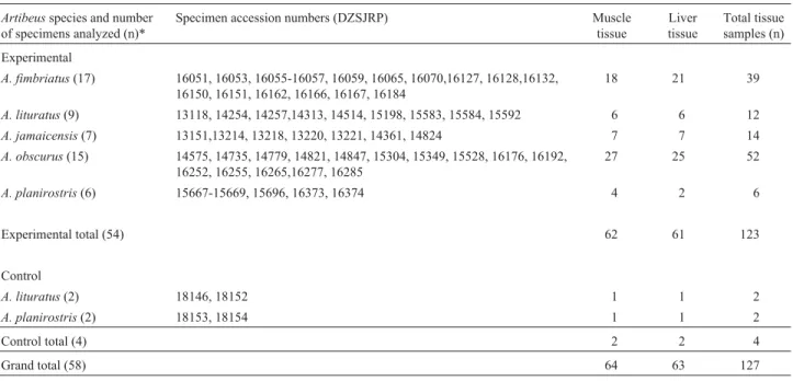
Table 1- Number of preserved voucher specimens (n) ofArtibeusfrugivorous bats and liver and muscle samples analyzed. The experimental group tis-sues were formalin-fixed while the tistis-sues in the control group were fresh-frozen.
Artibeusspecies and number of specimens analyzed (n)*
Specimen accession numbers (DZSJRP) Muscle
tissue
Liver tissue
Total tissue samples (n)
Experimental
A. fimbriatus(17) 16051, 16053, 16055-16057, 16059, 16065, 16070,16127, 16128,16132, 16150, 16151, 16162, 16166, 16167, 16184
18 21 39
A. lituratus(9) 13118, 14254, 14257,14313, 14514, 15198, 15583, 15584, 15592 6 6 12
A. jamaicensis(7) 13151,13214, 13218, 13220, 13221, 14361, 14824 7 7 14
A. obscurus(15) 14575, 14735, 14779, 14821, 14847, 15304, 15349, 15528, 16176, 16192, 16252, 16255, 16265,16277, 16285
27 25 52
A. planirostris(6) 15667-15669, 15696, 16373, 16374 4 2 6
Experimental total (54) 62 61 123
Control
A. lituratus(2) 18146, 18152 1 1 2
A. planirostris(2) 18153, 18154 1 1 2
Control total (4) 2 2 4
Grand total (58) 64 63 127
washed with HBSS, cut into small pieces and placed in a 15 mL sterile polypropylene tube containing 1 mL of HBSS. No treatment was applied prior to DNA extraction.
To extract DNA the suspensions of the formalin-fixed and fresh samples were centrifuged (Eppendorf Scientific, Hamburg, Germany) for five minutes at 3000 g and room temperature (27 °C to 35 °C), the supernatant discarded and the precipitate transferred to an autoclaved microtube con-taining 300 µL of lysis buffer (containing: Tris-HCl, 10 mM; EDTA, 1 mM; NaCl, 100 mM; sodium dodecyl sulfate (SDS, Synth, Brazil) 2% (v/v); and proteinase K, 4 mg mL-1(Invitrogen, USA) and then incubated at 56 °C overnight, with 1µL of 10µg mL-1RNAse solution (Invi-trogen, USA) being added one hour prior to the conclusion of incubation. Extraction and purification of DNA was per-formed by adding 50% by volume of 5 M potassium acetate solution, followed by of vigorous mixing and centrifu-gation at 3000 g and room temperature (27 °C to 35 °C) for 10 min. After centrifugation, the supernatant was removed and transferred to an autoclaved microtube containing 2.5 volumes of iced absolute ethanol to precipitate the DNA, which was pelleted by centrifugation at 12,000 g and room temperature (27 °C to 35 °C). The supernatant was discarded and the precipitate dried and then dissolved in 100µL to 200µL of TE buffer. The concentration of DNA was estimated by electrophoreses on 1.5% agarose gel con-taining 0.15µg mL ethidium-bromide using 100V, 1XTAE buffer, pH 8.0 (containing: Tris, 4.84 g; EDTA, 0.5 M; and 1.14 mL glacial acetic acid) and a 100 bp molecular marker ladder (Biolabs, England).
Each DNA extract was used as a template for PCR amplification using five primer pairs (Invitrogen, São Pau-lo) of different lengths, four amplifying mtDNA segments (12/16S 1-2, 1500 bp; 12/16S 3-4, 500 bp; CITB 4-5, 460 bp; and CITB 14-17, 460 bp) and a nuclear segment (RAG2, 1470 bp). Three different Taq DNA polymerases were tested, TaqGen polymerase (Biosystems), Platinum Taq DNA polymerase High Fidelity (Invitrogen) and Plati-num Taq DNA polymerase (Invitrogen). The PCR reaction was carried out in a total volume of 15µL containing as template 100 ng of sample DNA diluted in 200µL of TE buffer, 1.5 units of Taq DNA polymerase, 3 mM MgCl2,
7.5 ng of bovine serum albumin (BSA) and 0.2 mM each of dATP, dCTP, dGTP, and dTTP plus 1X enzyme mix (200 mM Tris-HCl, 500 mM KCl, 50 mM MgCl2).
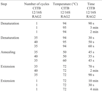
Ampli-fication was carried out in a GeneAmp PCR System 2400 (Perkin Elmer), with the PCR protocol being varied accord-ing the specificity of each primer pair. In brief, the basic protocol consisted of an initial denaturation at 94 or 95 °C, followed by 35 variable cycles plus a final extension at 72 °C. The modifications required by different primers are presented in Table 2. Amplifications were repeated for each of the three different Taq DNA polymerases.
Initially the performance of the different enzymes was tested using 30 successfully genomic DNA samples and a primer pair (CITB 4-5) that amplified a 460 bp seg-ment, these tests indicating that the Platinum Taq polymer-ase (not the High Fidelity version) was the most suitable product in our system. Fresh-frozen tissue samples from two A. lituratus and two A. planirostrisspecimens were used as control samples (fresh tissue DNA) and served as parameters to verify the amplification reaction conditions and quality of amplified fragments in all reactions. In the subsequent experiments we used the Platinum Taq poly-merase and tested all the samples.
The reactions were first obtained with the primers that amplified the longest DNA fragments (12/16S 1-2 and RAG2). The sequencing reactions were conducted on the 27 best amplified samples, 19 from the formalin-fixed ex-perimental group and eight from the fresh-frozen control group, selected by analyzing the concentration of amplified DNA after purification by DNA Sequencing Services of the Human Genome Study Center, University of Sao Paulo, Sao Paulo state, Brazil, which also developed the sequenc-ing reactions. The amplified samples were initially purified with a GFX PCR DNA and Gel Band Purification Kit (Amersham), quantified with a Low DNA Mass Ladder (Invitrogen) and sequenced in a MegaBACE 1000 se-quencer (GE Healthcare).The sequencing procedure was conducted according to the MegaBACE 1000 protocol with a DYEnamic ET Dye Terminator Kit (Thermo Sequenase II DNA polymerase - Code: US81090) and the same primer pairs as were used in the PCR.
The PCR amplification and sequencing reaction re-sults were evaluated by electrophoreses on 1.5% agarose gel containing 0.15µg mL ethidium-bromide using 100 V, 1XTAE buffer, pH 8.0 (containing: Tris, 4.84 g; EDTA, 0.5 M; and 1.14 mL glacial acetic acid) and a 100 bp
molec-Table 2- Reaction parameters for the five primer pairs.
Step Number of cycles CITB 12/16S RAG2
Temperature (°C) CITB 12/16S
RAG2
Time CITB 12/16S RAG2
Denaturation 1
1 1
94 95 94
90 s 3 min 2 min
Denaturation 35 40 35
94 95 94
30 s 50 s 60 s
Annealing 35
40 35
50 50 60
45 s 35 s 45 s
Extension 35
40 35
72 72 72
70 s 2 min 90 s
Extension 1
1 1
72 72 72
10 min 30 s
ular marker ladder (Biolabs, England). The Sequence Analyser Program was utilized to produce the electro-pherograms. The sequences were designed in the BioEdit Sequence Alignment Editor (Hall, 1999) and the alignment was carried out with Clustal W v.1.81 (Thompsonet al., 1994) and Blast 2 sequences. The quality of the sequence was evaluated using the electropherograms and alignments. The suitability of formalin-fixed tissue collection ex-traction procedures for use in molecular studies was as-sessed based on three criteria, the concentration and length of recovered DNA fragments, the ability to amplify mtDNA and nuclear segments of various lengths and the quality of sequencing.
The number of samples used in each treatment and the number and percentage of samples from which DNA was successfully extracted (i.e., samples having a genomic DNA band with a high molecular weight) are shown in Ta-ble 3. Independent of tissue (muscle or liver) the best results for quality and concentration of extracted DNA were ob-tained from tissues which had been immersed in TE buffer for seven days, with genomic DNA being successfully ex-tracted from tissues in 49 (90%) of the 54 samples held in TE buffer for seven days (Table 3) as evidenced by satisfac-tory electrophoretic bands. The electrophoreses bands ob-tained using DNA from fresh-frozen liver tissue from
A. lituratus (C), formalin-fixed muscle tissue from
A. obscurus(lane 1) and formalin-fixed muscle and liver tissue from A. fimbriatus (lanes 7 and 8) are shown in Figure 1A. The results obtained with tissues which had been immersed in TE buffer for four days were also factory, with 60% of the ten samples tested showing satis-factory bands. However, tissues which had been immersed in TE buffer for only one day showed satisfactory genomic DNA banding in only 18% of the samples 22 samples tested, with most samples showing a very low DNA con-centration represented by degraded material appearing as smears in the agarose gel (Figure 1A, evincing DNA from
formalin-fixed muscle tissue fromA. obscurus- lanes 2 and 4, andA. fimbriatus- lane 6). The samples maintained in GN buffer for 20 min or 1 h produced highly degraded DNA, especially under the high temperature used for the 20 min experiments.
Several recent publications have reported the viabil-ity of the extraction of nucleic acids from formalin-fixed paraffin-embedded tissue (FFPE) (Coombs et al., 1999; Masudaet al., 1999; Shiet al., 2002, 2004). Among the methods proposed in these papers is that of heating tissues to high temperatures in alkaline solutions. Shiet al.(2002) argue that this treatment probably acts on tissues by of pro-tein denaturation and hydrolysis, causing the rupture of cel-lular membranes and promoting disruption of the cross-linked bonds formed by formalin. However, contrastingly, we found that heating archival tissues at 80 °C for 20 min pH 10.6 GN buffer, similar conditions to those used by Shi
et al.(2002), was not very efficient. Apparently, the combi-nation of high temperature and alkaline pH produced an in-crease in DNA degradation. Our results indicated that the most satisfactory methodology for minimizing the deleteri-ous effects of formalin on DNA was that in which the fixed tissues were treated for at least four days at 37 °C at a neu-tral pH of about pH 7.0 to pH 8.0. This contrast between our study and the studies cited above may have been due to the fact that although the tissues used by us had been forma-lin-fixed they had not been paraffin-embedded, paraffin acting as an additional barrier to the action of temperature and alkaline pH on tissues and molecules.
The incubation time in the buffer was the most impor-tant element in improving the quality of extracted DNA. However, fixation and preservation conditions, and the preservation time of specimens in different collections can affect the DNA extraction process. We recommend the run-ning of several pilot-tests in order to establish the most ap-propriate treatment for each kind of material or specimen used.
Table 3- Combined number of formalin-fixed muscle and liver samples from preserved voucher specimens ofArtibeusfrugivorous bats tested in each treatment and the number (% in parentheses) of successfully extracted samples. The samples were held in 5 mL pH 8.0 Tris-EDTA buffer (TE, contain-ing: 0.01 M Tris, 0.001 M EDTA), changed every 48 h, or 5 mL pH 10.6 glycine-NaOH (GN: 0.2 M glycine, 0.2 M NaOH). A dash (-) indicates that the specified time was not used for samples from that species.
Time in TE buffer at 37 °C and number of samples Time in GN buffer and number of samples Total number of samples
Artibeusspecies 1 day 4 days 7 days 20 min at 80 °C 60 min at 37 °C
A. fimbriatus 10 5 12 8 4 39
A. lituratus 2 - 8 1 1 12
A. jamaicensis - - 10 2 2 14
A. obscurus 10 5 20 15 2 52
A. planirostris - - 4 1 1 6
Total 22 10 54 27 10 123*
Successfully extracted samples
4 (18%) 6 (60%) 49 (90%) 2 (7%) 0 (0%) 61 (49%)
The customary quantification of extracted DNA by spectrophotometry, was not undertaken in our study be-cause sequence amplification by PCR is apparently most influenced by the quality of the DNA extracted from fixed tissues (Shiet al., 2002; Bareaet al., 2004).
In the analyses of the performance of the different en-zymes using the 30 samples and the primer pair CITB 4-5 (460 bp) the Platinum Taq polymerase High Fidelity (Invi-trogen) generated non-specific fragments as well as the tar-get 460 bp segment but TaqGen polymerase (Biosystems) and Platinum Taq polymerase (Invitrogen) amplified only the target segment (Figure 1B), with Platinum Taq poly-merase producing the best results in terms of the concentra-tion and purity of the amplified fragments (Figure 1B columns 5 to 8). Therefore, the comparisons of the quality of the five primer pairs were based on the amplifications by Platinum Taq Polymerase.
The reactions were first carried out with those primers that amplified the longest DNA fragments (12/16S 1-2 and
RAG2). The efficiency of amplification was only about 30% (17 samples) and the most amplified segments con-tained only degraded material. The primers expected to am-plify the shortest fragments (12/16S3-4 (500 bp), CITB 4-5 (460 bp) and CITB 17-14 (460 bp)) produced the best re-sults because taken together they generated only one PCR product in 44 (72%) out of the 61 DNA samples used for amplification. Although the expected band was always present, in most samples it was accompanied by smears, probably representing degraded DNA, RNA and residual proteins.
The four fresh-frozen samples (fresh tissue DNA) which served as controls for the amplification reaction con-ditions and quality of amplified fragments in all reactions, nearly always produced the expected amplified fragments with few smears, the exception being for fragments ampli-fied with the Platinum Taq polymerase High Fidelity which usually produced more than one amplified fragment.
Our results indicate that the quality of the amplifica-tion reacamplifica-tion product is related not only to the efficiency of the polymerase enzyme and to the size of the amplified fragment but also to the quality of the extracted DNA and its level of impurity and degradation. However, a more effi-cient enzyme does not necessarily improve the quality of the amplified sample, as observed with Platinum Taq poly-merase High Fidelity. Good results can still be obtained by altering some reaction parameters, such as the concentra-tion of reacconcentra-tion components (Meunier and Grimont 1993; Davin-Regliet al., 1995; Halldénet al., 1996) and the num-ber of amplification cycles (Shibataet al., 1994).
The concentration and degree of purity of amplified fragments are the most important prerequisites for success-ful sequencing reactions. Only 31 (45%) of the 69 reactions amplified (representing 65 samples of DNA: 61 of forma-lin-fixed tissue and 4 of fresh-frozen tissue) reached the minimum concentration (20 ng/µL) required by Sequenc-ing Services for a successful process. Two-way sequencSequenc-ing of the DNA band was conducted for 27 reactions (repre-senting 19 from the formalin-fixed experimental group and eight from the fresh-frozen control group), with each am-plified sample yielding two sequences for the reaction. The 27 samples generated 54 sequences, although 18 of these sequences were not of the expected size and showed a low identity of alignment. The remaining 36 sequences, con-sisting of 20 from muscle and liver formalin-fixed tissues and 16 from the fresh-frozen tissues, were reliable se-quences according to the electropherogram and alignment analyses, although there were more gaps and unidentified bases in the sequences obtained from formalin-fixed tis-sues.
The fact that the DNA of 19 samples from the forma-lin-fixed experimental group (representing the five investi-gated species) that were submitted to sequencing were amplified in both the PCR reaction and during sequencing indicate that the amplification of samples obtained from
formalin-fixed NeotropicalArtibeus bat muscle and liver tissue is reproducible. Our study demonstrated that by es-tablishing optimized parameters for purification and ampli-fication of DNA sequencing reactions can generate reliable products for use in phylogenetic analysis, despite the fact that a number of number of sequence errors occurred with DNA extracted from the formalin-fixed tissue when com-pared with the sequences obtained from fresh-frozen tissue. These results suggest that DNA extracted from forma-lin-fixed tissue can be used in molecular studies of Neo-tropicalArtibeus bats and that our methodology may be applicable to other groups of animals, although further studies are needed to confirm that our methodology is ap-plicable to other groups. The information contained in bio-logical collections could be useful for generating molecular data which may help clarify evolutionary and biogeo-graphic aspects of problematic groups, particularly those that are difficult to collect, threatened by extinction or al-ready extinct
Acknowledgments
The authors are grateful to Dr. Peter James Harris for suggestions regarding the English of the manuscript. We also thank the Brazilian agencies Coordenadoria de Aper-feiçoamento de Pessoal de Ensino Superior (CAPES) and Fundação para o Desenvolvimento da UNESP (Fundunesp) for financial support.
References
Barea JA, Pardini MIMC and Gushiken T (2004) Extração de DNA de materiais de arquivo e fontes escassas para a utili-zação em reação de polimeriutili-zação em cadeia (PCR). Rev Bras Hematol Hemoter 26:274-281 (Abstract in English). Coombs NJ, Gough AC and Primrose JN (1999) Optimisation of
DNA and RNA extraction from archival formalin-fixed tis-sue. Nucleic Acids Res 27:12-17.
Corthals A and Desalle R (2005) An application of tissue and DNA banking for genomics and conservation: The Ambrose Monell Cryo-Collection (AMCC). Sys Biol 54:819-823. Davin-Regli A, Abed Y, Charrel RN, Bollet C and de Mico P
(1995) Variations in DNA concentrations significantly af-fect the reproducibility of RAPD fingerprint patterns. Res Microbiol 146:561-568.
Dubeau L, Chandler LA, Gralow JR, Nichols PW and Jones PA (1986) Southern Blotting analysis of DNA extracted from formalin-fixed pathology specimens. Cancer Res 46:2964-2969.
Faulkner SW and Leigh D (1998) Universal amplification of DNA isolated from small regions of paraffin-embedded, for-malin-fixed tissue. BioTechniques 24:47-50.
Frank TS, Svoboda-Newman SM and His ED (1996) Comparison of methods for extracting DNA from formalin-fixed paraffin sections for nonisotopic PCR. Diag Mol Pathol 5:220-224. Goelz SE, Hamilton SR and Vogelstein B (1985) Purification of
DNA from formaldehyde-fixed and paraffin-embedded tis-sue. Biochem Biophys Res Commun 130:118-126.
Hall TA (1999) BioEdit: A user-friendly biological sequence alignment editor and analysis program for Windows 95/98/NT. Nucleic Acids Symp Ser 41:95-98.
Halldén C, Hansen M, Nilsson NO, Hjerdin A and Säll T (1996). Competition as a source of errors in RAPD analysis. Theor Appl Genet 93:1185-1192.
Hanner R, Corthals A and Dessauer H (2005) Salvage of geneti-cally valuable tissues following a freezer failure. Mol Phylo-genet Evol 34:452-455.
Masuda N, Ohnishi T, Kawamoto S, Monden M and Okubo K (1999) Analysis of chemical modification of RNA from for-malin-fixed samples and optimization of molecular biology applications for such samples. Nucleic Acids Res 27:4436-4443.
Meunier JR and Grimont PAD (1993) Factors affecting repro-ducibility of random amplified polymorphic DNA finger-printing. Res Microbiol 144:373-379.
Oliver I, Pik A, Britton D, Dangerfield JM, Colwell RK and Beat-tie AJ (2000) Virtual biodiversity assessment systems. Bio-science 50:441-450.
Shi SR, Key ME and Kalra KL (1991) Antigen retrieval in forma-lin-fixed, paraffin-embedded tissues: An enhancement method for immunohistochemical staining based on micro-wave oven heating of tissues sections. J Histochem Cytochem 39:741-748.
Shi SR, Cote RJ and Taylor CR (2001) Antigen retrieval tech-niques: Current perspectives. J Histochem Cytochem 49:931-937.
Shi SR, Cote RJ, Wu L, Liu C, Datar R, Shi Y, Liu D, Lim H and Taylor CR (2002) DNA extraction from archival forma-lin-fixed, paraffin-embedded tissue sections base on the an-tigen retrieval principle: Heating under the influence of pH. J Histochem Cytochem 50:1005-1011.
Shi SR, Datar R, Liu C, Wu L, Zhang Z, Cote RJ and Taylor CR (2004) DNA extraction from archival formalin-fixed, paraf-fin-embedded tissues: Heat-induced retrieval in alkaline so-lution. Histochem Cell Biol 122:11-218.
Shibata D (1994) Extraction of DNA from paraffin-embedded tis-sue for analysis by polymerase chain reaction: New tricks from an old friend. Hum Pathol 25:561-563.
Thompson JD, Higgins DG and Gibson TJ (1994) Clustal W: Im-proving the sensivity of progressive multiple sequence aligment through sequence weighting, position-specific gap penalties and weight matrix choice. Nucleic Acids Res 22:4673-4680.
Yamashita K, Yoshida T, Shinoda H and Okayasu I (2001) Novel method for simultaneous analysis of p53 and K-ras muta-tions and p53 protein expression in single histologic sec-tions. Arch Pathol Lab Med 125:347-352.
Internet Resources
Blast 2 Sequences (www.ncbi.nlm.nih.gov/blast/bl2seq/wblast2. cgi).
Centro de Estudos do Genoma Humano (http://genoma.ib.usp.br/ servicos/seq-produtos_pcr.php).
Tatusova TA and Madden TL (1999) Blast 2 sequences - a new tool for comparing protein and nucleotide sequences, FEMS. Microbiol Lett 174:247-250, http: www.ncbi.nlm. nih.gov/blast/bl2seq/wblast2.cgi.