c-kit by Stromal Cell Derived Factor-1
a
Zhongpu Chen, Xiaodong Pan, Yuyu Yao, Fengdi Yan, Long Chen, Rong Huang, Genshan Ma*
Department of Cardiology, Zhongda Hospital, Medical School of Southeast University, Nanjing, Jiangsu, China
Abstract
Background:Cardiac progenitor cells (CPCs) have been proven suitable for stem cell therapy after myocardial infarction, especially c-kit(+)CPCs. CPCs marker c-kit and its ligand, the stem cell factor (SCF), are linked as c-kit/SCF axis, which is associated with the functions of proliferation and differentiation. In our previous study, we found that stromal cell-derived factor-1a(SDF-1a) could enhance the expression of c-kit. However, the mechanism is unknown.
Methods and Results:CPCs were isolated from adult mouse hearts, c-kit(+) and c-kit(2) CPCs were separated by magnetic beads. The cells were cultured with SDF-1aand CXCR4-selective antagonist AMD3100, and c-kit expression was measured by qPCR and Western blotting. Results showed that SDF-1a could enhance c-kit expression of c-kit(+)CPCs, made c-kit(2)CPCs expressing c-kit, and AMD3100 could inhibit the function of SDF-1a. After the intervention of SDF-1a and AMD3100, proliferation and migration of CPCs were measured by CCK-8 and transwell assay. Results showed that SDF-1a
could enhance the proliferation and migration of both c-kit(+) and c-kit(2) CPCs, and AMD3100 could inhibit these functions. DNA methyltransferase (DNMT) mRNA were measured by qPCR, DNMT activity was measured using the DNMT activity assay kit, and DNA methylation was analyzed using Sequenom’s MassARRAY platform, after the CPCs were cultured with SDF-1a. The results showed that SDF-1astimulation inhibited the expression of DNMT1 and DNMT3b, which are critical for the maintenance of regional DNA methylation. Global DNMT activity was also inhibited by SDF-1a. Lastly, SDF-1a
treatment led to significant demethylation in both c-kit(+) and c-kit(2) CPCs.
Conclusions: SDF-1a combined with CXCR4 could up-regulate c-kit expression of c-kit(+)CPCs and make c-kit(2)CPCs expressing c-kit, which result in the CPCs proliferation and migration ability improvement, through the inhibition of DNMT1 and DNMT3bexpression and global DNMT activity, as well as the subsequent demethylation of the c-kit gene.
Citation:Chen Z, Pan X, Yao Y, Yan F, Chen L, et al. (2013) Epigenetic Regulation of Cardiac Progenitor Cells Marker c-kit by Stromal Cell Derived Factor-1a. PLoS ONE 8(7): e69134. doi:10.1371/journal.pone.0069134
Editor:Adriano Marchese, Loyola University Chicago, Stritch School of Medicine, United States of America
ReceivedApril 9, 2013;AcceptedJune 12, 2013;PublishedJuly 24, 2013
Copyright:ß2013 Chen et al. This is an open-access article distributed under the terms of the Creative Commons Attribution License, which permits unrestricted use, distribution, and reproduction in any medium, provided the original author and source are credited.
Funding:This work was supported by the National Natural Science Foundation of China (No. 81270204). The funders had no role in study design, data collection and analysis, decision to publish, or preparation of the manuscript.
Competing Interests:The authors have declared that no competing interests exist. * E-mail: [email protected]
Introduction
Ischemic heart disease remain the leading causes of mortality and morbidity worldwide, and stem cell therapy may regenerate cardiac tissue directly by inducing neovasculogenesis and cardio-genesis. In 2003, cardiac progenitor cells (CPCs) were first reported to reside in the adult heart [1–3]. Resident CPCs may be particularly suitble for resurrecting dead myocardium because they are endogenous components of the adult heart and appear to be respnsible for the physiological and pathological turnover of cardiac myocytes and other cardiac cells. The heart have several populations of CPCs, which are self-renewing, clonogenic, multi-potent and have the ability to proliferate and differentiate into functional cardiomyocytes, smooth muscle cells, and other kinds of cells [2,4–6]. Among these CPCs, c-kit(+)CPCs are especially suitable in cell therapy for the recovery of injured cardiomyocytes [2,4]. c-kit(+)CPCs have the larger numbers than other types of CPCs, and have stronger proliferation and differentiation ability to repair the injured myocardium [7].
c-kit is a proto-oncogene and a tyrosine kinase growth factor receptor, expressed on several types of cells, including CPCs [8–
11], with the stem cell factor (SCF) as its ligand. c-kit expression is related to the regulation of cell proliferation, and migration [12– 15]. Stromal cell-derived factor-1a (SDF-1a) is a member of the CXC chemokine family, and CXCR4 is its receptor, which are expressed in a variety of cell types, including CPCs [16]. SDF-1a expression has been reported to increase after an acute myocardial infarction [17]. SDF-1a/CXCR4 axis could prompt stem cell homing to damaged cardiac tissue [16,18]. AMD3100 is a specific antagonist to SDF-1a, which binds to CXCR4 competitively for preventing the combination of SDF-1a and CXCR4. Recent studies have indicated that SDF-1a/CXCR4 and c-kit/SCF axes are closely linked [19]. Our study found that SDF-1a could enhance c-kit expression. However, limited information is known on the regulation of SDF-1aon c-kit.
patterns during DNA replication, whereas DNMT3a and DNMT3b are essential for de novo methylation [20–25]. DNA methylation is an important method in the regulatory mechanisms of gene expression [20–29]. In several diseases such as cancer, gene promoter CpG islands result in abnormal silencing [27–29]. A recent study has found that TGFb1 could regulate CD133 expression through the inhibition of DNMT1 and DNMT3b expressions, and subsequently, the demethylation of promoter-1 [30]. However, the influence of SDF-1aon the expression of c-kit by DNA methylation is unknown.
The present study demonstrates that SDF-1a combined with CXCR4 could up-regulate c-kit expression of c-kit(+)CPCs and make c-kit(2)CPCs expressing c-kit, which result in the CPCs proliferation and migration ability improvement, through the inhibition of DNMT1 and DNMT3b expression and global DNMT activity, as well as the subsequent demethylation of the c-kit gene.
Materials and Methods
Ethics Statement
All animal studies were carried out using a method approved by the the Care of Experimental Animals Committee of the Southeast University, and conform with the guidelines of the National Research Council (approval ID: SYXK-2010.3908).
Isolation and culture of CPCs
For the isolation and culture of CPCs in our laboratory [31], CPCs were acquired from the hearts of two-month-old wild-type male C57BL/6 mice (Yangzhou Laboratory Animal Center). The hearts were acquired using a method approved by the Care of Experimental Animals Committee of the Southeast University, Nanjing, China (Laboratory Animal Center of Southeast University). CPCs were isolated following the standard method described previously. After one or two weeks of growth, a layer of fibroblast-like cells were generated from the adherent myocardial tissue. On these fibroblast-like cells, several small, round, and phase-bright cells emerged, which were collected by the digestion of accutase enzyme, which did not affect the cell surface markers (at room temperature, under direct visualization, for a maximum of 3 min). The obtained cells were separated by magnetic-activated c-kit cell sorting magnetic beads (Miltenyi Biotec Inc., GER) following the instructions of the manufactur-ers. Cells were seeded at 26104 cells/ml on poly-D-lysine (Sigma, USA) coated dishes in cardiosphere growing medium (CGM; 35% IMDM/65%DMEM-Ham’s F-12 [Hyclone, USA] mixture containing 10% fetal calf serum [Hyclone, USA], 2 mmol/L L-glutamine [Hyclone, USA], 0.1 mmol/L 2-mecrap-toethanol [Sigma, USA], 2% B27 [Gibco, USA], 5 ng/ml basic fibroblast growth factor (bFGF) [R&D, USA], 10 ng/ml epider-mal growth factor (EGF) [Peprotech, USA], 40 nmol/L cardio-trophin-1 [Peprotech, USA], 1 unit/ml thrombin [Sigma, USA],
Figure 1. Characterization of cultured CPCs.(A) Cells (small, round, and phase-bright) migrated from the cardiac explants, and aggregated and proliferated on the fibroblast layer after 10 days of culture (6100 magnification). (B) Representative clone generated by CPCs (6100 magnification). (C) and (D) Representative flow cytometric analyses of c-kit(+)CPCs and c-kit(2)CPCs for the expression of the cell surface markers, namely, c-kit, and Sca-1.
100U/ml penicillinG [Hyclone, USA], and 100mg/ml strepto-mycin [Hyclone, USA]).
Characterization of CPCs
CPCs were characterized by phase-contrast microscopy that evaluates morphology and flow cytometric analysis to examine the expression of stem cell surface markers. In the flow cytometric analysis, CPCs were trypsinized and re-suspended in phosphate buffered saline (PBS) and blocked with 3% FBS for 15 min. CPCs were then labeled with PE-conjugated rat anti-mouse c-kit. FITC-conjugated rat anti-mouse Sca-1 (BD Biosciences, USA) was obtained at 4uC in a dark room for 30 min and then washed twice with cold PBS. Data were collected from 16105 cells on a FACSCalibur flow cytometer (BD Biosciences, USA) and analyzed using the WinMDI software.
Quantitative real-time PCR
Trizol reagent (Invitrogen, CA) was used to isolate total RNA from cells, according to the instructions of the manufacturer. First Strand cDNA was obtained by reverse transcription using the cDNA synthesis kit (Fermentas, CA), according to the instructions manufacturer. The cDNA were stored at 220uC until use. Quantitative real-time PCR (qPCR) was performed using IQ SYBR Green Supermix (Bio-Rad, USA). qPCR experiments were also performed using BIO-RAD MJ Mini Opticon Real-Time PCR System, and the matching analysis software was the BIO-RAD CFX Manager. Relative gene expression levels were calculated by normalization to GAPDH. Sequences of each primer were designed as follows: GAPDH forward primer: CAAGGTCATCCATGACAACTTTG and reverse primer: GTCCACCACC- CTGTTGCTGTAG, c-kit forward primer: ACATCGCCAGAGCCAACG and reverse primer: ATCCACTTTAATTTCGGGTCAA, DNMT1 forward primer: GAGCCCA- GCAAAGAGTAT and reverse primer: ATGGTAGAAGGAGGAACAG, DNMT3a forward primer: CTGTCCCATCCAGGCAGTAT and reverse primer; CTTAGCGG- TGTCTTGGAAGC, DNMT3bforward primer: AGATGATGGGAATGGCTCTG and reverse primer: TGCTGAAGATGATGCTCGAC.
Western blotting
Western blotting was performed as described in the following sentences. An equal amount of cell lysates (40mg protein) was denatured in 26SDS-PAGE sample buffer and electrophoresed for 3 h at 20 mA on 10% polyacrylamide gels. The separated proteins were then transferred into polyvinylidene difluoride (PVDF) membranes blocked by TBST solution (10 mM Tris-HCl, 150 mM NaCl, and 0.05% Tween-20), containing 5% nonfat dry milk for 4 h at room temperature. Subsequently, the proteins were incubated with primary antibodies (Santa Cruz, 1:1000 dilution), and placed on a rocker at 4uC overnight. Afterward, the proteins were washed three times with TBST for 15 min, and mixed with IgG-HRP (Santa Cruz, 1:5000) for 2 h at room temperature, which was also washed three times with TBST for 15 min. GAPDH was used as loading control (Santa Cruz, 1:1000). The membranes were incubated in an enhanced chemiluminescence detection system for 5 min, and imaged using a five- minute exposure film. Protein expression was quantified by scanning densitometry.
CPCs proliferation and migration
Cell proliferation assay was performed using CCK-8 kit (cell counting kit-8) (Dojido, Japan). According to the manufacture’s instruction, 5,106103 cells were seeded into 96-well culture plates. Adhesion was verified once (about 12 h later), the cells were incubated with SDF-1a (100 ng/ml for 48 h, Sigma) and AMD3100 (5mg/ml for 48 h, Sigma). Next, cells in each well were incubated with 10ml of CCK-8 at 37uC for 2 h. Then the optical density (OD) for each well was measured at 450 nm using a microplate reader (Bio-Rad Model 550, CA).
A cell migration assay was performed in 24-well Transwell plates (8.0mm, pore size) (Millipore, Billerica). The cells after intervented with SDF-1a and AMD3100 were seeded into the upper chamber of the transwell system at a concentration of 26104cells/well in 100ml medium, and the lower chamber was filled with 100 ng/ml SCF (Sigma) in 600ml medium. After 6 h of incubation at 37uC, 5% CO2, the upper sides of the filters were carefully washed with PBS, and cells remaining were removed with a cotton wool swab. The cells that migrated to the
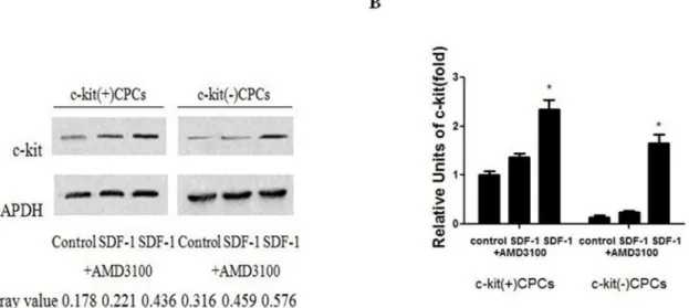
Figure 2. SDF-1aup-regulation on the expression of c-kit.(A) CPCs were stimulated with 100 ng/ml SDF-1aand 5mg/ml AMD3100 for 48 h. Western blotting was performed to analyze the protein expressions of c-kit and GAPDH. (B) qPCR was performed to analyze the mRNA expression of c-kit. Data were obtained from three independent experiments and expressed as mean6SD. n = 3. *P,0.05 versus the control group.
bottom side of the filter were fixed with 4% paraformaldehyde and stained using 0.1% crystal violet. The number of migrated cells were manually counted in three random fields per filter at 6200 magnification by a phase contrast microscope.
Nuclear DNMT Activity Assay
CPCs were stimulated with 100 ng/ml SDF-1a for 48 h. Nuclear protein was extracted using a nuclear extraction kit (Epigentek, Brooklyn, NY), Approximately 5mg of nuclear protein was applied for the DNMT activity assay, which was performed using an EpiQuik DNMT activity assay kit (Epigen-tek), according to the instructions of the manufacturer.
Bisulfite sequencing analysis
Bisulfite treatment: Genomic DNA sodium bisulfate conver-sion was performed using the EZ-96 DNA methylation kit (Zymo Research). The instruction of the manufacturer was followed, using 1mg of genomic DNA and the alternative conversion method (a two-temperature DNA denaturation).
Methylation analysis: Quantitative methylation analysis was carried out with Sequenom’s MassARRAY platform, using
MALDI-TOF mass spectrometry in combined with RNA base-specific cleavage (MassCLEAVE). When feasible, amplicons were designed to cover CpGs in the same region as the 59untranslated regions (59UTR). PCR primers were designed using Methprimer (www.urogene.org/methprimer/). For each reverse primer, an additional T7 promoter tag forin vivo transcription was added, whereas a 10-meter tag on the forward primer was used to adjust melting-temperature differences. MassCLEAVE biochemistry was performed as described previously [32]. Mass spectra was acquired by a MassARRAY Compact MALDI-TOF (Sequenom) and their methylation ratios were generated using the Epityper software v1.0 (Sequenom).
Statistical analysis
Statistical analysis was performed using SPSS (v 11.5, SPSS Inc.). All values were presented as mean6SD. The differences between the two groups were analyzed using the student’s T-test. All tests were two tailed and statistical significance was accepted ifP,0.05.
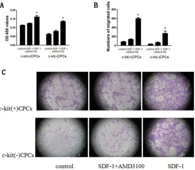
Figure 3. SDF-1aenhances the proliferation and migration of.(A) CPCs were stimulated with 100 ng/ml SDF-1aand 5mg/ml AMD3100 for 48 hours. And CCK-8 assay was used to determine the proliferation. (B) Quantitative analysis of migrated cells. (C) Representative migrated CPCs (stained with crystal violet) are shown (6200 magnification). Data were obtained from three independent experiments and are expressed as mean6 SD.n = 3.rol group.n of cit and GAPHD.0.Western blot wa c-kit at both protein and mRNA level *P,0.05 vs. control group.
Results
CPCs generation and phenotypic characterization CPCs were acquired from the hearts of adult C57BL/6 mouse by mild enzymatic digestion. c-kit(+)CPCs and c-kit(2)CPCs were separated by magnetic-activated cell sorting. After approximately 10 days of culture, a layer of fibroblast-like cells emerged from adherent myocardial tissues, followed by small, round and phase-bright cells (Figures 1A). The inverted phase-contrast microscope examinations showed that CPCs presented clone-like proliferation (Figure 1B). c-kit(+)CPCs and c-kit(2)CPCs were characterized by flow cytometric analysis of the cell surface markers, namely, c-kit and Sca-1 (Figure 1C and 1D).
SDF-1aup-regulates c-kit expression in CPCs
C-kit-positive CPCs were divided into three groups, namely, control, SDF-1a(treated with 100 ng/ml SDF-1a for 48 h), and SDF-1a + AMD3100 groups (treated with 100 ng/ml SDF-1a and 5mg/ml AMD3100 for 48 h). The groups were analyzed using western blotting and qPCR to identify protein and mRNA level. We found that SDF-1a could up-regulate c-kit expression of c-kit(+)CPCs and make c-kit(2)CPCs expressing c-kit at both
protein and mRNA levels, whereas AMD3100 could inhibit this function (Figures 2A and 2B).
SDF-1a enhances proliferation and migration of CPCs To determine whether SDF-1a will influence CPCs prolifera-tion and migraprolifera-tion toward SCF, we performed anin vitroCCK-8 assay and migration assay, and CPCs were placed under SDF-1a with or without the CXCR4 specific antagonist AMD3100. The results indicated that c-kit(+)CPCs proliferation rates of SDF-1a group (0.16260.008 OD) increase significantly, compared with that of control group (0.11460.002 OD) and SDF-1a+AMD3100group (0.12560.003 OD), and c-kit(2) CPCs proliferation rates of SDF-1agroup (0.13560.004 OD) increase significantly, compared with that of control group (0.06360.004 OD) and SDF-1a+AMD3100group (0.08060.006 OD) (Figure 3A). And c-kit(+)CPCs migration rates of SDF-1agroup (SCF+SDF-1a) (602.3620.0 cells) also increase significantly, compared with that of control group (with SCF, without SDF-1a) (85.0611.8 cells), and inhibition group (with SCF+ SDF-1a+AMD3100) (138.7614.6 cells), and c-kit(-)CPCs migration rates of SDF-1a group (SCF+SDF-1a) (272.0650.7 cells) also increase signicantly, compared with that of control group (with SCF, without SDF-1a) (37.065.0 cells), and inhibition group (with Figure 4. SDF-1ainhibition on the expression and activity of DNMT. (A) CPCs were stimulated with 100 ng/ml SDF-1aand 5mg/ml AMD3100 for 48 h. qPCR was used to determine the relative DNMT1 mRNA levels. (B) qPCR was used to determine the relative DNMT3amRNA levels. (C) qPCR was used to determine relative DNMT3bmRNA levels. (D) EpiQuik DNMT activity assay kit was used to analyze the global DNMT activity. Data were obtained from three independent experiments and expressed as mean6SD. n = 3.rol group.n of cit and GAPHD.0.Western blot wa c-kit at both protein and mRNA level *P,0.05 versus the control group.
SCF+SDF-1a+AMD3100) (67.3611.4 cells) (Figure 3B, and 3C). (n = 3, mean6SD, P,0.05).
SDF-1ainhibits DNMT1 and DNMT3bexpression CPCs were divided into three groups, namely, control, SDF-1+AMD3100 and SDF-1a groups to examine whether DNMT regulates c-kit expression through methylation of the promoter. DNMT1, DNMT3a, and DNMT3bexpression was examined in each group. The results showed that DNMT expression, include DNMT1, DNMT3a, and DNMT3b, was significantly higher in c-kit(2)CPCs compared to c-kit(+)CPCs, and SDF-1a combined with CXCR4 was able to reduce the DNMT1 and DNMT3b expression (Figures 4A, 4B and 4C). Subsequently, global DNMT activity in c-kit(+)CPCs nuclei was significantly reduced, from
0.3360.02mmol/hr/mg in the untreated cells to 0.1060.03mmol/ hr/mg, and SDF-1a+AMD3100 group was 0.2660.15mmol/ hr/mg, while, in c-kit(2)CPCs, from 0.7260.04mmol/hr/mg in the untreated cells to 0.4760.04mmol/hr/mg, and SDF-1a+AMD3100 group was 0.6660.03mmol/hr/mg (n = 3, mean6SD, P,0.05) (Figure 4D).
SDF-1a induces demethylation of c-kit promoter With the suppression of DNMT mRNA expression and activity by SDF-1a, we investigated whether SDF-1a induces c-kit expression through promoter demethylation. A total of 16 CpG sites in the promoter were divided into 11 CpG sites. c-kit forward primer: aggaagagagTTAATAGGAATAGAAA-TAAATGTTGGGG and reverse primer: cagtaatacgactcactatagg-Figure 5. Induction of SDF-1aon demethylation of the c-kit promoter in CPCs.(A) Profiling of the site-specific methylation of CpG sites in the c-kit promoter region. Each line represents a CpG methylation profile of the c-kit promoter region from the control (PC1 to PC3) and the SDF-1 (PS1 to PS3) samples. The colors of each circle represent the methylation level of each corresponding CpG unit. The white circles represent the missing data at a given CpG site. (B) CPCs were stimulated with 100 ng/ml SDF-1afor 48 h. Genomic DNA was extracted and subjected to Bisulfite sequencing analysis. The data represent the percentage of methylation at corresponding CpG sites, with CpG site number corresponding to the sites identified in the schematic diagram. Data were obtained from three independent experiments and are expressed as mean6SD. n = 3.rol group.n of cit and GAPHD.0.Western blot wa c-kit at both protein and mRNA level *P,0.05 versus the control group.
gagaaggctCTATACCCTCTAAAACCAA- AAACCC. CPCs were then divided into two groups, namely, control and SDF-1agroups. Genomic DNA was extracted and subjected to Bisulfite sequencing analysis. We found that SDF-1a could significantly reduce the methylation percentage of c-kit(+)CPCs in five out of seven, and all of seven for c-kit(2)CPCs, analyzed CpG sites after incubation. (Figures 5A and 5B).
Discussion
The key findings in our study are listed as follows: (1) An adequate amount of CPCs could be obtained from adult mouse heart tissue using enzymatic digestion, and c-kit(+)CPCs and c-kit(2)CPCs could be separated by magnetic-activated cell sorting, (2) SDF-1a combined with CXCR4 could up-regulate c-kit expression of c-kit(+) CPCs and make c-kit(2)CPCs expressing c-kit, this regulation could be suppressed by ADM3100, an inhibitor of SDF-1a, (3) SDF-1a combined with CXCR4 could enhance the proliferation and migration abilities of CPCs, and these fuctions could be suppressed by AMD3100, (4)SDF-1a combined with CXCR4 could up-regulated c-kit expression through the inhibition of DNMT1 and DNMT3b expression and global DNMT activity, as well as through subsequent demethylation of the c-kit gene.
In regenerating the functional cardiac tissue, stem cell therapy is an effective method for the recovery of the injured myocardium. We selected CPCs because of their incomparable cardiac regenerative capacity [33–34]. Presently, CPCs are based exclu-sively on the expression of a stem cell-related surface antigen, namely, c-kit and Sca-1. CPCs are multipotent, self-renewing, and clonogenic, which have the ability to transform cardiomyocytes in normal, as well as in aging and diseased hearts. CPCs apparently have a mixture of different subpopulations. Among these CPCs, c-kit(+)CPCs showed better cardiac repair capability thus are considered as the most suitable cells for myocardial regeneration therapies [35]. Hence, in this study, we selected the marker, c-kit, as the identification index of CPCs, as observed in the heart. Our study verified that, an adequate amount of CPCs could be acquired from an adult mouse heart through enzymatic digestion, c-kit(+)CPCs and c-kit(2)CPCs could be separated by magnetic-activated cell sorting. CPCs within 10 generations, which we had actually examined for c-kit expression, were used for these experiments. Researches showed that c-kit (+)CPCs could be passaged to the 40 generation, and still had kept the stem cell surface markers [36].
c-kit is a transmembrane tyrosine kinase factor receptor. Its ligand, SCF, is an early hemopoietic growth factor. c-kit/SCF axis supports the proliferation and migration of multiple hemopoietic lineages [37–39]. SDF-1abelongs to the CXC subfamily, which has the ability to facilitate the transmigration of hematopoietic cells through endothelial cell barriers [40]. CXCR4 is its receptor, a seven- transmembrane G protein-coupled receptor. SDF-1a expression is aimed to protect against myocardial ischemic injury [41], which is critical in progenitor cell tissue retention, trafficking, and homing [42]. SDF-1aexpression has been shown to enhance the survival of progenitor cells in several stimuli such as in ischemia/reperfusion injury [43–44], serum withdrawal and apoptotic cell death, through interaction with CXCR4 [45]. AMD3100 is a specific antagonist to SDF-1a, which competitively binds to CXCR4 to prevent the combination of SDF-1a and CXCR4, effectively blocking.90% of binding SDF-1a [46]. A recent study showed that AMD3100 with the concentration of 5mg/ml could efficiently prevent the SDF-1a/CXCR4 axis [47]. In our study, we found that SDF-1acombined with CXCR4 could
up-regulate c-kit expression of c-kit(+)CPCs and make c-kit(2)CPCs expressing c-kit, which result in the CPCs proliferation and migration abilities improvement. Research showed VEGFMSCs could induced SDF-1a and CXCR4 expression, and promoted CSCs proliferation and migration, whereas blockade of SDF-1a or its receptor CXCR4 by RNAi or antagonist significantly diminished these beneficial effects of VEGFMSCs [48]. Our results were similar to these results, and the conclusion was that SDF-1a/CXCR4 axis could affect CSCs proliferation and migration. However, the mechanism is not quite clear.
DNA methylation is an important mechanism for gene transcriptional silencing. CpG hypermethylation in DNA pro-moter regions is responsible for gene silencing [49–51]. DNA methylation status was regulated by DNMT, which hasde novo methylation activity. We found that SDF-1a combined with CXCR4 could inhibit global DNMT activity. Furthermore, DNMT expression, include DNMT1, DNMT3a, and DNMT3b, was significantly higher in c-kit(2)CPCs compared to c-kit(+)CPCs, and DNMT1 and DNMT3b expression was suppressed by the stimulation of SDF-1a combined with CXCR4. Therefore, DNMT1 and DNMT3b are critical enzymes in the mechanism of SDF-1a combined with CXCR4 induced c-kit expression. Meanwhile, Bisulfite sequencing anal-ysis was chosen to quantify the promoter methylation degree in multiple CpG sites. Our data demonstrated that SDF-1a significantly reduces c-kit promoter methylation of c-kit(+)CPCs in five out of seven CpG sites, and all of seven CpG sites for c-kit(2)CPCs. Therefore, the 7th and 15th CpG sites probably play an important role in the expression of kit gene in c-kit(2)CPCs. Although the effect of SDF-1a on methylation in individual CpG sites is relatively small, the overall effect of accumulated demethylation induced by SDF-1ain multiple CpG sites has significant influence on c-kit transcription. Therefore, SDF-1a induced demethylation in the c-kit promoter is important in the regulation of c-kit transcription.
Our study demonstrated that SDF-1acombined with CXCR4 could regulate c-kit expression, which result in the CPCs proliferation and migration ability improvement, and this function could be suppressed by ADM3100. Furthermore, the up-regulation of c-kit expression by SDF-1acombined with CXCR4 is through the inhibition of DNMT1 and DNMT3b expression and the global DNMTs activity, as well as through the subsequent demethylation of the c-kit gene. In summary, this study described a mechanism by which SDF-1acombined with CXCR4 regulates c-kit expression through promoter demethylation. However, no clue about the mechanism of the inhibiting effect of SDF-1a combined with CXCR4 to DNMT was found so far. We will continue to search for the possible mechanism. Our current findings provided a novel strategy for stem cell therapy in recovering damaged myocardium through the modification of the status of c-kit promoter methylation by potentially targeting SDF-1aor DNMTs.
Acknowledgments
We are grateful to Dr. Yao Liang Tang for his valuable technical assistance.
Author Contributions
References
1. Oh H, Bradfute SB, Gallardo TD, Nakamura T, Gaussin V, et al. (2003) Cardiac progenitor cells from adult myocardium: homing, differentiation, and fusion after infarction. Proc Natl Acad Sci USA 100: 12313–12318. 2. Beltrami AP, Barlucchi L, Torella D, Baker M, Limana F, et al. (2003) Adult
cardiac stem cells are multipotent and support myocardial regeneration. Cell 114: 763–776.
3. Urbanek K, Quaini F, Tasca G, Torella D, Castaldo C, et al. (2003) Intense myocyte formation from cardiac stem cells in human cardiac hypertrophy. Proc Natl Sci USA 100: 10440–10445.
4. Dawn B, Stein AB, Urbanek K, Rota M, Whang B, et al. (2005) Cardiac stem cell delivered intravascularly traverse the vessel barrier, regenerate infarcted myocardium, and improve cardiac function. PNAS 102: 3766–3771. 5. Davis DR, Zhang Y, Smith RR, Cheng K, Terrovitis J, et al. (2009) Validation
of the Cardiosphere Method to Culture Cardiac Progenitor Cells from Myocardial Tissue. PLoS One 4: e7195.
6. Messina E, De Angelis L, Frati G, Morrone S, Chimenti S, et al. (2004) Isolation and expansion of adult cardiac stem cells from human and murine heart. Circ Res 95: 911–921.
7. Linke A, Mu¨ller P, Nurzynska D, Casarsa C, Torella D, et al. (2005) Stem cells in the dog heart are self-renewing, clonogenic, and multipotent and regenerate infarcted myocardium, improving cardiac function. Proc Natl Sci USA 102: 8966–8971.
8. Teyssier-Le Discorde M, Prost S, Nandrot E, Kirszenbaum M (1999) Spatial and temporal mapping of c-kit and its ligand, stem cell factor expression during human embryonic haemopoiesis. Br J Haematol 107: 247–253.
9. Kunisada T, Yoshida H, Yamazaki H, Miyamoto A, Hemmi H, et al. (1998) Transgene expression of steel factor in the basal layer of epidermis promotes survival, proliferation, differentiation and migration of melanocyte precursors. Development 125: 2915–2923.
10. Anversa P, Kajstura J, Nadal-Ginard B, Leri A (2003) Primitive cells and tissue regeneration. Circ Res 92: 579–582.
11. Urbanek K, Cesselli D, Rota M, Nascimbene A, De Angelis A, et al. (2006) Stem cell niches in the adult mouse heart. Proc Natl Sci USA 103: 9226–9231. 12. Kitamura Y, Hirotab S (2004) Kit as a human oncogenic tyrosine kinase. Cell
Mol Life Sci 61: 2924–2931.
13. Miettinen M, Lasota J (2005) KIT (CD117): a review on expression in normal and neoplastic tissues, and mutations and their clinicopathologic correlation. Appl Immunohistochem Mol Morphol 13: 205–220.
14. Cheng M, Zhou J, Wu M, Boriboun C, Thorne T, et al. (2010) CXCR4-Mediated Bone Marrow Progenitor Cell Maintenance and Mobilization Are Modulated by c-kit Activity. Circ Res 107: 1083–1093.
15. Naqvi N, Li M, Yahiro E, Graham RM, Husain A (2009) Insights into the Characteristics of Mammalian Cardiomyocyte Terminal Differentiation Shown Through the Study of Mice with a Dysfunctional c-Kit. Pediatr Cardiol 30: 651– 658.
16. Tang YL, Zhu W, Cheng M, Chen L, Zhang J, et al. (2009) Hypoxic preconditioning enhances the benefit of cardiac progenitor cell therapy for treatment of myocardial infarction by inducing CXCR4 expression. Circ Res 104: 1209–1216.
17. Ma J, Ge J, Zhang S, Sun A, Shen J, et al. (2005) Time course of myocardial stromal cell-derived factor 1 expression and beneficial effects of intravenously administered bone marrow stem cells in rats with experimental myocardial infarction. Basic Res Cardiol 100: 217–223.
18. Mouquet F, Pfister O, Jain M, Oikonomopoulos A, Ngoy S, et al. (2005) Restoration of cardiac progenitor cells after myocardial infarction by self-proliferation and selective homing of bone marrow-derived stem cells. Circ Res 97: 1090–1092.
19. Cheng M, Zhou J, Wu M, Boriboun C, Thorne T, et al. (2010) CXCR4-mediated bone marrow progenitor cell maintenance and mobilization are modulated by c-kit activity. Circ Res 107: 1083–1093.
20. Miranda TB, Jones PA (2007) DNA methylation: the nuts and bolts of repression. J Cell Physiol 213: 384–390.
21. Okano M, Bell DW, Haber DA, Li E (1999) DNA methyltransferases Dnmt3a and Dnmt3b are essential for de novo methylation and mammalian development. Cell 99: 247–257.
22. Leonhardt H, Page AW, Weier HU, Bestor TH (1992) A targeting sequence directs DNA methyltransferase to sites of DNA replication in mammalian nuclei. Cell 71: 865–873.
23. Liu Y, Oakeley EJ, Sun L, Jost JP (1998) Multiple domains are involved in the targeting of the mouse DNA methyltransferase to the DNA replication foci. Nucleic Acids Res 26: 1038–1045.
24. Prokhortchouk E, Defossez PA (2008) The cell biology of DNA methylation in mammals. Biochim Biophys Acta 1783: 2167–2173.
25. Plachot C, Lelie`vre SA (2004) DNA methylation control of tissue polarity and cellular differentiation in the mammary epithelium. Exp Cell Res 298: 122–132. 26. Mann J, Oakley F, Akiboye F, Elsharkawy A, Thorne AW, et al (2007) Regulation of myofibroblast transdifferentiation by DNA methylation and MeCP2: implications for wound healing and fibrogenesis. Cell Death Differ 14: 275–285.
27. Poplawski T, Tomaszewska K, Galicki M, Morawiec Z, Blasiak J (2008) Promoter methylation of cancer-related genes in gastric carcinoma. Exp Oncol 30: 112–116.
28. Lee MP, Dunn BK (2008) Influence of genetic inheritance on global epigenetic states and cancer risk prediction with DNA methylation signature: challenges in technology and data analysis. Nutr Rev 66: S69–S72.
29. Ze-Jun-L, Masato M (2002) Gene promoter methylation – a potential cancer marker. Adv Clin Path 6: 43–46.
30. You H, Ding W, Rountree CB (2010) Epigenetic regulation of cancer stem cell marker CD133 by transforming growth factor-beta. Hepatology 51: 1635–1644. 31. Yan F, Yao Y, Chen L, Li Y, Sheng Z, et al. (2012) Hypoxic preconditioning improves survival of cardiac progenitor cells: Role of stromal cell derived factor-1a– CXCR4 axis. PLoS One 7: e37948.
32. Ehrich M, Nelson MR, Stanssens P, Zabeau M, Liloglou T, et al. (2005) Quantitative high-throughput analysis of DNA methylation patterns by base-specific cleavage and mass spectrometry. Proc Natl Acad Sci USA 102: 15785– 15790.
33. Rota M, Padin-Iruegas ME, Misao Y, De Angelis A, Maestroni S, et al. (2008) Local Activation or Implantation of Cardiac Progenitor Cells Rescues Scarred Infarcted Myocardium Improving Cardiac Function. Circ Re 103: 107–116. 34. Padin-Iruegas ME, Misao Y, Davis ME, Segers VF, Esposito G, et al. (2009)
Cardiac progenitor cells and biotinylated insulin-like growth factor-1 nanofibers improve endogenous and exogenous myocardial regeneration after infarction. Circulation 120: 876–887.
35. Kawaguchi N, Smith AJ, Waring CD, Hasan MK, Miyamoto S, et al. (2010) c-kitpos GATA-4 high rat cardiac stem cells foster adult cardiomyocyte survival through IGF-1 paracrine signalling. PLoS One 5: e14297.
36. Miyamoto S, Kawaguchi N, Ellison GM, Matsuoka R, Shin’oka T, et al (2010) Characterization of long-term cultured c-kit+cardiac stem cells derived from adult rat hearts. Stem Cells Dev 19: 105–116.
37. Martin FH, Suggs SV, Langley KE, Lu HS, Ting J, et al. (1990) Primary structure and functional expression of rat and human stem cell factor DNAs. Cell 63: 203–211.
38. Anderson DM, Lyman SD, Baird A, Wignall JM, Eisenman J, et al. (1990) Molecular cloning of mast cell growth factor, a hematopoietin that is active in both membrane bound and soluble forms. Cell 63: 235–243.
39. Huang E, Nocka K, Beier DR, Chu TY, Buck J, et al. (1990) The hematopoietic growth factor KL is encoded by the Sl locus and is the ligand of the c-kit receptor, the gene product of the W locus. Cell 63: 225–233.
40. Mo¨hle R, Moore MA, Nachman RL, Rafii S (1997) Transendothelial migration of CD34+and mature hematopoietic cells: an in vitro study using a human bone marrow endothelial cell line. Blood 89: 72–80.
41. Kortesidis A, Zannettino A, Isenmann S, Shi S, Lapidot T, et al. (2005) Stromal-derived factor-1 promotes the growth, survival, and development of human bone marrow stromal stem cells. Blood 105: 3793–3801.
42. Kucia M, Reca R, Miekus K, Wanzeck J, Wojakowski W, et al. (2005) Trafficking of normal stem cells and metastasis of cancer stem cells involve similar mechanisms: pivotal role of the SDF-1-CXCR4 axis. Stem Cells 23: 879– 894.
43. Hu X, Dai S, Wu WJ, Tan W, Zhu X, et al. (2007) Stromal cell derived factor-1 alpha confers protection against myocardial ischemia/reperfusion injury: role of the cardiac stromal cell-derived factor-1a-CXCR4 axis. Circulation 116: 654– 663.
44. Askari AT, Unzek S, Popovic ZB, Goldman CK, Forudi F, et al. (2003) Effect of stromal-cell-derived factor 1 on stem-cell homing and tissue regeneration in ischemic cardiomyopathy. Lancet 362: 697–703.
45. Tachibana K, Hirota S, Iizasa H, Yoshida H, Kawabata K, et al. (1998) The chemokine receptor CXCR4 is essential for vascularization of the gastrointes-tinal tract. Nature 393: 591–594.
46. Fricker SP, Anastassov V, Cox J, Darkes MC, Grujic O, et al. (2006) Characterization of the molecular pharmacology of AMD3100: a specific antagonist of the G-protein coupled chemokine receptor, CXCR4. Biochem Pharmacol 72: 588–596.
47. Stamatopoulos B, Meuleman N, De Bruyn C, Pieters K, Mineur P, et al. (2012) AMD3100 disrupts the cross-talk between chronic lymphocytic leukemia cells and a mesenchymal stromal or nurse-like cell -based microenvironment: preclinical evidence for its association with chronic lymphocytic leukemia treatments. Haematologica 97: 608–615.
48. Tang JM, Wang JN, Zhang L, Zheng F, Yang JY, et al. (2011) VEGF/SDF-1 promotes cardiac stem cell mobilization and myocardial repair in the infarcted heart. Cardiovasc Res 91: 402–411.
49. Ikeda JI, Morii E, Kimura H, Tomita Y, Takakuwa T, et al. (2006) Epigenetic regulation of the expression of the novel stem cell marker CDCP1 in cancer cells. J Pathol 210: 75–84.
50. Huang Y, Song H, Hu H, Cui L, You C, et al (2012) Trichosanthin inhibits DNA methyltransferase and restores methylation-silenced gene expression in human cervical cancer cells. Mol Med Report 6: 872–878.