A
DRIANA
R
AHAL
R
EBOUÇAS DE
C
ARVALHO
Preferência mastigatória em pacientes com paralisia
facial periférica flácida de duração igual ou superior a
seis meses: estudo clínico e eletromiográfico
Tese apresentada à Faculdade de Medicina da Universidade de São Paulo para obtenção do título de Doutor em Ciências
Área de concentração: Otorrinolaringologia Orientadora: Profª. Drª. Maria Valéria Schmidt
Goffi-Gomez
Dados Internacionais de Catalogação na Publicação (CIP)
Preparada pela Biblioteca da
Faculdade de Medicina da Universidade de São Paulo
Óreprodução autorizada pelo autor
Carvalho, Adriana Rahal Rebouças
Preferência mastigatória em pacientes com paralisia facial periférica flácida de duração igual ou superior a seis meses : estudo clínico e eletromiográfico / Adriana Rahal Rebouças de Carvalho. -- São Paulo, 2008.
Tese(doutorado)--Faculdade de Medicina da Universidade de São Paulo. Departamento de Oftalmologia e Otorrinolaringologia.
Área de concentração: Otorrinolaringologia. Orientadora: Maria Valéria Schmidt Goffi-Gomez.
iii
Você nunca saberá o que é suficiente se não se permitir
saber o que é mais que suficiente. Algumas vezes na
vida é preciso correr o risco de ir longe demais.
v
DEDICATÓRIA
A meu marido, Luis Eduardo, meu amor querido, grande companheiro e incentivador em todos os momentos de nossas vidas.
Aos meus filhos Carlos Eduardo (Cadu) e Gustavo (Gu), que de maneira muito especial sempre estiveram ao meu lado em todo esse longo caminho, torcendo pelo bom sucesso.
Aos meus pais Fares e Samira, meus grandes exemplos de vida.
Aos meus irmãos, Renata, Patricia, Nemer e Flávia, meus cunhados, Luiz Antonio, Fernando, Marcelo e Lucia pelo apoio, incentivo e torcida, por serem verdadeiros irmãos.
AGRADECIMENTOS
Agradeço aos participantes deste sonho.
A Profª. Drª. Maria Valéria Schmidt Goffi-Gomez, orientadora e grande amiga, por acreditar e depositar toda sua confiança em mim, sempre com colocação serena e adequada nos momentos críticos.
Ao Prof. Dr. Ricardo Bento, Professor Titular do Departamento de Otorrinolaringologia da Universidade de São Paulo, ícone da Otorrinolaringologia, grande responsável pela pujança do Departamento de Otorrinolaringologia da USP e pela oportunidade de realizar esta pós-graduação.
Ao Prof. Dr. Luiz Ubirajara Sennes, Professor Livre Docente Doutor, Diretor da Pós-Graduação do Departamento de Otorrinolaringologia da
Universidade de São Paulo, “motor” da pós-graduação.
Ao Prof. Dr. Fares Rahal, Professor Livre Docente, pai, grande incentivador e exemplo de perseverança.
Ao Prof. Dr. Victor Pereira, por todo empenho, dedicação e ajuda na realização deste trabalho. Todo o meu respeito e admiração.
vii
À Fonoaudióloga Daniele Fontes Bernardes, amiga e companheira em toda essa batalha.
A Profª. Drª. Maria Cecília Lorenzi e Dr Raimar Weber, pelo trato estatístico deste trabalho.
A Profª. Drª. Lucia Garcez Lohmann, pelo olhar atento durante a análise dos resultados e ajuda contínua durante essa pesquisa.
As Fonoaudiólogas, Laura Garcia Espartosa Vasconcelos e Maria Flavia Bonadia Bueno de Moraes pela ajuda e torcida constante.
À Marilede Alves, secretária da Pós-Graduação do Departamento de Otorrinolaringologia da Universidade de São Paulo, por seu trabalho árduo, responsável e eficaz de ajudar a organizar a vida dos pós graduandos do Departamento de Otorrinolaringologia.
À Jacira Freire Silva, bibliotecária do Departamento de Otorrinolaringologia da Universidade de São Paulo, por sua dedicação e disponibilidade para ajudar em todos os tipos de pesquisas.
À Márcia Alves e Luci Vânia Lima da Silva, secretárias da Clínica de Otorrinolaringologia da Universidade de São Paulo, grandes incentivadoras e sempre disponíveis em orientar todas as dúvidas.
A Fonoaudióloga e amiga Deborah Basile Rolim por todo o apoio.
Esta tese está de acordo com as seguintes normas, em vigor no momento desta publicação:
Referências: adaptado de International Committee of Medical Journals Editors (Vancouver)
Universidade de São Paulo. Faculdade de Medicina. Serviço de Biblioteca e Documentação.
Guia de apresentação de dissertações, teses e monografias. Elaborado por Anneliese Carneiro
da Cunha, Maria Julia de A. L. Freddi, Maria F. Crestana, Marinalva de Souza Aragão, Suely Campos Cardoso, Valéria Vilhena. 2ª ed. São Paulo: Serviço de Biblioteca e Documentação; 2005.
ix
SUMÁRIO
Normas da revista Artigo
Lista de abreviaturas e siglas Lista de gráficos
Lista de figuras Lista de tabelas Resumo
Summary
1 INTRODUÇÃO ... 01
2 OBJETIVOS ... 09
3 MÉTODOS ... 11
3.1 Casuística ... 12
3.1.1 Critérios de inclusão ... 12
3.1.2 Critérios de exclusão ... 13
3.2 Métodos ... 14
3.2.1 Anamnese ... 14
3.2.2 Exame clínico ... 15
3.2.3 Avaliação eletromiográfica de superfície ... 16
3.2.3.1 Provas eletromiográficas ... 18
3.2.4 Critérios de interpretação ... 19
3.3 Análise Estatística ... 24
4RESULTADOS ... 26
4.1. Anamnese ... 27
4.2 Avaliação Clínica ... 28
4.2.1 Análise da característica do estado clínico dos dentes e da oclusão dentária ... 28
4.2.2 Avaliação clínica da musculatura orofacial ... 29
4.2.3 Avaliação da mastigação ... 30
4.3 Avaliação Eletromiográfica ... 33
5 DISCUSSÃO ... 36
6 CONCLUSÃO ... 50
7 ANEXOS ... 52
NORMAS DA REVISTA
Author Guidelines (For a printer friendly version, please click here.)Otolaryngology-Head and Neck Surgery is an international, peer-reviewed journal. We invite submission of articles on topics pertaining to the science and art of medicine that help fulfill the Journal's mission of publishing "contemporary, ethical, clinically relevant information in otolaryngology, head and neck surgery (ear, nose, throat, head, and neck disorders) that can be used by otolaryngologists, scientists, and related specialists to improve patient care and public health." Articles are published because of scientific merit and are not to be considered general practice guideline standards.All manuscripts and editorial communications should be sent online, via Editorial Manager (EM), to: Richard M. Rosenfeld, MD, MPH, Editor in Chief, Otolaryngology- Head and Neck Surgery. Do not submit paper manuscripts.
Hard copy/print versions will not be accepted. Please see Manuscript Submission, below, and go to
http://otohns.edmgr.com for directions. The editor may not consider your manuscript for publication if authors do not comply with the following instructions.
Submissions not in compliance with these instructions will be returned to the author by the editorial office, and a corrected version must be resubmitted within 30 days. Papers not resubmitted within that time will be withdrawn from consideration.
In addition, accepted manuscripts sent to the publisher (Elsevier) will be typeset and proofs will then be sent electronically to the corresponding author. If proofs are not approved and received by Elsevier within 30 days, the article will not be published.
EDITORIAL POLICIES
All original and review articles are assessed by 2 or more reviewers who rate the manuscript based on the following criteria: 1. Relevance to mission: Can the information in this manuscript be used to improve patient care and public
health?
2. Internal validity: Are the study design, conduct, and analysis described in a manner that is unbiased, appropriate, and reproducible?
3. External validity: Was the study sample chosen appropriately and described in adequate detail for results
to be generalized?
4. Level of evidence: Does this manuscript significantly improve the knowledge base beyond what is already published on this topic?
5. Ethical conduct: Is the manuscript original, approved by an institutional review board (if applicable), and
free of undisclosed conflicts of interest?
The reviewers are also asked to provide comments that assist the editor to decide if the manuscript should be accepted, revised, or rejected.
ARTICLE CATEGORIES
Otolaryngology-Head and Neck Surgery publishes the types of articles defined below. When submitting your
manuscript, please follow the instructions relevant to the applicable article category. Please check Manuscript Preparation and Submission for further details.
xi
affiliations, and locations. Designate ONE author as the corresponding author. Also indicate where the paper was presented, if applicable.
A structured Abstract of up to 200 words with the headings: Objective, Study Design, Subjects and Methods, Results, and Conclusion.
A brief Introduction outlining the wider context that generated the study and the specific issues or
hypotheses the study addresses.
A Methods section with enough detail to ensure reproducibility of the research, including statistical methods and sample size calculation. All procedures must list approval by the local IRB.
A Results section that uses appropriate descriptive and analytic statistics to summarize data.
A Discussion section that summarizes key findings, highlights antecedent literature on the topic, explains what the current study adds to existing knowledge, and details the strengths and limitations of the current research.
A one-paragraph Conclusion summarizing the impact of the study and suggesting areas that might benefit
from further research.
Manuscript length of no more than 3,000 words (exclusive of the title page and abstract,) with up to 20 references, and a total of 10 images (figures and/or tables).
Adherence to the CONSORT statement (www.consort-statement.org) when reporting a randomized trial.
Reviews: Scholarly reviews of the literature on important subjects in otolaryngology-head and neck surgery that challenge readers to expand existing information. Systematic reviews that reduce bias with explicit procedures to select, appraise, and analyze studies are highly preferred over traditional narrative reviews. The components of a review article are:
A title page, including the manuscript title and all authors' full names, academic degrees, institutional
affiliations, and locations. Designate ONE author as the corresponding author. Also indicate where the paper was presented, if applicable.
A structured Abstract of up to 250 words with the headings: Objective, Data Sources, Review Methods, Results, and Conclusion.
An Introduction outlining the explicit clinical problem, rationale for the intervention (if applicable), and the rationale for conducting the review.
A Methods section that specifies the information sources, search strategy, inclusion and exclusion criteria
for articles, criteria and process used for validity assessment (if none, so state), process for data abstraction, and statistical methods for summarizing data.
A Results section that describes study selection, study characteristics, and, when applicable, uses statistical methods to summarize data and to assess heterogeneity.
A Discussion section that summarizes key findings, makes clinical inferences based on validity, interprets results in light of the total available evidence, and lists potential biases in the review process.
A one-paragraph Conclusion summarizing the impact of the study and suggesting areas that might benefit
from further research.
Manuscript length of no more than 4,500 words (exclusive of the title page and abstract), with up to 100 references and a total of 15 images (figures and/or tables).
Adherence to the QUOROM statement (www.consort-statement.org/QUOROM.pdf) when reporting a systematic review of randomized trials.
Commentaries: Communication of a novel, scientifically based opinion or insight as an independent contribution, or regarding a manuscript published in the Journal within the past 6 months. Commentaries should contain a title page, abstract of up to 150 words, a main point and supporting discussion, and may be authored by an individual, group, society, or committee with an important concern of interest to readers.
Manuscript length: no more than 1,800 words (exclusive of title page and abstract), with up to 10 references, and a total of 3 images (figures and/or tables).
Short Scientific Communications: Quick communication of scientific research that is not yet ready for presentation in full form. Such research should have the potential to stimulate communications among researchers and clinicians that may lead to new concepts and supportive work. This section will also include communication of important, innovative, or technical developments. Selected manuscripts will receive an expedited review and publication. Manuscript length: Submissions must have a title page, a structured abstract of 150 words, a maximum length of no more than 900 words, 5 references, and a total of 2 images (figures and/or tables).
Clinical Techniques and Technology: A short report of unique or original methods for: (1) surgical techniques or medical management: OR (2) new devices or technology. Submissions must have a title page and no abstract. Manuscript length: no more than 900 words, 5 references, and a total of 2 images (figures and/or tables).
Case Reports: Report of a truly unique, highly relevant, and educationally valuable case. Submissions should have a title page, NO abstract, and include an Introduction and Discussion. Do not combine case reports with a review of the literature. Manuscript length: no more than 750 words, 5 references, and a total of 2 images (figures and/or
tables). IRB approval is required for all Case Reports.
will be considered, although this will require reducing the text by 150 words.
Letters to the Editor: Brief letters to the editor regarding published material or information of timely interest. If the letter is related to a previously published article it must be submitted within 6 months of publication, and those authors will be invited to reply. The letter should be titled and double-spaced. It should be brief and to the point, with no more than 400 words, 5 references, and only figure or table.
Book Reviews: Authors who wish to have their book considered for review by the Journal should send the book to the editorial office with a cover letter so stating, and including the author's e-mail address. (Journal Editorial Office: One Prince St, Alexandria, VA 22314-3357.)
Supplements: Supplements to the Journal are considered for publication on the basis of importance of topic, expertise of participants, and scientific quality of the articles presented. All supplements undergo peer review. Private funding for supplements is encouraged. Contact the Managing Editor of the Journal (otomanager@entnet.org) for further information and an application form, which must be returned before a supplement can be scheduled.
MANUSCRIPT PREPARATION
Correct preparation of the manuscript will expedite the review and publishing process. Manuscripts must conform to acceptable English usage. Spell out any abbreviations the first time they appear in the text and indicate the abbreviation immediately afterward in parentheses. Use all abbreviations consistently throughout the manuscript. For further questions concerning style, consult a recent issue of this Journal.
Title: Do not exceed 15 words. Identify all animal research as such in the title.
Title Page:The corresponding author must be the same in both the online (EM) submission and the title page. Include the submission title and all authors' full names, academic degrees, institutional affiliations, and locations. Designate ONE author as the corresponding author, and provide a complete address, e-mail address, and phone/fax numbers. The corresponding author will receive all correspondence regarding the manuscript, as well as proof pages and reprint requests. Also indicate where the paper was presented, and if applicable, provide a brief acknowledgment of any grants and/or other assistance received.
Abbreviations: Do not use abbreviations in the title or abstract. When using abbreviations in the text, indicate the abbreviation parenthetically after the first occurrence and use the abbreviation alone for all subsequent occurrences.
Text: Do not use the "Track Changes" feature of any word processing program. If this feature has been used for any portion of the manuscript, all changes must be accepted before building a .pdf submission. Do not use "Endnotes" or similar programs for entering references. The Editorial Office will not edit or process submissions containing this formatting. When preparing the text:
See ARTICLE CATEGORIES for length requirements.
Number all pages beginning with the title page as #1.
Include the Abstract as page #2.
Use only a 10- or 12-point font in either Arial, Times New Roman, or Century styles.
Double-space the manuscript (including references, figure legends, and tables) with minimum 1-inch margins.
Use generic drug and equipment names when possible; cite the proprietary names in parentheses after first mention, if desired. Identify equipment by manufacturer name and location.
State all measurements in metric units, and if desired, add English units in parentheses.
Begin references on a separate page after acknowledgments.
Source of Funding: All sources of funding should be declared in an acknowledgment at the end of the text. In addition, for all Original Research and Review Articles add a subhead, "Role of the funding source," to the end of the Methods section and describe the role of the study sponsor(s), if any, in study design; in the collection, analysis, and interpretation of data; in the writing of the report; and in the decision to submit the paper for publication. If the funding source had no such involvement, the authors should so state.
Acknowledgements: All papers prepared in consultation with a writer, statistician, or any other contributor who is not a coauthor must contain an acknowledgment, following the text, indicating full name(s), degrees, and explicit role(s) in the design, conduct, analysis, or presentation of the research. As noted above, any funding sources should also be declared in this section.
References: Authors are responsible for the completeness, accuracy, and format of their references.
Do not use "Endnotes" or similar programs for entering references.
Cite references in the text by number in the form of a superscript.
xiii
footnotes in the text.
List only the first 3 authors, and add et al after the third author.
Abbreviate journal titles as shown in the Cumulative Index Medicus. Translate any article titles that are not in English.
Examples of correct forms of references are:
Journal reference: Tarasidis G, Watanabe O, Mackinnon SE, et al. End-to-side neurorraphy: a long-term study of neural regeneration in a rat model. Otolaryngol Head Neck Surg 1998;119:337-341.
Book reference: McMinn RMH, Hutchings RT, Logan BM. Color atlas of head and neck anatomy. 1st ed.
Chicago: Year Book Medical Publishers; 1982. p. 10-25.
Chapter reference: Oppenheim JJ, Schecter B. Lymphocyte transformation. In: Friedman R, Friedman H, editors. Manual of clinical immunology. 2nd ed. Washington (DC): American Society for Microbiology; 1980. p. 233-45.
Internet reference: Fredrickson, BL. (2000, March 7). Cultivating positive emotions to optimize health and well-being. Prevention & Treatment, 3, Article 0001a. Retrieved November 20, 2000, from
http://journals.apa.org/prevention/volume3/pre0030001a.html.
Legends for Figures: Provide a legend for each figure. List the legends (double-spaced) on a separate text page, after the reference page. Because all figures will be printed in black and white unless selected by the Editor for color reproduction, please refrain from using color descriptors in the legend.
Tables: Data appearing in tables should supplement, not duplicate, the text. Tables should contain at least 2 columns of data, and should not list qualitative information or single-column numeric data that can be easily described in the Results section. Put tables on separate pages and number them in order of their mention in the text. Place tables after the figure legend page and after the list of references. Provide a brief title for each table, and define any abbreviations in table footnotes. Tables must be no more than 7 inches (18 cm) wide, and should use, at minimum, a 10-point font in either Arial, Times New Roman, or Century styles.
Figures: Must be submitted in electronic format, preferably in TIF or JPEG format. Figures must be entered separately into EM, including the number of the figure in the description box (e.g., Figure 1).
Figures should be created using graphics software such as Photoshop or Illustrator. DO NOT USE
PowerPoint, Corel Draw, or Harvard Graphics. Do not put your figures in Microsoft Word documents.
Color figures are encouraged whenever possible for contrast, though they may not necessarily be selected for publication. COLOR figures submitted with the manuscript may appear in black and white in print, unless selected by the Editor, but will appear on the website in color at no extra charge. When color images appear in print in black and white, the black and white contrast will diminish, so choose distinct color contrasts and/or patterns for best conversion to black and white images.
If a color image is accepted for print, it must meet the following specifications: CMYK at least 300 DPI. Gray scale images should be at least 300 DPI. Combinations of gray scale and line art should be at least 600 DPI. Line art (black and white or color) should be at least 1200 DPI.
If figures have multiple parts (e.g., A, B, C, D), each part must be counted as a separate image in the total number allowed.
Appendices: Will only be published online, not in the print Journal, and may include additional figures or tables that enhance the value of the manuscript. Appendices must be submitted online with the rest of the manuscript and labeled as such. Questionnaires will be considered as Appendices only.
Video Clip Submission: Video Clips may be submitted to enhance the value and impact of any article type listed above, but should not be submitted as stand-alone contributions. They can be submitted only after a manuscript has been accepted for publication. Instructions for submission of video clips are available online at
http://journals.elsevierhealth.com/periodicals/ymhn/content/videosubmission.
MANUSCRIPT SUBMISSION
Before starting the submission process, TWO disclosure forms must be completed for all authors. A) The Electronic Disclosure/Authorship Form (online only) will be electronically uploaded for inclusion in
the manuscript so that reviewers have access to disclosure information. (A manuscript cannot be submitted without including this form). This 3-column disclosure form can be downloaded from the EM website. Go to
http://otohns.edmgr.com - then to "User Instructions" and "Mandatory Items." It requires information on (1) potential conflicts of interest that are upcoming or existed in the past 24 months (if none, state explicitly), and (2) each author's role in creating the final version of the manuscript (e.g., design, conduct, analysis, or presentation of the research). This form is in addition to, not in place of, the complete Copyright/Disclosure Form (described next) that must be submitted off-line independently by each author.
mail to: Editorial Office, Otolaryngology-Head and Neck Surgery, One Prince St, Alexandria, VA 22314.
Go to http://otohns.edmgr.com for directions in using Editorial Manager (EM), the online submission and review system. EM recommends using the most current version of Firefox, Internet Explorer, Safari, or as browsers.
To use Editorial Manager, you must have Adobe Acrobat Reader (a PDF reader) 5.0 or later installed on your system. If you need to install this software, you can download the free Adobe Acrobat Reader at the following address: http://www.adobe.com/products/acrobat/readstep2.html If you experience difficulty installing or using this software, contact your IT department for assistance.
Authors should first read the User Instructions and then, as a first-time user, register in Editorial Manager. Do not use accented letters or symbols in your registration. If you have already registered and received a user name and password, you should not register again. Instead, click Login in the bar at the top of the page. The system will take you through the process step by step. There are tutorials for both authors and reviewers.
You will be guided to fill in the necessary information, then upload your manuscript text (including references, figure legends, tables, etc.) and figures (see above for further information on figures). The Abstract must be included twice--once alone, where indicated, and once as a part of the whole manuscript. It is advisable to save the complete manuscript as a word-processing document, then upload it into EM.
ETHICAL CONCERNS
Conflict of Interest: A conflict of interest exists when an author or the author's institution has financial or personal relationships with other people or organizations that inappropriately influence his or her actions. Financial relationships are easily identifiable, but conflicts can also occur because of personal relationships, academic competition, or intellectual passion. A conflict can be actual or potential, and full disclosure to the Editor is the safest course. Failure to disclose conflicts may lead to publication of an Erratum. All submissions must include BOTH FORMS listed above (Electronic Disclosure/Authorship Form, and the paper Copyright/Disclosure Form), with disclosure of all relationships that could be viewed as presenting a potential conflict of interest. The Editor may use such information as a basis for editorial decisions, and will publish such disclosures if they are believed to be important to readers in judging the manuscript.
Patient Confidentiality: For manuscripts that contain photographs of a person, submit a written release from the person or guardian, or submit a photograph that will not reveal the person's identity (eye covers are inadequate to protect patient identity).
IRB Policy and Animal Studies: Following the mandate of the Declaration of Helsinki (Sec. B, para.13 , World Medical Association Declaration of Helsinki, amended 2004), all medical procedures must be approved by an Institutional Review Board (IRB).Therefore, any such procedures discussed in submissions to Otolaryngology-Head and Neck Surgery must list approval by the local IRB. For experiments involving animals, state the animal-handling protocol in the Methods section including approval by an institutional board. If a review board does not exist in the authors' institution, authors must include a statement describing what you did, using words similar to this: "An IRB is not available in our institution, but this is how we obtained proper consent from the patients, in keeping with the mandate of the Declaration of Helsinki (or, 'ethical handling of animals')." Then add a description of how you complied with this.
Duplicate or Redundant Submission: Manuscripts are considered with the understanding that they have not been published previously and are not under consideration by another publication. If the author explicitly wishes the journal to consider duplicate publication, he or she must submit the request, in writing, to the Editor with appropriate justification. Submit written permission from the copyright holder (normally the publisher) for all PREVIOUSLY PUBLISHED material, including tables, figures, and direct quotations longer than 100 words.
NIH Public Access Policy:
Special Subject Repositories Certain repositories such as PubMed Central ("PMC") are authorized under special
arrangement with Elsevier to process and post certain articles. The following agreements have been established for authors whose articles have been accepted for publication in an Elsevier journal and whose underlying research is supported by one of the following funding bodies:
National Institutes of Health: Elsevier will send a version of the author's accepted manuscript that includes author revisions following peer-review for public access posting 12 months after final publication. Because the NIH "Public Access" policy is voluntary, authors may elect not to deposit such articles in PMC. If you wish to "opt out" and NOT deposit to PMC, you must indicate this by sending an e-mail to NIHauthorrequests@elsevier.com. More information regarding the agreement between Elsevier and the National Institutes of Health can be found at http://www.elsevier.com/wps/find/authorsview.authors/nihauthorrequest.
xv
ARTIGO
Clinical and electromyographic study of lateral preference in mastication in patients
with long-standing peripheral facial paralysis
Adriana Rahal1
Maria Valéria Schmidt Goffi-Gomez2
1 Ms, Departmentof Otorhinolaryngology, Hospital of the University of São Paulo Medical
School (HCFM/USP) SP, Brazil
2Phd, Departmentof Otorhinolaryngology, Hospital of the University of São Paulo Medical
School (HCFM/USP) SP, Brazil
Correspondence: Ms Adriana Rahal, Departamento de Otorrinolaringologia, Faculdade de
Medicina da Universidade de São Paulo, Rua Dr. José Rodrigues Alves Sobrinho, 89/9ª,
Abstract
Introduction: Peripheral facial paralysis (PFP) usually affects the facial nerve in part or in
whole on one side of the face. Most patients with acute PFP find it difficult to chew on the
paralyzed side, especially due to compromised buccinator function. In addition, the sagging
of the ipsilateral lip commissure tends to compromise lip competence.
Objective: to evaluate the impact of long-standing PFP upon mastication, regarding to
clinical mastication preference besides clinical and electromyographic activity of the
masseters.
Method: The study included 27 male and female subjects aged 16−69 years with permanent
natural dentition and long-standing PFP. Patients answered questions on their mastication
habits before and after onset of PFP and were submitted to clinical myofunctional
examination and electromyographical tests of the masseters during clenching and habitual
mastication.
Results: According to the anamnesis, 77,8 % claimed to prefer chewing on the unaffected
side. Clinically, 70% presented a lateral preference in mastication. In the clinical evaluation,
the buccinators and orbicularis oris differed significantly (p=0.025) between the healthy and
the paralyzed side. Only 22.2% of the patients presented increased thickness of the
contralateral masseters. No statistical significant electromyographic difference was observed
between the affected and unaffected side masseters.
Conclusion: In general, subjects with flaccid-stage PFP for 6 months or longer preferred to
masticate on the unaffected side. No significant clinical or electromyographic differences
were found between the affected and unaffected side masseter in this patient population.
xvii Introduction
In bilateral mastication the masticatory effort is distributed evenly on the teeth stabilizing the
periodontal tissues and synchronizing the activity of masticatory muscles innervated by the
fifth cranial nerve. Any disorder affecting the complex interaction between the masticatory
muscles, the teeth and the temporomandibular joints required to grind or pulverize food may
seriously compromise mastication1.
Peripheral facial paralysis (PFP) usually affects the facial nerve in part or in whole on one
side of the face2. Two stages may be recognized, one flaccid, marked by the absence of
nerve impulses, the other with incomplete recovery and anomalous reinnervation,
characterizing its sequelae.
In the flaccid stage, due to muscle incompetence on the paralyzed side of the face, patients
display sagging facial muscles and loss of facial expression, while wrinkles tend to smoothen
out3. Once movement on the paralyzed side has been compromised, the patient cannot
protrude, retract or close the lips properly, nor fully inflate the cheeks4.
Clinically, few patients with PFP can chew on the paralyzed side because the affected
buccinator muscle no longer retains the food between the teeth. In addition, the sagging of
the ipsilateral lip commissure tends to compromise oral competence.
When the flaccid stage of PFP exceeds six months, patients are at risk of chronic muscle
and facial asymmetry5. Subjective orofacial evaluation may not reveal real cause and can
lead to misdiagnosis. However, technological advances have made new and more objective
methods available (such as surface electromyography) for both diagnostic purposes and
myofunctional therapy6.
Objective
The objective of the present study was to evaluate the impact of longterm flaccid peripheral
facial nerve paralysis on mastication, especially with regard to:
clinical masseter activity during food bolus formation
electromyographic activity of the masseters.
Methods
Subjects were recruited among patients referred to myofunctional treatment by the Facial
Nerve Paralysis Team at the Division of Clinical Otorhinolaryngology, Hospital of the
University of São Paulo Medical School (HCFM/USP).
Inclusion criteria:
Age ≥ 16 years
Flaccid unilateral peripheral facial nerve paralysis for ≥ 6 months
Grade House- Brackmann V -VI paralysis7.
Permanent natural dentition, whether complete or not
Presence of premolars and/or molars in all half-arches
PFP of idiopathic, iatrogenic or traumatic origin (e.g. from gunshot or cranial fracture)
Exclusion criteria:
Previous PFP
Congenital PFP
Congenital facial asymmetry
Fewer than 8 teeth in each arch
History of speech therapy associated with orofacial motricity disorder
xix
Twenty seven patients, 16 female with ages ranging from 16 to 61 and 11 male with ages
ranging from 19 to 69 were selected during a period from July 2006 and December 2007.
Procedure
The evaluation was carried out in two steps: 1) anamnesis and clinical evaluation of
mastication, and 2) electromyographic testing. The anamnesis included open questions
about the time of onset and cause of PFP, mastication preference prior to PFP, pain during
mastication, retention of residues in the oral cavity and mastication difficulties associated
with PFP. Subsequently, patients were examined clinically for bite marks, dental condition
and orofacial muscle function according to a standard protocol.
Orbicularis oris: lip protrusion and closure
Buccinators: alternated cheek inflation
Masseters: clenching during maximum intercuspidation
Disposable silicone gloves, wooden spatulas and split loaves of French bread were used in
the clinical habitual mastication study, when checking for8.
Lateral preference in mastication
Pain
Presence of residues after swallowing
Use of hand to support cheek during mastication
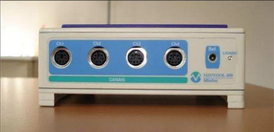
The electromyographic evaluation of the surface bundles of the right and left masseter was
performed with disposable bipolar electrodes (Ag-AgCI double Hal) attached to a Miotec®
a disposable unipolar reference electrode (200 Medi Trace Foam). After the skin had been
rubbed with 70° alcohol to improve action potential conduction and system impedance,
electrodes filled with conductive gel were attached longitudinally along the muscle bundles
(to avoid interference from adjacent muscles) on the anterior right side of the neck and on
the thickest part of the masseters over the goniac angle of the jaw9.
The electromyographic tests consisted of 1) clenching during maximum intercuspidation, and
2) habitual mastication (movements repeated during 15 seconds). In the former test, patients
were asked to clench their teeth with maximum effort during three seconds and then relax.
Three repetitions were performed within 15 seconds. In the habitual mastication test,
patients were asked to chew comfortably on three seedless raisins.
The following evaluation criteria were used:
Clinical observation:
Angle´s bite classification system. Class I = normal; Class II and III = abnormal.
Dental condition: absence of caries at visual inspection = good dental condition;
presence of caries at visual inspection = poor dental condition.
Evaluation of orofacial muscle function:
o Orbicularis oris
Protrusion was considered symmetrical when centralized, and assymetrical when
diverted. Diversion towards the healthy side (H) occured due to increased activity on this
side.
Lip closure with inflated cheeks was considered symmetrical when maintained for 10
xxi o Buccinators:
Alternated inflation of cheeks to detect asymmetry. The healthy side (H) is the most
active; the paralyzed side (P) is the most inflated, showing the lack of resistance.
o Masseters:
Considered symmetrical when the muscle bulk was similar on both sides upon palpation;
asymmetrical when masseter differed in thickness.
Evaluation of mastication:
Lateral preference: “yes” was checked if the patient chewed on only one side during
habitual mastication. The preferred side was registered (H for “healthy”; P for
“paralyzed”). “No” was checked if mastication was bilateral or alternated.
Pain: the presence (“yes”) or absence (“no”) of pain and the side of occurrence (H or P)
were registered as reported by the patient.
Residues: “Yes” was checked if residues were present in the oral cavity after swallowing.
“No” was checked if not. The side (H or P) on which residues were observed was also
registered.
Use of hand to support cheek during mastication: “Yes” was checked if the patient used
the hand to support the cheek during mastication. “No” was checked if not. The
supported side (H or P) was also registered.
Electromyographic tests:
The Miograph® software with Miotool inferface provided by the device manufacturer was
used to calculate the root mean square (RMS) of the electrical muscle potentials from both
Teeth clenching: mean isometric contraction (in µV) of three 3-second clenching cycles.
Habitual mastication: mean isotonic contraction (in µV) of 15 seconds.
The electromyographic data of the PFP patients was statistically compared to a control
group of non-facial paralysis subjects. The control group was composed of sixty subjects,
thirty male and thirty female, with ages ranging from 25 to 45. Control data consisted of
retrospective EMG masticatory assessment which had already been performed for another
study10.
EMG procedure was the same for both groups. The EMG equipment used in the control
group was K6 I, Myotronics with disposable bipolar electrodes (Ag-AgCI double Hal) in the
right and left masseter and a disposable unipolar reference electrode (200 Medi Trace
Foam) in the right sternocleidomastoid muscle.
Statistical analysis
The masseter activity index (MAI) between the two sides was calculated from the
electromyographic data. In PFP patients and controls with confirmed lateral preference in
mastication, MAI was obtained by dividing the electromyographic activity on the
non-preferred side by the electromyographic activity on the non-preferred side. In patients with no
lateral preference, MAI was calculated by dividing the electromyographic activity on the
paralyzed side by the electromyographic activity on the unaffected side (PFP patients) or by
dividing activity on the right side by activity on the left side (controls). The MAI values were
then compared for PFP patients and controls according to the presence of lateral preference
in mastication.
The comparison between the experimental group and the control group data was possible,
despite de use of different equipment because we did not consider the absolute RMS values,
xxiii
were described in terms of frequency while qualitative variables were expressed in mean values
and standard deviation. Odds ratios were calculated for associations between categorical
variables. When applicable, mean values were analysed by Student´s t test and paired t test. All
statistical tests were two-tailed, and the level of significance was set at p<0.05.
Results
According to the anamnesis (Table 1), most patients (77.8%) reported chewing on both sides
before onset of PFP. On the other hand, more than half the subjects denied having
mastication difficulties or aiding mastication with the hand (55.6% and 62.9%, respectively),
at least 6 months after the onset of the PFP.
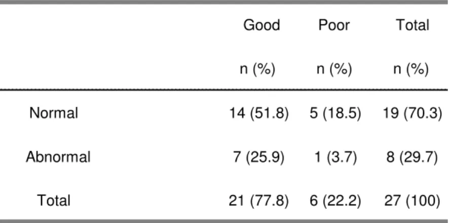
As shown in Table 2, most patients presented good dental condition (21/27) and normal bite
(14/27). Patients had an average of 12 teeth ± 1.20 on each side of the mouth.
Table 3 shows that of 19 (70%) patients with Class I bite, only 12 (44%) reported preferring
to chew on the healthy side. All patients with Class II bite preferred the healthy side.
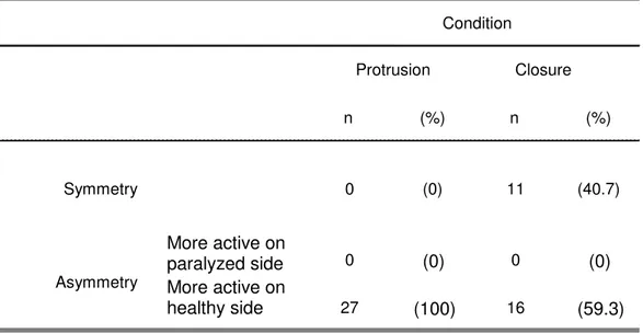
Although all patients (100%) had asymmetrical lips upon protrusion (Table 4), as many as
40.7% presented adequate lip closure.
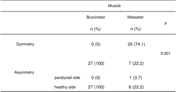
As shown in Table 5, 100% of the patients had asymmetrical buccinators with greater activity
on the healthy (H) side, whereas 25.9% presented asymmetrical masseters (22.2% with
greater activity on the healthy side).
The clinical study revealed that 85.2% of the subjects had a lateral preference in mastication
(H = 77.8%). Pain and hand-aided mastication were infrequently observed (7.7% and 14.8%,
respectively). Residues were seen in 63% of cases (P = 100%) (Table 6).
Distribution patients according to clinically evaluated lip condition and lateral preference in
mastication during the habitual mastication test (Table 7).
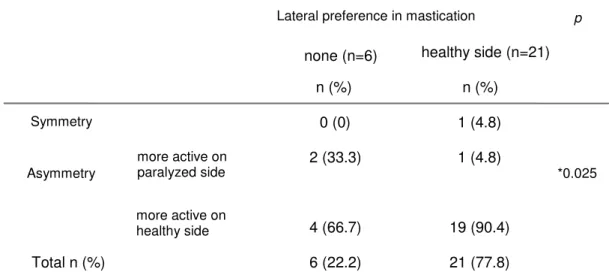
Patients with and without clinically evaluated lateral preference in mastication differed
significantly during habitual mastication with regard to the clinical condition of the buccinators
Lateral preference in mastication occurred more frequently among PFP patients than among
controls (p<0.001), as shown in Figure 1. The odds ratio of a subject with lateral preference
in mastication belonging to the group of PFP patients was 7.6 times greater than that of
belonging to the group of controls (CI: 95%; range: 2.6–21.6).
Comparison between facial paralysis group (PFP) and control group regarding the MAI
(masseter activity index) during clenching and habitual mastication, in those patients who did
not show any preference and in those who showed lateral preference during mastication
(Figures 2, 3)
Discussion
Patients with flaccid PFP are often referred to a speech of physical therapist in order to
stimulate orofacial muscle function. In fact, studies on treatment of PFP have described the
difficulties of PFP patients to perform daily actions such as eating, chewing, speaking and
socializing, along with some of the emotional problems that may ensue from this condition11.
Masseter function was evaluated electro myographically by Rahal and Goffi-Gomez (2007)12
during mastication in six subjects with flaccid unilateral PFP for over six months. They found
no significant difference in electromyographic activity between the masseters on the two
sides of the face. Due to the small sample size, the study did not propose to compare
electromyographic findings with clinical mastication outcomes.
Our findings show that 100% of the subjects (27/27) presented asymmetrical buccinator
function with greater muscle action on the unaffected side. Likewise, in all patients (27/27)
the orbicularis oris was more competent on the healthy side upon lip protrusion while 60%
(16/27) displayed insufficient lip closure on the paralyzed side. Tomiyama et al. (2003)13
found that duration and amplitude of the facial muscle movements depend on mastication
cycles and on the contact between the upper and lower lip. This would explain why PFP
xxv
buccinator and masseter function were statistically significant in our study. Our findings
suggest that in flaccid PFP patients lateral preference in mastication may be directly
associated with loss of buccinator function, rather than masseter thickness. Not even was
related to its increase in thickness This appears to contradict the electromyographic and
ultrasonographic findings published by Georgiaki et al. (2007)14 on lateral preference in
mastication in women showing a direct relation between masseter thickness and
myoelectrical activity during clenching. In addition, according to those authors, lateral
preference in mastication was directly associated with masseter bulk.
Moreover, dental condition and changes in occlusion are known to be important factors
directly related to adequate mastication and lateral preference15. Only 22% (6/27) of our
patients presented poor dental condition (observed on both sides), and none of these
manifested any lateral preference.
In the our study, 70% (19/27) of the subjects presented normal Class I occlusion while 30%
(8/27) were rated as Class II. None were assigned to Class III. It should be remembered that
dental occlusion influences masseter strength.
Of 19 patients (70%) rated as Class I, 12 (44%) preferred to chew on the healthy side, 1
(4%) preferred the paralyzed side and 6 (22%) presented no lateral preference. All Class II
patients in our study (n=8; 30%) preferred to masticate on the unaffected side. Nevertheless,
considering Class II are antero-posterior deviations, there would be no influence in the
masticatory preference.
Prior to onset of PFP and regardless of dental abnormalities, 22% (6/27) reported chewing
on one side only (3 on the healthy side and;3 on the paralyzed side). Our results lend
support to the findings of Nissan et al.16 who demonstrated a relation between lateral
preference and brain hemisphere dominance. Likewise, these authors found lateral
preference to be unrelated to tooth loss, implants and full dentition.
The anamnesis and the clinical evaluation revealed that subjects with PFP tend to masticate
on the unaffected side, especially due to the inability of the buccinator to eliminate residues
upon clinical evaluation 77.8% (21/27) were observed to prefer masticating on the healthy side.
Residues were found after swallowing in 63% (17/27), invariably on the affected side. Being
previously warned to the use of the hands as a helpful tool during mastication, 37% (10/27)
claimed mastication improved when the hand was pressed against the cheek. We, however,
observed this practice in only 14.8% (4/27) of the subjects. Even fewer (7.4%; n=2) reported
pain in the masseter on the affected side during the clinical evaluation.
When submitting 30 healthy subjects with clinically normal occlusion and temporomandibular
joints to electromyographical testing of the masseters during clenching and habitual
mastication, Rahal and Goffi-Gomez (submitted)17 observed significant average differences
between the right and left side (clenching: 24%; habitual mastication: 27%). In the present
study, the corresponding figures were much lower (2% and 3%, respectively).
EMG activity during clenching and habitual mastication in patients with PFP and in the
control group of both sexes was compared. We divided them into two subgroups according
to their masticatory lateral preference. Among those subjects who did not show any
masticatory preference there was no statistical difference of the masseter activity index
(MAI) between the control and the PFP group in both tests. Among those subjects who
showed lateral masticatory preference there was a statistical signficant difference between
the two groups in both tests. This fact shows that masticatory preference is accompanied by
higher masseter activity. However, in the PFP group, this was not observed. We might think
that this was due to a stronger action of the buccinator muscle.
Oncins et al.18 performed electromyographic and electrognathographic tests of the temporal
muscles and masseters of 26 healthy volunteers during mandibular rest and habitual
mastication of raisins. The study revealed that 65.4% preferred masticating on one side,
even in the absence of anatomical changes.
On the other hand, in a study involving 30 healthy subjects with full dentition and clinically
normal occlusion and temporomandibular joints, observed similar electromyographic
xxvii
Patients with and without lateral preference in mastication did not differ significantly with
regard to lip condition. It may thus be concluded that the orbicularis oris was not a major
determinant in lateral preference. However, the same two groups of patients differed
significantly with regard to buccinator activity, which was invariably greater on the unaffected
side in patients preferring to masticate on this side.
Study compared the clinical findings of mastication with carrots and parafilm to
electromyographic findings of the masseters in 29 healthy subjects with full dentition and
clinically normal temporomandibular joints. Mastication preference was assumed to differ
between the left and right masseter by 20%. With an 88.8% agreement between clinical and
electromyographic findings, the authors concluded that 72.4% of the subjects had a lateral
preference in mastication20.
Our study have shown that even though unaffected and affected masseter thickness and
electromyographic activity were similar, patients still preferred to masticate on the unaffected
side. Thus, neither dental condition, nor occlusion, pain in the temporomandibular joints or lip
incompetence could explain the mastication preference of patients with PFP. It follows that
buccinator dysfunction, compromising the maintenance of the food between the teeth and
causing residues to remain after swallowing, is the major factor determining lateral
preference in mastication in patients with PFP.
Conclusion
In general, subjects with flaccid-stage PFP for 6 months or longer preferred to masticate on
the unaffected side. No significant clinical or electromyographic differences were found
between the affected and unaffected side masseter in this patient population.
References
1. Pereira LJ, Duarte Gavião MB, et al. Influence of oral characteristics and food
2. Alonso-Navarro H, Zurdo-Hernández JM, Ortí-Pareja M, et al. Familial recurring
peripheral facial palsy. Rev. Neurol 2005; 40(1):61
3. Bento RF, Goffi-Gomez MVS, Bogar P, et al. Evaluating the contribution of
myofunctional therapy the recovery of idiopathic facial palsy. Rev Bras
Otorrinolaringologia.1996;62(4): 322-330.
4. Goffi-Gomez MVS, Vasconcelo LGE, Moraes MFBB. Myofunctional therapy in facial
palsy. Arq Fund Otorrinol. 1999;3(1):30-34.
5. Sinsel NK, Guelinckx PJ. Effect of unilateral partial facial paralysis on periosteal
growth at the muscle-bone interface of facial muscles and facial bones. Plast Reconstr
Surg. 2003; 111(4): 1432-45.
6. Hanawa S, Tsuboi A, Watanabe M, et al study for perioral facial muscles function
during mastication. J. Oral Rehab 2008;35:159-170.
7. House JW, Brackmann DE. Facial nerve grading system. Otolaryngol Head and Surg.
1985; 93:146-7.
8. Marchesan IQ. When, why and who starts working with swallowing. In: Marchesan IQ,
(org). Tratamento da Deglutição – The Expertise in different countries.São José dos
Campos: Pulso Editorial; 2005:15-32.
9. Rahal A, Pierotti S. Electromyography and cephalometry in speech therapy. In:
Ferreira LP, Befi-Lopes DM, Limongi SCO, (org). Tratado de Fonoaudiologia 2004.
São Paulo: Roca; 2004: 512-26.
10. Rodrigues KA, Ferreira LP. Masseter muscles electromyography study of individuals
with and without malocclusion during dental clenching. Electromyography and Clinical
neurophysiology. 2004; 44(5): 271-75.
11. Novak CB. Rehabilitation Strategies for Facial nerve Injuries. Seminars in Plastic
xxix
13. Tomiayama N, Ichida T, Yamaguchi K. Electromyographic activity of lower lip
muscles when chewing with the lips in contact and apart. The Angle Orthodontist.
2003;74(1) 31-36
14. Georgiaki I, Tortopidis D, Garefis P, et al. Ultrasonographic thickness and
electromyographic activity of masseter muscle of human females. J Oral Rehabil.
2007;34(2):121-28.
15. Bianchini EMG. Chewing and ATM: assessment and therapy. In: Marchesan IQ (org)
Fundamentos em Fonoaudiologia- aspectos clínicos da motricidade oral. 2ª ed. Rio de
Janeiro: Guanabara Koogan, 2005; 45-58.
16. Nissan J, Gross MD, Shifman A, et al. Chewing side preference as a type of
hemispheric laterality. Journal of Oral Rehabilitation 2004 31; 412-16
17. Rahal A, Goffi-Gomez MVS. Electromyographic study of the masseter during the
clench and habitual mastication in adults with normal dental occlusion. Submitted in
Revista Sociedade brasileira de Fonoaudiologia. 2008.
18. Oncins MC, Freire RMAC, Marchesan IQ. Mastication: analysis by electromyography
and electrognathographic. Its use in clinical speech pathologist. Distúrbios da
comunicação. 2006; 18:155-66.
19. Bérzin F. Surface eletromiography in the diagnosis of syndromes of the
crânio-cervical pain Braz. J. Oral Sci 2004; 3(10): 484-491.
20. Pignataro NG, Bérzin F, Rontani RMP. Identification of the preferred side of
mastication through electromyographic examination compared to the visual. Rev.
Table 1: Distribution of 27 patients with PFP exceeding 6 months according to time of onset.
Total No n (%)
Paralyzed side Healthy side
Unilateral mastication prior to PFP 3 (11.1) 3 (11.1) 6 21 (77.8) Present toothache 2 (7.4) 0 (0) 2 25 (92.6) Present mastication difficulties 6 (22.2) 6 (22.2) 12 15 (55.6) food left after swallowing 5 (18.5) 14 51.9) 19 8 (29.6)
Mastication aided with hand 5 (18.5) 5 (18.5) 10 17 (62.9) Yes n (%)
PFP=peripheral facial paralysis
Table 2: Distribution of 27 patients with PFP for 6 months or longer according to dental
condition (good vs. poor) and bite (normal vs. abnormal).
Good Poor Total
n (%) n (%) n (%)
Normal 14 (51.8) 5 (18.5) 19 (70.3)
Abnormal 7 (25.9) 1 (3.7) 8 (29.7)
Total 21 (77.8) 6 (22.2) 27 (100)
xxxi
Table 3: Distribution of 27 patients with PFP for 6 months or longer according to type of
occlusion and lateral mastication preference.
Dental occlusion Healthy side preferred n (%)
Paralyzed side preferred
n (%)
No preference
n (%) Total Class I 13 (44) 0 (0) 6 (22) 19 (70) Class II 8 (30) 0 (0) 0 (0) 8 (30)
n = total number of patients
Table 4: Distribution of 27 patients with PFP for 6 months or longer according to
myofunctional condition of the orbicularis oris.
n (%) n (%)
Symmetry 0 (0) 11 (40.7)
More active on
paralyzed side 0 (0) 0 (0)
More active on
healthy side 27 (100) 16 (59.3)
Closure Protrusion
Condition
Asymmetry
Tabela 5: Distribution of 27 patients with PFP for 6 months or longer according to clinical
condition of buccinator and masseter bulk.
Symmetry paralyzed side healthy side 0 (0) 27 (100) 1 (3.7) 6 (22.2) Asymmetry
n (%) n (%)
0 (0) 20 (74.1)
27 (100)
0.001 Muscle
Buccinator Masseter
7 (22.2)
p
n = total number of patients
Table 6: Distribution of 27 patients with PFP for 6 months or longer according to clinical
evaluation of mastication.
Parameters Total No
Paralyzed
side Healthy side
n (%) n (%) n (%) n (%)
Lateral
preference 0 (0) 21 (77.8%) 21 (77.8%) 6 (22.2)
Pain 2 (7.4) 0 (0) 2 (7.4) 25 (92.6)
Food remaining
after swallowing 17 (63) 0 (0) 17 (63) 10 (37) Aided by hand 4 (14.8) 0 (0) 4 (14.8) 23 (85.2)
Yes
xxxiii
Table 7: Distribution of 27 patients with PFP for 6 months or longer according to clinically
evaluated lip condition and lateral preference in mastication during the habitual mastication test.
p
none (n=6) healthy side (n=21)
n (%) n (%)
adequate 3 (50) 8 (38.1)
inadequate 3 (50) 13 (61.9)
Lateral preference in mastication
Lip closure 0.129
n = total number of patients
Table 8: Distribution of 27 patients with PFP for 6 months or longer according to the clinical
condition of the buccinators and clinically evaluated lateral preference in mastication during
habitual mastication.
none (n=6) healthy side (n=21) p
n (%) n (%)
Symmetry 0 (0) 1 (4.8)
more active on
paralyzed side 2 (33.3) 1 (4.8)
more active on
healthy side 4 (66.7) 19 (90.4)
Total n (%) 6 (22.2) 21 (77.8)
Asymmetry *0.025
Lateral preference in mastication
n = total number of patients
Figure 1: Lateral preference in mastication occurred more frequently among PFP patients
than among controls (p<0.001). The odds ratio of a subject with lateral preference in
mastication belonging to the group of PFP patients was 7.6 times greater than that of
belonging to the group of controls (CI: 95%; range: 2.6–21.6).
Figure 2. Comparison between facial paralysis group (PFP) and control group regarding the
MAI (masseter activity index) during clenching in those patients who did not show any
preference and in those who showed lateral preference during mastication
Figure 3. Comparison between facial paralysis group (PFP) and control group regarding the
MAI (masseter activity index) during habitual mastication in those patients who did not show
xxxv Figure 1
xxxvii
xxxix
LISTA DE ABREVIATURAS E SIGLAS
PFP Paralisia facial periférica
CAPEPesq Comissão de Ética para Análise de Projetos de Pesquisa HCFMUSP Hospital das Clínicas da Faculdade de Medicina da
Universidade de São Paulo lado P Lado da paralisia facial
lado S Lado são
LISTA DE GRÁFICOS
Gráfico 1 - Amostra dos 27 indivíduos segundo sexo ... 14 Gráfico 2 - Representação dos valores eletromiográficos nas
provas de aperto dentário e mastigação habitual entre os lados sadio e o paralisado em 27 indivíduos com PFP em fase flácida com tempo de instalação igual ou
superior a seis meses ... 33 Gráfico 3 - Razão entre os lados da atividade eletromiográfica em
(µV) durante aperto dentário entre grupo com PFP e
grupo controle ... 34 Gráfico 4 - Razão entre os lados da atividade eletromiográfica em
(µV) durante mastigação habitual entre grupo com PFP
xli
LISTA DE FIGURAS
Figura 1 - Material utilizado para avaliação clínica ... 16 Figura 2 - Equipamento para avaliação eletromiográfica ... 17 Figura 3 - Representação de paciente com os eletrodos nos
músculos masseteres direito e esquerdo e no músculo
esternocleidomastóideo direito ... 17 Figura 4 - Traçado eletromiográfico da prova de três apertos
dentários em janela de 15 segundos... 18 Figura 5 - Traçado eletromiográfico da prova de mastigação
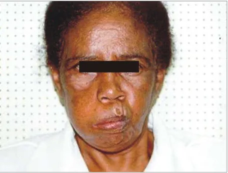
habitual em janela de 15 segundos ... 19 Figura 6 - Foto de paciente inflando a bochecha do lado da
paralisia (lado direito da face). A atividade maior é do
bucinador do lado sadio (lado esquerdo da face) ... 20 Figura 7 - Representação do traçado eletromiográfico da prova
de aperto dentário com os valores em V ... 22 Figura 8 - Representação do traçado eletromiográfico da prova
LISTA DE TABELAS
Tabela 1 - Distribuição de 27 indivíduos com PFP em fase flácida com tempo de instalação igual ou superior a seis meses, de acordo com as respostas às questões
da anamnese ... 27 Tabela 2 - Distribuição de 27 indivíduos com PFP em fase
flácida, há seis meses ou mais, pelo estado clínico dos dentes (bom x ruim) em relação à mordida
(normal e alterada) ... 28 Tabela 3 - Distribuição de 27 indivíduos com PFP em fase
flácida, com tempo de instalação igual ou superior a seis meses, conforme o tipo de oclusão dentária e
preferência mastigatória ... 29 Tabela 4 - Distribuição de 27 indivíduos com PFP em fase
flácida, com tempo de instalação igual ou superior a seis meses, há seis meses ou mais, conforme a
avaliação miofuncional do músculo orbicular da boca ... 29 Tabela 5 - Distribuição de 27 indivíduos com PFP em fase
flácida, com tempo de instalação igual ou superior a seis meses, pela avaliação clinica da massa muscular
dos músculos bucinadores e masseteres ... 30 Tabela 6 - Distribuição de 27 indivíduos com PFP em fase
flácida, com tempo de instalação igual ou superior a seis meses, conforme dados da avaliação clínica da
mastigação ... 30 Tabela 7 - Condição clínica de lábios comparada à preferência
clínica mastigatória na prova de mastigação habitual em 27 indivíduos com PFP em fase flácida com
tempo de instalação igual ou superior a seis meses ... 31 Tabela 8 - Condição clínica dos músculos bucinadores
comparada à preferência clínica mastigatória na prova de mastigação habitual em 27 indivíduos com PFP em fase flácida com tempo de instalação igual
xliii
Tabela 9 - Distribuição quanto à preferência mastigatória entre o grupo controle com 60 indivíduos e 27 indivíduos com PFP em fase flácida com tempo de instalação igual
ou superior a seis meses ... 32 Tabela 10 - A atividade eletromiográfica (em V) com intervalo de
confiança (25% a 75%) entre os grupos com PFP e grupo controle nas provas de aperto dentário e mastigação habitual divididos em dois grupos sem e
RESUMO
Carvalho ARR. Preferência mastigatória em pacientes com paralisia facial periférica flácida de duração igual ou superior a seis meses: estudo clínico e eletromiográfico [tese]. São Paulo: Faculdade de Medicina, Universidade de São Paulo; 2008. 72p.
Introdução: a paralisia facial periférica (PFP) é caracterizada por lesão geralmente unilateral do nervo facial em qualquer parte de seu trajeto. Na paralisia total há perda dos movimentos de todos os segmentos da hemiface ipsilateral à lesão. Clinicamente observa-se que a maioria dos pacientes com PFP em fase flácida apresenta dificuldade para mastigar do lado paralisado, pois a manutenção dos alimentos entre as arcadas dentárias está comprometida pela falta de participação do músculo bucinador. Aliada a isso pode ocorrer incompetência labial devido à flacidez da hemiface afetada em conseqüência à queda da comissura labial ipsilateral. Objetivo: verificar as conseqüências da PFP unilateral na fase flácida, com duração de pelo menos seis meses, na função mastigatória quanto a preferência clínica mastigatória e diferença eletromiográfica entre os masseteres.Casuística e Método: foram selecionados 27 indivíduos de ambos os gêneros, com PFP em fase flácida com pelo menos seis meses de paralisia, com idade entre 16 anos e 67 anos, com dentição natural permanente, selecionados por um protocolo específico, complementado com exame clínico miofuncional e avaliação eletromiográfica de superfície nos músculos masseteres nas provas de aperto dentário e mastigação habitual. Resultados: de acordo com as respostas da anamnese, 77,8% dos pacientes referiram mastigar preferencialmente do lado sadio. Clinicamente, 70% apresentaram preferência mastigatória. A atividade muscular dos bucinadores e orbicular da boca foi estatisticamente significante (p = 0,025) entre os lados sadio e paralisado. Apenas, 22,2% dos pacientes apresentou diminuição de massa do masseter do lado paralisado. Não houve diferença eletromiográfica estatisticamente significante entre os lados sadio e paralisado nos masseteres. Conclusão: no presente estudo, pacientes com PFP unilateral na fase flácida, com duração de pelo menos seis meses, apresentaram preferência clínica mastigatória pelo lado sadio. Não houve diferença clínica e eletromiográfica entre os lados paralisado e sadio nos músculos masseteres.