Yingying Han1,2, Qilong Wang1, Ping Song1, Yi Zhu2, Ming-Hui Zou1*
1Department of Biochemistry and Department of Medicine, University of Oklahoma Health Science Center, Oklahoma City, Oklahoma, United States of America,
2Department of Physiology and Pathophysiology, Peking University Health Science Center, Beijing, China
Abstract
Redox state is a critical determinant of cell function, and any major imbalances can cause severe damage or death.
Objectives: The aim of this study is to determine if AMP-activated protein kinase (AMPK), a cellular energy sensor, is activated by oxidants generated by Berberine in endothelial cells (EC).
Methods:Bovine aortic endothelial cells (BAEC) were exposed to Berberine. AMPK activity and reactive oxygen species were monitored after the incubation.
Results:In BAEC, Berberine caused a dose- and time-dependent increase in the phosphorylation of AMPK at Thr172 and acetyl CoA carboxylase (ACC) at Ser79, a well characterized downstream target of AMPK. Concomitantly, Berberine increased peroxynitrite, a potent oxidant formed by simultaneous generation of superoxide and nitric oxide. Pre-incubation of BAEC with anti-oxidants markedly attenuated Berberine-enhanced phosphorylation of both AMPK and ACC. Consistently, adenoviral expression of superoxide dismutase and pretreatment of L-NG-Nitroarginine methyl ester (L-NAME; a
non-selective NOS inhibitor) blunted Berberine-induced phosphorylation of AMPK. Furthermore, mitochondria-targeted tempol (mito-tempol) pretreatment or expression of uncoupling protein attenuated AMPK activation caused by Berberine. Depletion of mitochondria abolished the effects of Berberine on AMPK in EC. Finally, Berberine significantly increased the phosphorylation of LKB1 at Ser307 and gene silencing of LKB1 attenuated Berberine-enhanced AMPK Thr172 phosphorylation in BAEC.
Conclusion:Our results suggest that mitochondria-derived superoxide anions and peroxynitrite are required for Berberine-induced AMPK activation in endothelial cells.
Citation:Han Y, Wang Q, Song P, Zhu Y, Zou M-H (2010) Redox Regulation of the AMP-Activated Protein Kinase. PLoS ONE 5(11): e15420. doi:10.1371/ journal.pone.0015420
Editor:Aimin Xu, University of Hong Kong, China
ReceivedAugust 20, 2010;AcceptedSeptember 21, 2010;PublishedNovember 5, 2010
Copyright:ß2010 Han, et al. This is an open-access article distributed under the terms of the Creative Commons Attribution License, which permits unrestricted use, distribution, and reproduction in any medium, provided the original author and source are credited.
Funding:This study was supported by National Institutes of Health (NIH) grants (HL079584, HL080499, HL074399, HL089920, HL096032, and HL105157), and a Research Award from the American Diabetes Association, and funds from the Travis Endowed Chair of the University of Oklahoma Health Science Center (all to Dr. Zou). Dr. M.H. Zou is a recipient of Established Investigator Award of American Heart Association. Part of this work was also supported by an international cooperation grant from the Chinese National Science Foundation. The funders had no role in study design, data collection and analysis, decision to publish, or preparation of the manuscript.
Competing Interests:The authors have declared that no competing interests exist. * E-mail: ming-hui-zou@ouhsc.edu
Introduction
AMP-activated protein kinase (AMPK), a heterotrimeric complex comprised of a catalytic subunit a and two regulatory subunits,bandc, is an evolutionarily conserved serine/threonine protein kinase that is ubiquitously expressed. It was initially characterized as a ‘‘fuel gauge’’, modulating cellular energy flux in eukaryotic cells in response to changes of AMP:ATP ratios [1,2,3]. AMPK can be activated by conditions that increase intracellular AMP such as exercise [4], hypoxia [5], hypoglycemia [6], as well as certain cytokines and drugs such as leptin [7], adiponectin [8], metformin [9], statins [10] and rosiglitazone [11]. The primary mechanism responsible for AMPK activation involves phosphor-ylation of AMPK at Thr172 residue located within the activation loop ofasubunits [12]. Activation of AMPK has been linked to numerous energy-conserving cellular processes, such as promotion of glucose uptake and glucose transportation [13]; stimulation of glycogen, cholesterol, fatty acid, triacylglycerol synthesis, glucose and fatty acid oxidation [14]; and acceleration of mitochondrial biogenesis [15]. In addition to its roles in energy homeostasis,
more recent studies have identified some broader protective roles for AMPK in atherosclerosis [16] and inflammation [17] as well as some beneficial angiogenic effects [18,19].
AMPK. One recent study reported Berberine activated AMPK by inhibiting respiratory complex I, a similar effect to metformin and rosiglitazone [24]. However, the precise mechanism by which Berberine activates AMPK has not been established. The aim of our study was to identify the underlying mechanism of Berberine-induced AMPK activation.
Reactive oxygen and nitrogen species (ROS and RNS) such as superoxide anions (O22), hydrogen peroxide (H2O2), and
peroxynitrite (ONOO2) are small and highly reactive molecules having both physiological and pathological effects [25]. Normal cellular ROS concentrations play important roles in cell signaling pathways and are vital for physiological functions. Peroxynitrite (ONOO2) is a highly reactive ROS formed by O22 and NO. It
has been demonstrated that metformin inhibits complex I of the respiratory chain to generate mitochondrial O22, and then
ONOO2, which leads to AMPK activation via a c-Src and PI3K-dependent pathway [26]. Our data indicate that Berberine-induced AMPK activation is mediated by mitochondria-derived superoxide and peroxynitrite in endothelial cells (EC).
Materials and Methods
Materials
Human umbilical vein endothelial cells (HUVEC) and bovine aortic endothelial cells (BAEC) were purchased from American Type Culture Collection (Rockville, MD), cell culture media were purchased from Lonza Group Ltd. (Switzerland). Berberine, ethidium bromide, sodium pyruvate, uridine, and L-nitroarginine methyl ester (L-NAME) were obtained from Sigma-Aldrich Inc. (St. Louis, MO). Dihydroethidium (DHE) and dihydrorhodamine-123 (DHR) were obtained from Invitrogen Corporation (Carlsbad, CA).
Antibodies against phospho-AMPK a (Thr172), AMPK a,
phospho-Acetyl-CoA carboxylase (ACC) (Ser79) andb-actin were from Cell Signaling (Beverly, MA). Antibody raised against SOD was obtained from Santa Cruz Biotechnology Inc (Santa Cruz, CA). Antibody against UCP2 was from Alpha Diagnostic Intl. Inc. (San Antonio, TX). RNeasy Mini Kit and RNA UltraSense One-Step RT-PCR System were obtained from Qiagen Inc (Valencia, CA).
Cell culture
HUVEC were cultured in endothelial basal media (EBM) with EGMTMSingleQuots from LONZA (Walkersville, MD) [27]. The medium was changed every 2 days before the cells reached confluence. Cells were incubated in a humidified atmosphere of
5% CO2/95% air at 37uC. BAEC were grown in EBM
supplemented with 2% fetal bovine serum. To generate mito-chondria-depleted BAEC (ru cells), wild-type BAEC were incubated in medium containing ethidium bromide (50 ng/ml), sodium pyruvate (1 mM), and uridine (50mg/ml) for 3 weeks, as described previously [26]. Therustatus of cells was confirmed by the absence of cytochrome c oxidase subunit II by RT-PCR.
Measurement of cellular ATP, ADP, and AMP
After being treated with Berberine or vehicle, HUVEC (100-cm2 dishes) were immediately washed with ice-cold PBS, then scraped with HClO4, and centrifuged at 4uC (5 min, 14,000 rpm).
After centrifugation, the supernatants were neutralized by KOH. The supernatants were detected by HPLC to determine the contents of ATP, ADP, and AMP.
Detection of superoxide anions
Intracellular O22 was measured using the dihydroethidium
(DHE) fluorescence/high-performance liquid chromatography (HPLC) assay with minor modifications [28]. Briefly, BAEC were
incubated with 0.5mM DHE for 30 min, and then extracted with 30% methanol. Oxyethidium (a product of DHE and O22) and
ethidium (a product of DHE auto-oxidation) were separated and quantified using a C-18 HPLC column (mobile phase: gradient of acetonitrile and 0.1% trifluoroacetic acid). O22 production was
determined by the conversion of DHE into oxyethidium.
Measurement of peroxynitrite
ONOO2 generation in BAEC after Berberine treatment was determined using chemiluminescence detection by formation of the fluorescent dye rhodamine 123 (RH) from dihydrorhodamine 123 (DHR). 5mM DHR was added into culture medium, after 1 hour incubation, 200ml medium was added into 96-well plate, and fluorescence was measured at 570 nm with excitation at 500 nm. Since DHR slowly auto oxidizes to RH, background fluorescence in medium alone was subtracted from the measured values with ONOO2.
Adenovirus infection
BAEC or HUVEC were infected in EBM with EGMTM
SingleQuots for 48 hours with adenovirus encoding green fluorescence protein (GFP), superoxide dismutase 1 (SOD1) or catalase. Adenoviruses encoding GFP served as control, and transfection efficiency was typically.80% as determined by GFP expression.
Western blot
Western blotting assays were done as described previously [29]. The primary antibodies were polyclonal and raised against p-AMPK-Thr172 (1:1000), p-ACC-Ser79 (1:1000), AMPK (1:1000), SOD1 (1:1000), UCP2 (1:1000), and b-actin (1:3000). The intensity (area density) of the individual bands on Western blots was measured by densitometry (model GS-700, Imaging Densi-tometer; Bio-Rad). The background was subtracted from the calculated area.
Reverse transcription–polymerase chain reaction (RT-PCR)
Total cellular RNA was isolated from treated HUVEC with the total RNA isolation protocol for the RNeasy Mini Kit RNA. The procedures for semi-quantitative reverse transcription-polymerase chain reaction (RT-PCR) were performed by using
forward (59-GCCATGCAGTACTTCACCAA-39) and reverse
(59-AGGCTTCTGTGATGGCCACCG-39) primers
correspond-ing to bovine cytochrome c oxidase subunit II mRNA accordcorrespond-ing to the manufacturer’s recommendations. Reactions were run for 20 cycles at conditions as follows: denaturation for 30 seconds at 94uC, annealing for 30 seconds at 55uC, and extension for 40 seconds at 72uC. Constitutively expressed glyceraldehyde-3-phosphate dehydrogenase (GAPDH) mRNA was amplified as internal control.
Statistical analysis
Statistical comparisons were performed using a Student’sttest or one-way ANOVA with Bonferroni’s procedure for post hoc analysis. Values ofp,0.05 were considered significant.
Results
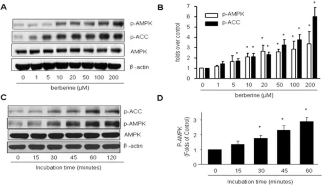
Berberine causes a dose- and time-dependent increase in AMPK in EC
confluent BAEC were treated with different concentrations of Berberine (0, 1, 5, 10, 20, 50, 100, 200mM) for 2 h. AMPK activation was evaluated by monitoring the phosphorylation of AMPK at Thr172 and ACC at Ser79. As shown in Fig. 1A and B, Berberine increased the phosphorylation of AMPK-Thr172 and ACC-Ser79 in a dose-dependent manner. Furthermore, activation
of AMPK by Berberine was also time-dependent. 10mM
Berberine increased the phosphorylation of AMPK-Thr172 and ACC-Ser79 as early as 30 minutes, reaching a peak value at about 60 minutes without affecting the total content of AMPK (Fig. 1C). Because 10mM of Berberine caused AMPK activation dramati-cally, we used this concentration of the compound for subsequent experiments.
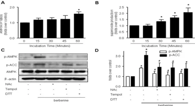
Effects of Berberine on AMP/ATP ratios
We next determined if AMPK activation by Berberine was due to the changes of AMP/ATP ratio. As depicted in Fig. 2A, the AMP/ATP ratio didn’t change until 60 min incubation of Berberine (10mM), suggesting changes in AMP/ATP ratios occur only at late stage of Berberine incubation (.1 h).
Berberine increases intracellular superoxide
Our previous studies have shown that redox signaling plays an important role in AMPK activation [26,30,31]. In order to investigate whether endogenous superoxide was also involved in AMPK activation caused by Berberine, the generation of intracellular superoxidewas assessed by using DHE/HPLC assay. As shown in Fig. 2B, exposure of BAEC to Berberine (10mM), a concentration at which AMPK was activated, also significantly increased DHE fluorescence as early as 30 min after treatment and superoxide release continued to rise when incubation prolonged (Figure 2B), indicating that Berberine triggered intracellular superoxide generation.
Anti-oxidants attenuate Berberine-induced AMPK activation in EC
We next evaluated if antioxidants altered Berberine-induced AMPK activation. Pre-incubation with antioxidant reagents (NAc, DTT and tempol) significantly attenuated AMPK activation caused by Berberine (Fig. 2C and 2D). This implies that reactive oxygen species are involved in the AMPK activation process induced by Berberine.
Activation of AMPK by Berberine is peroxynitrite-dependent
Since simultaneous generation of both O22and NO increases
the formation of ONOO2 and our previous studies [30,31,32] have also demonstrated that ONOO2activates AMPK, we sought to investigate if endogenous ONOO2was involved in this process. To this end, the effects of Berberine on AMPK activity were monitored under conditions where ONOO2 production was inhibited. SOD1 was overexpressed in BAEC (to scavenge O22) or
pre-treated with L-NAME (1 mM; to inhibit NO production). Adenoviral expression of SOD1 dramatically increased the levels of SOD1 in BAEC compared with GFP infection (Figure 3A). Interestingly, adenoviral overexpression of SOD1 markedly attenuated Berberine-induced AMPK Thr172 phosphorylation (Fig. 3A and 3B). In contrast, adenoviral overexpression of catalase, an enzyme detoxifying H2O2, had no effects on
Berberine-enhanced AMPK activation in BAEC (Fig. 3A and 3B), suggesting that H2O2is not involved in AMPK activation by
this compound. Similarly, pre-treatment with L-NAME, a non-selective NOS inhibitor, did not alter the basal levels of AMPK phosphorylation at Thr172 (Fig. 3C and 3D), but L-NAME abolished the effects of Berberine on the phosphorylation of AMPK at Thr172 and ACC at Ser79 (Fig. 3C and 3D).
Figure 1. Effects of Berberine on the phosphorylation of AMPK-Thr 172 and ACC-Ser79, AMP/ATP ratios, and superoxide production in BAEC.A & B, Dose-dependent increase of of AMPK-Thr172 and Ser79 phosphorylation by berberine in BAEC. Confluent BAEC were incubated with different doses of Berberine (1mM to 200mM) for 2 h. The blot is representative of three individual experiments. n = 3 *p,0.05,
control versus Berberine-treated.C & D, Time-dependent increase of AMP-Thr172 and Ser79 by Berberine (10mM) in BAEC. The blot is representative
We next determined if scavenging ONOO2altered the effects of Berberine. Uric acid is a widely used scavenger of ONOO2. The production of intracellular ONOO2was directly inhibited by this
scavenger and AMPK-Thr172 phosphorylation was detected under this condition (Fig. 3E and 3F). The data show AMPK phosphorylation at Thr172 was inhibited by uric acid pretreatment. Figure 2. Activation of AMPK by Berberine is mediated by reactive oxygen species (ROS).A, Time-course of Berberine (10mM) on AMP/
ATP ratios in BAEC. After exposure to Berberine (10mM) for the time indicated, BAEC were subjected to HPLC assays to measure the AMP/ATP ratio.
n = 5, *p,0.05, control versus Berberine-treated.B, Time-dependent increase of superoxide anions caused by Berberine (10mM) in BAEC. After
incubation with Berberine (10mM) for the time indicated, intracellular ROS were detected by the DHE fluorescence/HPLC assay as described in
‘‘Materials and Methods.’’ n = 5, *p,0.05, control versus Berberine-treated.C, BAEC were pre-treated with anti-oxidants tempol (10mM), NAc (2 mM),
or DTT (1 mM) for 30 min, and then treated with Berberine for 2 h.D, Summary data for the effects of anti-oxidants n = 5 *p,0.05, control versus Berberine-treated,#p,0.05, Berberine alone versus Berberine with one or the other of the two anti-oxidants.
doi:10.1371/journal.pone.0015420.g002
Figure 3. Activation of AMPK by Berberine is ONOO2-dependent.A & B, Effects of adenoviral overexpression of SOD and catalase on
Berberine-induced phosphorylation of AMPK-Thr172 and ACC-Ser79 in BAEC. BAEC were transfected with adenovirus overexpressed with SOD, catalase or GFP (50 MOI) for 24 h. n = 4, *p,0.05 control versus Berberine-treated).C & D, Effects of L-NAME on Berberine-induced phosphorylation of AMPK-Thr172 and ACC-Ser79 in BAEC. BAEC were pre-treated with the eNOS inhibitor L-NAME (1 mM) for 30 min. Thereafter BAEC were treated with Berberine (10mM) for 2 h. n = 3, *p,0.05;E & F, Effects of uric acid on Berberine-induced phosphorylation of AMPK-Thr172 and ACC-Ser79 in BAEC.
BAEC were pretreated with the ONOO2scavenger, uric acid (0.5 mM) for 30 min, and then treated with Berberine (10
mM) for 2 h. n = 4, *p,0.05,
control versus Berberine-treated,#p,0.05, Berberine alone versus Berberine plus uric acid.G, Berberine increases the formation of ONOO2in BAEC.
After 10mM Berberine treatment for the time indicated and DHR (5mM) incubation for 1 hr, the production of ONOO2was determined by DHR
oxidation. n = 4 *p,0.05 control versus Berberine-treated.G. Effects of adenoviral overexpression of SOD or L-NAME on the formation of ONOO2
caused by berberine in BAEC. After Berberine (10mM) treatment for the time indicated and DHR (5mM) incubation for 1 hour, the production of
ONOO2was determined by DHR oxidation. n = 4 *p
To further confirm the role of ONOO2, its synthesis was measured after incubation with Berberine. ONOO2 production was increased significantly as early as 30 min following incubation (Fig. 3G). Further, either adenoviral overexpression of SOD or L-NAME administration of L-L-NAME, ablated berberine-induced ONOO- formation in EC (Figure 3H). Taken together, our result suggest that berberine increased the formation of ONOO2.
Identification of mitochondria as the source of superoxide anions
We next identified the source of oxidants by which Berberine activated AMPK in BAEC. Increasing evidence suggests that NAD(P)H oxidases, xanthine oxidase, and mitochondria are major sources of superoxide in endothelial cells [33]. Pre-incubation of allopurinol and oxypurinol (two potent inhibitors for xanthine oxidase) did not alter the effects of Berberine (Figure 4A). Similarly, ovexpression of p47phox dominant negative mutants (p47-DN),a cytosolic and membrane subunit of NAD(P)H oxidase, didn’t alter AMPK phosphorylation at Thr172 in EC (Figure 4C). This result was further confirmed by the inability of overexpression of p67phoxdominant negative mutants, another subunit of NAD(P)H oxidase 9 (data not shown). Taken together, our results exclude the possibility of NAD(P)H oxidase as a potential source of oxidants provoked by Berberine treatment. In contrast, pre-incubation of mitochondria-targeted tempol, which inhibits oxidant formation from mitochondria, attenuated Berberine-enhanced phosphoryla-tion of AMPK at Thr172 and ACC at Ser79 in BAEC (Figure 4C and 4D), implying that mitochondria might a source of superoxide in Berberine-treated EC.
Mitochondrial uncoupling protein 2 (UCP2), an inner mem-brane mitochondrial protein, has been implicated in superoxide modulation. It was interesting to test if gene silencing of UCP2 accentuated the effects of Berberine on AMPK. BAEC were transfected with UCP2 siRNA or control siRNA. As depicted in
Fig. 4E and 4F, gene silencing of UCP2 increased the phosphorylation of AMPK at Thr172. Importantly, transfection of UCP2-specific siRNA but not control siRNA, accentuated the effects of Berberine on AMPK phosphorylation at Thr172 (Figure 4E and 4F). Conversely, adenoviral overexpression of UCP-2 accentuated Berberine-enhanced AMPK phosphorylation at Thr172 in EC (Figure 4G). Taken together, these results support a role of mitochondrialsuperoxidein the effects of Berberine on AMPK.
Berberine fails to activate AMPK in mitochondria-deleted ru-BAEC
To further confirm if mitochondrialsuperoxidewas required for these Berberine-induced effects, we created BAEC without functional mitochondria (ru cells). After BAEC were incubated with mitochondria deleting conditional medium for 3 weeks, BAEC dramatically reduced the expression of cytochrome c oxidase subunit II at the mRNA levels detected by RT-PCR (Fig. 5A), confirming a deficiency of mitochondria inruBAEC. Subsequently we tested if Berberine increases intracellular superoxide in ru-BAEC. We confirmed our expectation in the demonstration that Berberine did increasesuperoxideproduction in wild-type BAEC, but failed to do so in mitochondria-deletedru -BAEC, as assayed by DHE staining (Figure 5B). Further, as predicted, Berberine also increased the phosphorylation of AMPK at Thr172 and ACC at Ser79 in BAEC. However, it failed to increase AMPK phosphorylation inru-BAEC (Fig. 5C and D).
It was provocative to determine if AMPK inruBAEC could be activated by exogenous ONOO2. Both BAEC andruBAEC were therefore exposed to chemically synthesized ONOO2(50mM) for 5 min. Figure 5C show that ONOO2caused activation of AMPK in both BAEC andruBAEC to a similar degree, suggesting that
the response to ONOO2 in ru BAEC remained unchanged
compared to BAEC. Thus, the decrease of AMPK activation inru
Figure 4. Reactive oxygen species (ROS) involved in Berberine induced AMPK activation are derived from mitochondria.A, Effects of xanthine oxidase inhibitors, allopurinol and oxypurinol, on Berberine-enhanced phosphorylation of AMPK-Thr172 and ACC-Ser79 in BAEC. BAEC were pre-treated with mitochondrial tempol (50mM) for 30 min, and then treated with Berberine (10mM) for 2 h. The blots are representative of three
independent experiments.B, Effects of adenoviral overexpression of p47phox dominant negative mutants on Berberine-enhanced phosphorylation of AMPK-Thr172 and ACC-Ser79 in BAEC. The blots are representative of three independent experiments.C & D, Effects of mitochondrial tempol (*p,0.05, control versus Berberine-treated,#p,0.05, Berberine-alone versus Berberine plus mitochondrial tempol).E & F, Effects of UCP-2 siRNA transfection on Berberine-enhanced phosphorylation of AMPK-Thr172 and ACC-Ser79 in BAEC. BAEC were transfected with UCP2 siRNA for 24 hr, and then treated with Berberine (10mM) for 2 hr. n = 3, *p,0.05, control versus Berberine-treated,#p,0.05, Berberine-alone versus Berberine plus UCP2 siRNA.G.Effects of adenoviral overexpression of UCP-2 on Berberine-enhanced phosphorylation of AMPK-Thr172 and ACC-Ser79 in BAEC. n = 3 *p,0.05, control versus UCP-2;#p,0.05, control verus Berberine-alone;+p,0.05 Berberine-treated versus Berberine plus UCP2.
BAEC is likely due to its attenuation of ONOO2production inru
BAEC in response to Berberine.
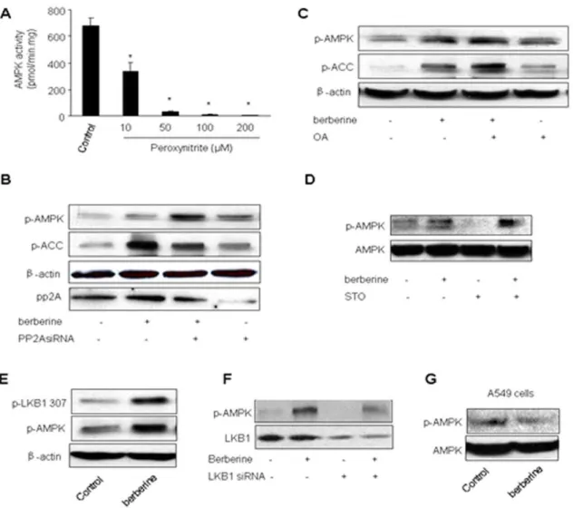
Exogenous ONOO2inhibits AMPK activity in vitro To exclude a direct effect of ONOO2on AMPK, recombinant AMPK was exposed to chemically synthesized ONOO2in vitro. After the treatment, AMPK activity was assayed. Unexpectedly, ONOO2 caused a dose-dependent inhibition of AMPK activity, suggesting a direct inhibitory effect of ONOO2 on AMPK (Figure 6A). This suggests that ONOO2 might exert opposite effects on AMPK activity, i.e., AMPK activation via upstream enzymes and AMPK suppression via an unknown posttransla-tional modification by ONOO2.
PP2A is not required for Berberbine-induced AMPK activation
PP2A is reported to regulate AMPK activation [34]. We next determined if PP2A inhibition with pharmacological or genetic means altered Berberine-induced AMPK phosphorylation. As depicted in Figure 3B, OA pretreatment had no effect on Berberine-induced phosphorylation of AMPK-Thr172 and ACC at Ser79. Concomitantly, transfection of PP2A siRNA didn’t attenuate the phosphorylation of AMPK or ACC, suggesting that the effect of Berberine was PP2A independent.
STO609 treatment does not alter Berberine-enhanced phosphorylation of AMPK and ACC in EC
Calcium calmodulin protein kinase II (CAMKK II) is reported to be an upstream kinase for nitric oxide-induced AMPK activation [35]. We next assayed the contribution of CaMKKII in berberine-induced AMPK activation. As shown in Figure 6D,
pretreatment of STO-609 (100 nM) had no effect on Berberine-enhanced AMPK phosphorylation in EC.
Berberine increases LKB1 phosphorylation at Serine 307 in EC
LKB1 is a known upstream kinase of AMPK. Phosphorylation of Ser307 is reported for metformin-induced AMPK activation in endothelial cells [36]. Thus, we next determined if Berberine altered LKB1 phosphorylation at Serine 307. As shown in Figure 6E, Berberine markedly increased the detection of LKB1 phosphorylation at Serine 307 (Figure 6E), which was paralleled with increased AMPK phosphorylation at Thr172 (Figure 6E). Further, transfection of LKB1 siRNA abolished Berberine-enhanced AMPK phosphorylation in EC (Figure 6F). Finally, Berberine had no effects on AMPK Thr172 phosphorylation in LKB1 deficient A549 cells (Figure 6G). Taken together, our results imply that LKB1 was required for Berberine-induced AMPK phosphorylation.
Discussion
In this study, we observed that mitochondria derived superoxide and peroxynitrite are required for Berberine-induced AMPK activation (Fig. 6). The key evidence can be summarized as follows: (1) Exposure of BAEC to Berberine at the dose required for AMPK-Thr172 phosphorylation significantly increased
intracel-lular O22 and ONOO2 production. Although Berberine
increased intracellular AMP/ATP ratio after 1 h of incubation, intracellular O22and ONOO2production was increased at about
30 min incubation, suggesting that this time frame adequately allows oxygen and nitrogen species to initiate AMPK activation. (2) Inhibition of ONOO2formation by overexpression of SOD1 Figure 5. Mitochondria depletion abolishes Berberine-induced AMPK phosphorylation in endothelial cells.Confluent monolayers of BAEC were incubated in EGM with 2% fetal bovine serum alone or inrumedium as described in ‘‘Materials and Methods.’’A, After incubation, BAEC were harvested, total RNA were extracted, and mRNA for cytochrome c oxidase subunit II (CytOxII) were estimated by RT-PCR. Data are representative of three independent experiments.B, BAEC andrucells were treated with Berberine (10mM) for 30 min; the production of ROS was detected by DHE/
HPLC assay.C&D. Effects of mitochondria depletion on AMPK-Thr172 phosphorylation caused by Berberine and ONOO-. BAEC andrucells were treated with Berberine (10mM) for 2 h or ONOO2(50mM) for 15 min, after treatment, cells were lysed and proteins were separated by SDS-PAGE and
(to scavenge O22), or incubation with L-NAME (to prevent NO
production), attenuated Berberine-induced AMPK phosphoryla-tion. (3) When BAEC were pre-treated with uric acid (an ONOO2 scavenger), the AMPK phosphorylation induced by Berberine was abolished. (4) Exposure of BAEC to mitochondrial tempol (which blocks O22release from mitochondria), also blocked
Bereberine-induced AMPK phosphorylation. In contrast, incubation with allopurinol, oxypurinol or overexpression with adenovirus encod-ing p47phox-DN, did not alter Berberine induced AMPK activation. These results suggest that mitochondria, rather than xanthine oxidase or NAD(P)H oxidase, were the major source of superoxide in cells exposed to Berberine. (5) When using UCP2 siRNA to knock down the expression of UCP2 (which uncouples mitochondrial oxidative phosphorylation from ATP production), Berberine-induced AMPK phosphorylation was further enhanced. (6) These results were further corroborated by the findings inru -BAEC lacking functional mitochondria. Exposure ofru- BAEC to
Berberine neither enhanced intracellular superoxide release nor AMPK phosphorylation. Cumulatively these results indicate that activation of AMPK by Berberine is mitochondrial superoxide dependent.
Another important finding in this study is that the phosphor-ylation of LKB1 at S307 is involved in LKB1-mediated AMPK activation induced by berberine. Our published study [36] has reported that in multiple cell types the signaling pathways engaged by several physiological stimuli converge upon LKB1 phosphor-ylation at S307, which directs the nucleocytoplasmic transport of LKB1 and consequent AMPK activation. However, further investigation of LKB1 Ser307 phosphorylation is warranted.
The traditional view has been that ROS exert toxic effects by damaging or killing cells under pathophysiological conditions. However, growing experimental evidence now suggests that ROS such as ONOO2are required in some physiological processes as well [25]. It is conceivable that sub-toxic concentrations of ROS/ Figure 6. Increased LKB1 Serine 307 phosphorylation by Berberine in BAEC.A.Inhibition of AMPK activity by ONOO2.Recombinant AMPKa1b1c1 was treated with chemically synthetized ONOO- at concentrations indicated for 5 minutes and AMPK activity was assayed as described in Methods and Materials. N = 5 *P,0.05; control verse ONOO2-treated;B. Effects of okadaic acid (OA) on Berberine-enhanced phosphorylation of
AMPK-Thr172 and ACC-Ser79 in BAEC. The blot is representative of three blots. from three individual experiments.C. Effects of transfection of PP2A siRNA on Berberine-enhanced phosphorylation of AMPK-Thr172 and ACC-Ser79 in BAEC. The blot is representative of three blots from three individual experiments. D. Effects of STO-609 on Berberine-enhanced phosphorylation of AMPK-Thr172 and ACC-Ser79 in BAEC. The blot is representative of three blots.E.Effects of berberine on the phosphorylation of LKB1 serine 307 in BAEC. The blot is representative of three blots from three individual experiments. F. Genetic inhibition of LKB1 abolishes Berberine-induced phosphorylation of AMPK-Thr172 in BAEC. The blot is representative of three blots from three individual experiments.GEffects of Berberine on AMPK phosphorylation at Thr172 in LKB1-deficient A549 cells.
RNS generated in the cell are critical in molecular transduction [26,30,37]. Our data demonstrate that Berberine evokes O22
release and O22or its derived oxidant, ONOO2, is required for
Berberine-enhanced AMPK activation. Berberine has been reported to exert therapeutic effects on diabetes, obesity and inflammation [17,38,39,40]. Thus, AMPK activation by Berberine might help explain the beneficial effects in these conditions.
Turner et al [24] reported that Complex I of the respiratory chain represents a major target of Berberine in its effect of improving insulin insensitivity (such as in type II diabetes) through increased AMPK activity. It has also been demonstrated that Berberine selectively accumulates in mitochondria on K1735-M2 melanoma cells to arrest cell proliferation, causes mitochondrial fragmentation and depolarization, and oxidative stress [41]. Berberine has been shown to inhibit mitochondrial respiration and decrease calcium loading capacity through induction of the mitochondrial permeability transition (MPT) via interactions with the adenine nucleotide translocator, (ANT) [41,42]. The antitu-mor effect of Berberine has also been demonstrated to possess a mitochondrial component [43]. Whether the mitochondrial complex I is involved in its anti-microbial, anti-arrhythmic or other pharmacological properties remains unknown. Previous work from our laboratory has demonstrated that complex I associated ROS/RNS production is related to many protective effects observed in therapy using metformin and the statin drugs [26,30]. Recently, it has also been reported that Berberine
enhances AMPK activity in a variety of cells, such as adipocytes, tumor cells, macrophages, smooth muscle cells and endothelial cells [17,23,40,44]). In addition, other investigators have also shown that AMPK is a major intermediate in facilitating some beneficial effects of Berberine [17,38,39,45]. Here we have demonstrated an important role of mitochondria derived super-oxide and ONOO2 in Berberine induced AMPK activation. However, the possibility that this compound might exert its beneficial metabolic and cardiovascular properties through an ONOO2dependent pathway similar to metformin and the statins [30], needs to be further investigated.
In summary, we demonstrate for the first time that Berberine increases mitochondria-derived superoxide and peroxynitrite to activate AMPK. Activation of AMPK by Berberine can potentially expand the pharmaceutical role of the compound in the management of several diverse yet intertwined human pathologies.
Acknowledgments
We are grateful to Dr. Najeeb Shirwany, University of Oklahoma Health Science Center for his editorial assistance.
Author Contributions
Conceived and designed the experiments: MZ. Performed the experiments: YH QW PS. Analyzed the data: YH QW PS. Wrote the paper: YH YZ MZ.
References
1. Kemp BE, Stapleton D, Campbell DJ, Chen ZP, Murthy S, et al. (2003) AMP-activated protein kinase, super metabolic regulator. Biochem Soc Trans 31: 162–168.
2. Long YC, Zierath JR (2006) AMP-activated protein kinase signaling in metabolic regulation. J Clin Invest 116: 1776–1783.
3. Foretz M, Taleux N, Guigas B, Horman S, Beauloye C, et al. (2006) [Regulation of energy metabolism by AMPK: a novel therapeutic approach for the treatment of metabolic and cardiovascular diseases]. Med Sci (Paris) 22: 381–388. 4. Coven DL, Hu X, Cong L, Bergeron R, Shulman GI, et al. (2003) Physiological
role of AMP-activated protein kinase in the heart: graded activation during exercise. Am J Physiol Endocrinol Metab 285: E629–636.
5. Fukuyama Y, Ohta K, Okoshi R, Suehara M, Kizaki H, et al. (2007) Hypoxia induces expression and activation of AMPK in rat dental pulp cells. J Dent Res 86: 903–907.
6. McCrimmon RJ, Fan X, Ding Y, Zhu W, Jacob RJ, et al. (2004) Potential role for AMP-activated protein kinase in hypoglycemia sensing in the ventromedial hypothalamus. Diabetes 53: 1953–1958.
7. Minokoshi Y, Kim YB, Peroni OD, Fryer LG, Muller C, et al. (2002) Leptin stimulates fatty-acid oxidation by activating AMP-activated protein kinase. Nature 415: 339–343.
8. Zhou L, Deepa SS, Etzler JC, Ryu J, Mao X, et al. (2009) Adiponectin Activates AMP-activated Protein Kinase in Muscle Cells via APPL1/LKB1-dependent and Phospholipase C/Ca2+/Ca2+/Calmodulin-dependent Protein Kinase Kinase-dependent Pathways. J Biol Chem 284: 22426–22435.
9. Zhou G, Myers R, Li Y, Chen Y, Shen X, et al. (2001) Role of AMP-activated protein kinase in mechanism of metformin action. J Clin Invest 108: 1167–1174. 10. Kou R, Sartoretto J, Michel T (2009) Regulation of Rac1 by simvastatin in endothelial cells: differential roles of AMP-activated protein kinase and calmodulin-dependent kinase kinase-beta. J Biol Chem 284: 14734–14743. 11. Ceolotto G, Gallo A, Papparella I, Franco L, Murphy E, et al. (2007)
Rosiglitazone reduces glucose-induced oxidative stress mediated by NAD(P)H oxidase via AMPK-dependent mechanism. Arterioscler Thromb Vasc Biol 27: 2627–2633.
12. Johnson LN, Noble ME, Owen DJ (1996) Active and inactive protein kinases: structural basis for regulation. Cell 85: 149–158.
13. Xiao X, Su G, Brown SN, Chen L, Ren J, et al. (2009) Peroxisome proliferator-activated receptors gamma and alpha agonists stimulate cardiac glucose uptake via activation of AMP-activated protein kinase. J Nutr Biochem.
14. Hardie DG, Sakamoto K (2006) AMPK: a key sensor of fuel and energy status in skeletal muscle. Physiology (Bethesda) 21: 48–60.
15. Kukidome D, Nishikawa T, Sonoda K, Imoto K, Fujisawa K, et al. (2006) Activation of AMP-activated protein kinase reduces hyperglycemia-induced mitochondrial reactive oxygen species production and promotes mitochondrial biogenesis in human umbilical vein endothelial cells. Diabetes 55: 120–127. 16. Zang M, Xu S, Maitland-Toolan KA, Zuccollo A, Hou X, et al. (2006)
Polyphenols stimulate AMP-activated protein kinase, lower lipids, and inhibit
accelerated atherosclerosis in diabetic LDL receptor-deficient mice. Diabetes 55: 2180–2191.
17. Jeong HW, Hsu KC, Lee JW, Ham M, Huh JY, et al. (2009) Berberine suppresses proinflammatory responses through AMPK activation in macro-phages. Am J Physiol Endocrinol Metab 296: E955–964.
18. Nagata D, Mogi M, Walsh K (2003) AMP-activated protein kinase (AMPK) signaling in endothelial cells is essential for angiogenesis in response to hypoxic stress. J Biol Chem 278: 31000–31006.
19. Li X, Han Y, Pang W, Li C, Xie X, et al. (2008) AMP-activated protein kinase promotes the differentiation of endothelial progenitor cells. Arterioscler Thromb Vasc Biol 28: 1789–1795.
20. Bova S, Padrini R, Goldman WF, Berman DM, Cargnelli G (1992) On the mechanism of vasodilating action of berberine: possible role of inositol lipid signaling system. J Pharmacol Exp Ther 261: 318–323.
21. Choi MS, Yuk DY, Oh JH, Jung HY, Han SB, et al. (2008) Berberine inhibits human neuroblastoma cell growth through induction of p53-dependent apoptosis. Anticancer Res 28: 3777–3784.
22. Cui G, Qin X, Zhang Y, Gong Z, Ge B, et al. (2009) Berberine differentially modulates the activities of Erk, p38 MAPK and JNK to suppress Th17 and Th1 T cell differentiation in type 1 diabetic mice. J Biol Chem.
23. Yin J, Gao Z, Liu D, Liu Z, Ye J (2008) Berberine improves glucose metabolism through induction of glycolysis. Am J Physiol Endocrinol Metab 294: E148–156. 24. Turner N, Li JY, Gosby A, To SW, Cheng Z, et al. (2008) Berberine and its more biologically available derivative, dihydroberberine, inhibit mitochondrial respiratory complex I: a mechanism for the action of berberine to activate AMP-activated protein kinase and improve insulin action. Diabetes 57: 1414–1418. 25. Acker T, Fandrey J, Acker H (2006) The good, the bad and the ugly in
oxygen-sensing: ROS, cytochromes and prolyl-hydroxylases. Cardiovasc Res 71: 195–207.
26. Zou MH, Kirkpatrick SS, Davis BJ, Nelson JS, Wiles WGt, et al. (2004) Activation of the AMP-activated protein kinase by the anti-diabetic drug metformin in vivo. Role of mitochondrial reactive nitrogen species. J Biol Chem 279: 43940–43951.
27. Song P, Zhang M, Wang S, Xu J, Choi HC, et al. (2009) Thromboxane A2 Receptor Activates a Rho-associated Kinase/LKB1/PTEN Pathway to Attenuate Endothelium Insulin Signaling. J Biol Chem 284: 17120–17128. 28. Fernandes DC, Wosniak J, Jr., Pescatore LA, Bertoline MA, Liberman M, et al.
(2007) Analysis of DHE-derived oxidation products by HPLC in the assessment of superoxide production and NADPH oxidase activity in vascular systems. Am J Physiol Cell Physiol 292: C413–422.
29. Song P, Xie Z, Wu Y, Xu J, Dong Y, et al. (2008) Protein kinase Czeta-dependent LKB1 serine 428 phosphorylation increases LKB1 nucleus export and apoptosis in endothelial cells. J Biol Chem 283: 12446–12455.
31. An Z, Wang H, Song P, Zhang M, Geng X, et al. (2007) Nicotine-induced activation of AMP-activated protein kinase inhibits fatty acid synthase in 3T3L1 adipocytes: a role for oxidant stress. J Biol Chem 282: 26793–26801. 32. Zou MH, Hou XY, Shi CM, Nagata D, Walsh K, et al. (2002) Modulation by
peroxynitrite of Akt- and AMP-activated kinase-dependent Ser1179 phosphor-ylation of endothelial nitric oxide synthase. J Biol Chem 277: 32552–32557. 33. Mueller CF, Laude K, McNally JS, Harrison DG (2005) ATVB in focus: redox
mechanisms in blood vessels. Arterioscler Thromb Vasc Biol 25: 274–278. 34. Wu Y, Song P, Xu J, Zhang M, Zou MH (2007) Activation of protein
phosphatase 2A by palmitate inhibits AMP-activated protein kinase. J Biol Chem 282: 9777–9788.
35. Zhang J, Xie Z, Dong Y, Wang S, Liu C, et al. (2008) Identification of nitric oxide as an endogenous activator of the AMP-activated protein kinase in vascular endothelial cells. J Biol Chem 283: 27452–27461.
36. Xie Z, Dong Y, Zhang J, Scholz R, Neumann D, et al. (2009) Identification of the serine 307 of LKB1 as a novel phosphorylation site essential for its nucleocytoplasmic transport and endothelial cell angiogenesis. Mol Cell Biol 29: 3582–3596.
37. Zhang M, Dong Y, Xu J, Xie Z, Wu Y, et al. (2008) Thromboxane receptor activates the AMP-activated protein kinase in vascular smooth muscle cells via hydrogen peroxide. Circ Res 102: 328–337.
38. Lee YS, Kim WS, Kim KH, Yoon MJ, Cho HJ, et al. (2006) Berberine, a natural plant product, activates AMP-activated protein kinase with beneficial metabolic effects in diabetic and insulin-resistant states. Diabetes 55: 2256–2264.
39. Kim WS, Lee YS, Cha SH, Jeong HW, Choe SS, et al. (2009) Berberine improves lipid dysregulation in obesity by controlling central and peripheral AMPK activity. Am J Physiol Endocrinol Metab 296: E812–819.
40. Wang Y, Huang Y, Lam KS, Li Y, Wong WT, et al. (2009) Berberine prevents hyperglycemia-induced endothelial injury and enhances vasodilatation via adenosine monophosphate-activated protein kinase and endothelial nitric oxide synthase. Cardiovasc Res 82: 484–492.
41. Pereira GC, Branco AF, Matos JA, Pereira SL, Parke D, et al. (2007) Mitochondrially targeted effects of berberine [Natural Yellow 18, 5,6-dihydro-9,10-dimethoxybenzo(g)-1,3-benzodioxolo(5,6-a) quinolizinium] on K1735-M2 mouse melanoma cells: comparison with direct effects on isolated mitochondrial fractions. J Pharmacol Exp Ther 323: 636–649.
42. Pereira CV, Machado NG, Oliveira PJ (2008) Mechanisms of berberine (natural yellow 18)-induced mitochondrial dysfunction: interaction with the adenine nucleotide translocator. Toxicol Sci 105: 408–417.
43. Letasiova S, Jantova S, Miko M, Ovadekova R, Horvathova M (2006) Effect of berberine on proliferation, biosynthesis of macromolecules, cell cycle and induction of intercalation with DNA, dsDNA damage and apoptosis in Ehrlich ascites carcinoma cells. J Pharm Pharmacol 58: 263–270.
44. Mantena SK, Sharma SD, Katiyar SK (2006) Berberine, a natural product, induces G1-phase cell cycle arrest and caspase-3-dependent apoptosis in human prostate carcinoma cells. Mol Cancer Ther 5: 296–308.