Pedigree Reveals Two Novel Loci on Chromosome 7 for
Non-Syndromic Familial Premature Ovarian Failure
Sandrine Caburet1,2*, Petra Zavadakova3¤a, Ziva Ben-Neriah4, Kamal Bouhali5, Aure´lie Dipietromaria5, Ce´line Charon6, Ce´line Besse6, Paul Laissue7,8¤b¤c, Vered Chalifa-Caspi9, Sophie Christin-Maitre10, Daniel Vaiman7,8, Giovanni Levi5, Reiner A. Veitia1,2, Marc Fellous7,8*
1Institut Jacques Monod, Universite´ Denis Diderot, CNRS UMR7592, Paris, France,2Universite´ Paris Diderot-Paris VII, Paris, France,3Department of Medical Genetics, University of Lausanne, Lausanne, Switzerland,4Department of Genetics, Hadassah University Hospital, Jerusalem, Israel,5E´volution des Re´gulations Endocriniennes, CNRS UMR7221, Muse´um National d’Histoire Naturelle, Paris, France,6CEA/CNG, Institut de Ge´nomique, Evry, France,7Institut Cochin, Universite´ Paris Descartes, CNRS UMR 8104, Paris, France,8Inserm, U1016, Paris, France,9National Institute for Biotechnology in the Negev, Ben-Gurion University of the Negev, Beer-Sheva, Israel, 10Inserm U933 Ge´ne´tique de la Reproduction, Service d’Endocrinologie de la Reproduction, Hoˆpital Saint-Antoine, Universite´ Pierre-et-Marie-Curie, Paris, France
Abstract
Background:The human condition known as Premature Ovarian Failure (POF) is characterized by loss of ovarian function before the age of 40. A majority of POF cases are sporadic, but 10–15% are familial, suggesting a genetic origin of the disease. Although several causal mutations have been identified, the etiology of POF is still unknown for about 90% of the patients.
Methodology/Principal Findings: We report a genome-wide linkage and homozygosity analysis in one large consanguineous Middle-Eastern POF-affected family presenting an autosomal recessive pattern of inheritance. We identified two regions with a LODmax of 3.26 on chromosome 7p21.1-15.3 and 7q21.3-22.2, which are supported as
candidate regions by homozygosity mapping. Sequencing of the coding exons and known regulatory sequences of three candidate genes (DLX5, DLX6andDSS1) included within the largest region did not reveal any causal mutations.
Conclusions/Significance:We detect two novel POF-associated loci on human chromosome 7, opening the way to the identification of new genes involved in the control of ovarian development and function.
Citation:Caburet S, Zavadakova P, Ben-Neriah Z, Bouhali K, Dipietromaria A, et al. (2012) Genome-Wide Linkage in a Highly Consanguineous Pedigree Reveals Two Novel Loci on Chromosome 7 for Non-Syndromic Familial Premature Ovarian Failure. PLoS ONE 7(3): e33412. doi:10.1371/journal.pone.0033412
Editor:Andreas R. Janecke, Innsbruck Medical University, Austria
ReceivedSeptember 28, 2011;AcceptedFebruary 8, 2012;PublishedMarch 13, 2012
Copyright:ß2012 Caburet et al. This is an open-access article distributed under the terms of the Creative Commons Attribution License, which permits unrestricted use, distribution, and reproduction in any medium, provided the original author and source are credited.
Funding:SC, RAV and MF are supported by the University Paris Diderot-Paris7 (www.univ-paris-diderot.fr/). SC and RAV are supported by the Ligue contre le Cancer (www.ligue-cancer.net), the Foundation pour la Recherche Me´dicale (FRM, www.frm.org), and the Centre National de la Recherche Scientifique (CNRS, www.cnrs.fr). This project was also supported by the GIS-Institut des Maladies Rares (www.institutmaladiesrares.net). KB is a recipient of a doctoral fellowship from the French Ministry of Research. AD was supported by the Institut Emilie du Chatelet (www.institutemilieduchatelet.org). This research was partially supported by the EU Consortium IDEAL (HEALTH-F2-2011-259679) and by the Direction de la Recherche Clinique, Assistance Publique-Hoˆpitaux de Paris (PHRC AOR 016). The funders had no role in study design, data collection and analysis, decision to publish, or preparation of the manuscript.
Competing Interests:The authors have declared that no competing interests exist.
* E-mail: caburet.sandrine@ijm.univ-paris-diderot.fr (SC); marc.fellous@inserm.fr (MF)
¤a Current address: Division of Molecular Pediatrics, Centre Hospitalier Universitaire Vaudois, Lausanne, Switzerland
¤b Current address: Unidad de Gene´tica, Escuela de Medicina y Ciencias de la Salud, Universidad del Rosario, Bogota´, Colombia ¤c Current address: Gene´tica Molecular de Colombia, Departamento de Fertilidad Humana, Bogota´, Colombia
Introduction
Premature ovarian failure (POF; MIM 311360 [OMIM]), clinically characterized by the early loss of normal ovarian function, is a cause of infertility in women under the age of 40 [1,2,3,4]. POF can result from different ovarian defects, including early decrease in the primordial follicle pool, increased or accelerated follicular atresia and follicle growth blockade. The clinical characteristics of POF are a primary or secondary amenorrhea associated with elevated levels of circulating gonad-otropins LH and FSH [5].
Although POF may be due to metabolic, autoimmune, infectious or iatrogenic causes, compelling evidence suggests that
certain forms of the disease have a genetic etiology. Many POF cases are familial [6,7,8,9] and specific genetic alterations or mutations have been associated with syndromic or non-syndromic forms of the disease. The most frequent anomalies are X chromosome alterations, including X chromosome monosomy (Turner syndrome) [10], X deletions and translocations, mutations of the BMP15 gene [11,12,13], and premutations of the FMR1
gene (Fragile X syndrome) [3]. POF-associated mutations were also identified in autosomal genes, such asATM [14,15],FSHR
[16],GDF9[13,17],NOBOX [18,19],AIRE[20], StAR[21] and
FOXL2[22,23,24,25].
disease are highly heterogeneous [5]. Therefore, a comprehensive identification of POF-predisposing genes is necessary to acquire a more accurate vision of the genetic causes of the disease.
Genome-wide linkage analysis has been successfully used to identify causal mutations in human diseases, including POF, for which such approach led to the identification of a 15.8 Mb region on chromosome 5 [7] and a mutation in POF1B on the X chromosome [6]. In order to identify novel POF-associated loci, we analyzed a large, highly consanguineous, non-syndromic POF family using a combination of genome-wide linkage and homozygosity mapping.
Materials and Methods
Ethical Statement
The research protocol was approved by the Medical Center Ethical Committee of the Hadassah University Hospital (Jerusa-lem). Written informed consent was obtained from all individuals included in the study.
Participants and Data Collection
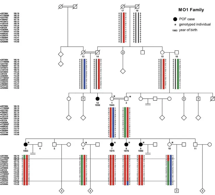
A disease gene mapping was initiated in a large highly consanguineous Middle Eastern family (MO1). The MO1 Palestinian family (pedigree in Figure 1), with 5 affected women, was recruited through the Genetic Department of Hadassah Hospital in Jerusalem. Medical reports were obtained and completed when possible. Blood samples were collected for hormonal, genomic and cytogenetic studies. Karyotypes and genomic DNA isolation were performed by standard procedures. The proband is the eldest of 6 sisters, 4 of them being affected with POF with primary amenorrhea. Their mother and father are first cousins, connected through at least 3 earlier consanguineous marriages. The non-affected mother and the affected maternal aunt were also born from a consanguineous marriage between first cousins, born from consanguineous marriages as well. The proband was diagnosed with POF at the age of 17 years because of amenorrhea, small and undeveloped breasts and streak gonads in ultrasound scan. The three other affected sisters were diagnosed with POF on the basis of a similar phenotype at ages between 18 and 20. All affected sisters presented with a normal uterus, but small ovaries with the appearance of ‘‘streak gonads’’ upon echography. Their height was within the normal range. Karyo-type, FMR1 premutation analysis and hormonal dosage were performed at the time of diagnosis (Table 1). No increased sedimentation rate or any other abnormal test for the presence of auto antibodies could be found in any patient. At the age of 19, the youngest POF patient suffered from a simultaneous bilateral ovarian cancer. After surgery, histological analysis revealed a gonadoblastoma on the right ovary, and a complex dysgermino-ma/embryonic carcinoma/choriocarcinoma in the tumor of the left ovary. In addition to these four cases, a maternal aunt (individual 12, Figure 1) was diagnosed with POF and primary amenorrhea (her DNA was not available for the genome-wide linkage analysis). The non-affected sisters of the proband had menarche at ages within the normal range, and conceived naturally 2 and 3 children, respectively.
Microsatellites genotyping and SNP genotyping
Genotyping was performed on anonymous samples without knowledge of any personal identifiers at the Genotyping platform from the Institut de Ge´nomique (Evry, France). After a genome-wide linkage analysis with 457 microsatellite markers (average autosomal marker spacing of 10 cM) that identified a significant linkage on chromosome 7, we performed a high-resolution
genome-wide scan, using the Affymetrix GeneChipH Human Mapping 250 K Nsp Array (Affymetrix Inc., Santa Clara, CA). On this array, bi-allelic markers are equally distributed across the genome with a median physical distance between SNPs of 4.8 kb, an average distance of 11.2 kb, and an average 0.30 of heterozygosity. The Affymetrix genome-wide genotyping was performed according to the standard manufacturer’s protocol (GeneChipHMapping 250 K Assay Manual Rev. 3/96-Well Plate Protocol), using 250 ng of genomic DNA per sample. Washing and staining steps were performed with Affymetrix GeneChipH Fluidics Station 450 and scanning step with the Affymetrix GeneChipHScanner 3000 7 G. Raw data were obtained with Affymetrix GeneChipH Operating Software (GCOS). Data were extracted with Affymetrix Gene-ChipH Genotyping Analysis Software (GTYPE) using BRLMM model as genotype calling method.
Linkage analysis and Homozygosity mapping
After classical filtering of SNP markers for genotyping quality, mendelian errors and verification of Hardy-Weinberg equilibrium, multipoint parametric linkage analysis was performed with Merlin under the assumption of recessive inheritance, with a disease allele frequency of 0.0001 and a penetrance of 0.99 [26]. The haplotypes in the identified 7q21-22 region were reconstructed with Merlin and presented graphically with HaploPainter v.1.043 [27]. The homozygous regions were confirmed and displayed using the dedicated software KinSNP (http://bioinfo.bgu.ac.il/ bsu/software/KinSNP/) [28]. POF patients in the MO1 family were searched for runs of shared homozygosity (ROSHs) with a minimum length of 1 cM according to the deCode genetic map. The degree of tolerance for genotyping errors within ROSHs was adjusted such that heterozygous calls surrounded by 15 or more shared homozygous SNPs were ignored.
Sequencing of candidate genes
Candidate genes were amplified by PCR from genomic DNA of one affected POF patient and one non-affected sister (details of primers and PCR conditions are provided in Document S1). PCR products were sequenced by standard Sanger sequencing.
Results and Discussion
Linkage analysis in a highly consanguineous POF family The highly consanguineous MO1 family (Figure 1) includes 5 POF patients in two generations (familial and clinical details in Material and Methods and Table 1). Although it was not possible to retrieve further familial information to assess earlier consan-guinity loops, the high degree of consanconsan-guinity in this family strongly suggested an autosomal recessive inheritance of the disease allele from a common ancestor. A first genome-wide linkage analysis with 457 microsatellite markers identified a 10.5 Mb-long region within 7q21-22 (maximum LOD-score (LODmax) of 3.8). A second whole-genome scan with a 250 K SNP array identified four chromosomal regions with significant linkage under a recessive model (LODmax= 3.26, Figure 2 and Table 2). It confirmed the significant linkage for a 9.96 Mb region on 7q21.3-22.2. It identified also an additional 2.56 Mb-long linkage region in 7p21.1, and two narrow segments, each spanning less than 0.2 Mb, within a short region of 6.6 Mb in 13q14. These regions had not been detected by the first genome-wide scan, since they were entirely framed by the analyzed microsatellites.
Table 1.Clinical and genetic data for the MO1 POF patients included in the study.
Individual Type of amenorrhea FSH (mIU/ml) Estradiol (pg/ml) LH (mUI/ml) Karyotype FMR1 premutation
MO1–3 primary 63.1 20.2 19.9 46,XX No
MO1–7 primary 51.4 12.1 21.1 46,XX No
MO1–8 primary 45.7 12.5 18.0 46,XX No
MO1–9 primary 55.3 22.0 18.0 46,XX No
Reference values 3–21 30–190 1.0–14.7
doi:10.1371/journal.pone.0033412.t001
Figure 1. Pedigree of the consanguineous MO1 family, with haplotype reconstruction of the 7q21-22 region.Squares indicate males and circles indicate females. Black symbols indicate women affected with Premature Ovarian Failure. Year of birth, when known, is indicated below the number of the individual. Double connecting lines indicate consanguineous marriages, all known to be between first cousins even when the precise consanguinity loop could not be established. Asterisks mark the individuals included in the microsatellite and/or the SNP whole-genome genotyping studies. The haplotypes for the large 7q21-22 region were reconstructed with Merlin [26] for the individuals genotyped on the SNP array, and visualized with HaploPainter. For the sake of simplicity, the number of depicted SNPs was reduced, to 3 SNPs at the border of the shared homozygous block (2 SNPs out and 1 SNP in) and to approximately 1 informative SNP/cM inside the block. A red bar represents the ancestral disease haplotype transmitted both by the mother 1 and the father 2, and the green and blue bars represent the wild-type haplotypes. Recombination events in individuals 3 and 8 delimit the region of interest between the external markers rs11768589 and rs2727744 (horizontal lines).
The four affected sisters shared a common disease haplotype, at the homozygous state because of the inheritance from a common ancestor through the paternal and maternal branches. Their affected sisters were either heterozygous (individual 6) or non-carrier (individual 10) for the disease haplotype (Figure 1). In addition, as the haplotypes confirmed that the affected sisters were not hemizygous for these regions, the mutation is not a large-scale deletion. Five larger ROSHs are detected by KinSNP, but in all these, the non-affected mothers and all the daughters shared the same haplotype at the homozygous state, which is incompatible with the transmission of the disease in the family. Therefore, the candidate region on the 7q21.3-22.2 is the largest homozygous stretch compatible with linkage.
The two peaks on chromosome 13 are not detected as ROSHs by KinSNP, due to the small size of the homozygous stretches.
However, a visual inspection of the haplotypes in the KinSNP output confirmed that these peaks indeed contain a range of SNPs that are homozygous by descent in the four affected sisters, and heterozygous in the non-affected sisters and the non-affected mother (data not shown). Additional linkage analysis along 0.2, 0.4, 0.6, 0.8 and 1 cM grids on chromosome 13 showed that the positive peaks disappeared only with the setting of 1 cM. Thus, those peaks probably do not result from linkage disequilibrium and are likely to reflect identity-by-state homozygosity.
In addition, the SNP-based linkage analysis identified a near-significant region of 0.19 Mb on Xp22.33, that spans the boundary between the pseudo-autosomal region and the X specific region (LOD = 2.94, figure 2). This peak is detected as a ROSH by KinSNP. However, the analysis of the haplotypes revealed that the non-affected mother, the healthy sisters, as well Figure 2. Whole genome linkage analysis for the MO1 family.The multipoint parametric LOD-score is plotted against the physical location of markers along the genome. The chromosome number is indicated in the upper part of the graph. Genotype analysis was performed on the Affymetrix GeneChipHHuman Mapping 250 KNspArray (Affymetrix Inc., Santa Clara, CA). A significant LOD-score was obtained for two regions on chromosome 7. The apparent single peak on chromosome 13 encompasses two distinct very small peaks.
doi:10.1371/journal.pone.0033412.g002
Table 2.Characteristics of significant LOD-score loci identified by genome-wide linkage in the MO1 family.
Chrom 7 7 13 13
Cytoband 7p21.1 7p15.3 7q21.3 7q22.2 13q14.12 13q14.3
LOD max 3.257 3.257 3.257 3.257
#of SNPs 297 643 25 27
Flanking SNP rs13222101 rs7806550 rs11768589 rs17152355 rs4245330 rs2248414 rs536338 rs797493
Position (cM) 31.654 34.696 106.189 115.436 47.21 47.495 53.166 53.377
Size (cM) 3.042 9.247 0.285 0.211
Position (bp, in GRCh37)
17723272 20278642 95615196 105575202 44661321 44798197 51103461 51261023
Size (Mb) 2.56 9.96 0.14 0.16
Homozyg. YES YES NO NO
#of genes 11+5 pseudogenes 166+39 pseudogenes 1 hypothetical 1 (2 terminal exons)
Candidate Genes SHFM1, DLX5, DLX6
The flanking SNPs are the first ones outside the LODmaxpeak, defined as having a LOD-score,LODmax-2 or LOD-score,LODmax-1. The Homozyg. line indicates whether
as the affected daughters, shared the same haplotype at the homozygous state for 15 of the 20 SNPs within the peak (data not shown). Therefore, it is highly unlikely that this locus contains the mutation responsible for the POF phenotype in this family.
Sequencing of POF candidate genes in the MO1 family In total, the two loci on chromosome 7 span 12.29 cM and 12.52 Mb, and contain 177 genes (details of positions, sizes and genes content for all peaks with a LOD-score above 3 are presented in Table 2, and a list of all the genes located in these regions in Table S1).
We cannot exclude the smaller loci on chromosome 13 as possibly containing the mutation involved in this familial POF, however they do not contain any evident functional candidate gene, if any gene at all. The first locus on chromosome 13 could possibly contain one gene (C13orf44), but the corresponding RefSeq sequences were permanently suppressed from the Gene database because there is currently insufficient support for the transcript and the protein (GeneID 79024). The second locus on chromosome 13 contains only the 2 terminal exons of two transcripts of the DLEU1 gene, a long non-coding RNA gene with multiple splicing variants implicated as a tumor suppressor in B-cell chronic lymphocytic leukemia.
Similarly, the region on 7p21.1 does not contain any obvious functional candidate genes.TWIST1was formerly known as the BPES3 locus, because eyelid features of some patients with Saethre-Chotzen syndrome, due to mutations inTWIST1, were similar to those presented by patients with BPES. Nevertheless, this specific phenotype observed in those patients affected with Saethre-Chotzen syndrome was later recognized to be due to phenotypic variability. In addition, patients with Saethre-Chotzen syndrome do not present any ovarian defects. ThereforeTWIST1
cannot be considered as a POF candidate gene.
On the contrary, the region on 7q21.3-22.3 includes at least three POF functional candidate genes.DLX5andDLX6code for two closely linked homeobox transcription factors, involved in the control of steroidogenesis [29]. The allelic reduction ofDlx5and
Dlx6in the mouse is associated with a POF-like phenotype, with an early reduction of fertility and a rapid and premature follicular depletion. This study also provided evidence supporting a reciprocal regulation betweenDlx5,Dlx6andFoxl2, a transcription factor essential for ovarian development and function, also implicated in POF [30,31,32]. Another potential candidate gene isSHFM1, located in close proximity of theDLX5/DLX6locus. Its nematode ortholog, dss-1, is functionally conserved during evolution and was shown to be required for oogenesis and normal female fertility inC. elegans(with no impact on gonad development or on male fertility) [33]. The murineDss1is expressed in the early genital tubercle during fetal development, and in the early genital bud of the newborn mice [34]. Although these three genes are implicated in the etiology of the Split Hand/Split Foot Malformation Type I syndrome (SHFM1, MIM%183600) [35], their possible function in the ovary prompted us to sequence them in the MO1 family. Sequencing of the coding, promoter and known enhancers regions ofDLX5and DLX6did not show any DNA variants segregating with the disease. Likewise, we did not find any variants in the promoter and coding sequence ofSHFM1. Conclusions
We have indentified two novel POF-associated loci on chromosome 7, that do not coincide with the regions identified by genome-wide linkage in the two previous studies of familial POF cases: the POF1B gene on the X chromosome, and a 15.8 Mb region in 5q14.1-q15 [6,7]. In addition, these regions on
chromosome 7 are not included and do not contain the CNVs that were detected as statistically different from controls in POF patients by Aboura and colleagues [36]. The identification of two other putative loci in our study highlights the important genetic heterogeneity of this disease.
The fact that the severe ovarian cancer affecting the youngest POF patient (individual 9) was bilateral and simultaneous strongly suggests a genetic origin. However, the association between POF and ovarian cancer is highly unusual and, to our knowledge, was never described before. Besides, a direct relation between POF and ovarian cancer is doubtful since only one POF-affected sister developed a cancer at a young age. Therefore it is more likely that the co-occurrence of these diseases in this patient could be related to the high familial consanguinity.
We could not find any sequence variant segregating with the disease in the coding and known regulatory sequences ofDLX5/6
and the coding sequence ofSHFM1. Although we cannot exclude the existence of still unknown regulatory features, possibly affecting both SHFM1 and DLX5/DLX6, which could harbor the causal mutation for POF in our family, the most likely hypothesis is that the mutation is present in the coding sequence of another gene located in the 7p or the 7q regions.
Among those genes, other possible candidates include
SMURF1, the CYP3 gene family and VGF. SMURF1 encodes an E3 ubiquitin ligase specific of regulatory SMAD proteins, that was shown in rat and human granulosa cell lines to ubiquitinylate R-Smad 1 and 5, two of the regulatory Smads activated by oocyte-secreted BMP15, a known POF gene [37]. The CYP3
gene cluster in 7q includes four genes, CYP3A43, CYP3A4,
CYP3A7 and CYP3A5, encoding cytochrome P450 enzymes, known to be implicated in drug metabolism and synthesis of cholesterol and steroids. Although some of the CYP3A genes appear to have a restricted expression in the liver and to be mainly responsible for drug detoxification, we cannot exclude a role in steroid synthesis in the ovary [38].VGFencodes a 68-kDa precursor of multiple bioactive peptides with diverse neuroen-docrine functions, expressed abundantly in the brain, and in peripheral endocrine tissues including the pituitary gland. In addition to a role in the regulation of energy homeostasis,VGF
could also regulate reproduction, since homozygous Vgf-null mice are infertile, presenting a delayed sexual maturation, incomplete mammary development and ovaries with only primary and atretic follicles, apparently due to an abnormal pitituary gonadotropin content [39].
As the two loci identified on chromosome 7 contain too many genes for a direct sequencing approach in search of the causal mutation, we plan to proceed to an exome-sequencing in the MO1 family. Hopefully, this study will enable us to identify the mutation implicated in Premature Ovarian Failure in this family and might lead to the identification of one or several genes involved in the control of ovarian function and development.
Supporting Information
Document S1 Primers and conditions of PCR used for sequencingSHFM1, DLX5andDLX6.
(DOC)
Table S1 List of genes present in the loci identified on chromosomes 7 and 13 by genome-wide linkage in the MO1 POF family.The lists were established using the NCBI Genome Viewer, using the rs number of the border SNPs, as given in Table 1. Pseudogenes are written in grey.
Acknowledgments
We are indebted to Jacques S. Beckmann for his great help in the analysis of the first genome scan, and his very helpful comments and suggestions all along the project. We thank Simon Heath (Institut de Ge´nomique, CEA, Evry, France) for the initial analysis of the genotyping data, Robert Ivanek (Friedrich Miescher Institute for Biomedical Research, Basel, Switzerland) for his help with data file preparation, Besma Lakhal (CHU Farhat-Hached, Sousse, Tunisia) for helping with the sequencing of the candidate genes, and Inbar Plaschkes (National Institute for Biotechnology in the Negev, Ben-Gurion University of the Negev, Beer-Sheva, Israel) for her
help with KinSNP analysis. In addition, we express our thanks to the affected patients and their relatives for participating in our study and for their keen interest in our results.
Author Contributions
Conceived and designed the experiments: SC MF. Performed the experiments: SC PL CB CC. Analyzed the data: SC PZ VCC DV KB AD GL SCM MF. Contributed reagents/materials/analysis tools: ZBN VCC. Wrote the paper: SC PZ GL RAV MF. Organized, supervised, and performed the phenotyping: ZBN.
References
1. Nelson LM, Covington SN, Rebar RW (2005) An update: spontaneous premature ovarian failure is not an early menopause. Fertil Steril 83: 1327–1332.
2. Coulam CB, Bustillo M, Schulman JD (1986) Empty follicle syndrome. Fertil Steril 46: 1153–1155.
3. Goswami D, Conway GS (2005) Premature ovarian failure. Hum Reprod Update 11: 391–410.
4. Christin-Maitre S, Pasquier M, Donadille B, Bouchard P (2006) [Premature ovarian failure]. Ann Endocrinol (Paris) 67: 557–566.
5. Nelson LM (2009) Clinical practice. Primary ovarian insufficiency. N Engl J Med 360: 606–614.
6. Lacombe A, Lee H, Zahed L, Choucair M, Muller JM, et al. (2006) Disruption of POF1B binding to nonmuscle actin filaments is associated with premature ovarian failure. Am J Hum Genet 79: 113–119.
7. Oldenburg RA, van Dooren MF, de Graaf B, Simons E, Govaerts L, et al. (2008) A genome-wide linkage scan in a Dutch family identifies a premature ovarian failure susceptibility locus. Hum Reprod 23: 2835–2841.
8. van Kasteren YM, Schoemaker J (1999) Premature ovarian failure: a systematic review on therapeutic interventions to restore ovarian function and achieve pregnancy. Hum Reprod Update 5: 483–492.
9. Vegetti W, Marozzi A, Manfredini E, Testa G, Alagna F, et al. (2000) Premature ovarian failure. Mol Cell Endocrinol 161: 53–57.
10. Sybert VP, McCauley E (2004) Turner’s syndrome. N Engl J Med 351: 1227–1238.
11. Di Pasquale E, Beck-Peccoz P, Persani L (2004) Hypergonadotropic ovarian failure associated with an inherited mutation of human bone morphogenetic protein-15 (BMP15) gene. Am J Hum Genet 75: 106–111.
12. Dixit H, Rao LK, Padmalatha VV, Kanakavalli M, Deenadayal M, et al. (2006) Missense mutations in the BMP15 gene are associated with ovarian failure. Hum Genet 119: 408–415.
13. Laissue P, Christin-Maitre S, Touraine P, Kuttenn F, Ritvos O, et al. (2006) Mutations and sequence variants in GDF9 and BMP15 in patients with premature ovarian failure. Eur J Endocrinol 154: 739–744.
14. Barlow C, Hirotsune S, Paylor R, Liyanage M, Eckhaus M, et al. (1996) Atm-deficient mice: a paradigm of ataxia telangiectasia. Cell 86: 159–171. 15. Barlow DH (1996) Premature ovarian failure. Baillieres Clin Obstet Gynaecol
10: 361–384.
16. Aittomaki K, Lucena JL, Pakarinen P, Sistonen P, Tapanainen J, et al. (1995) Mutation in the follicle-stimulating hormone receptor gene causes hereditary hypergonadotropic ovarian failure. Cell 82: 959–968.
17. Shimizu T, Miyahayashi Y, Yokoo M, Hoshino Y, Sasada H, et al. (2004) Molecular cloning of porcine growth differentiation factor 9 (GDF-9) cDNA and its role in early folliculogenesis: direct ovarian injection of GDF-9 gene fragments promotes early folliculogenesis. Reproduction 128: 537–543. 18. Qin Y, Choi Y, Zhao H, Simpson JL, Chen ZJ, et al. (2007) NOBOX
homeobox mutation causes premature ovarian failure. Am J Hum Genet 81: 576–581.
19. Qin Y, Shi Y, Zhao Y, Carson SA, Simpson JL, et al. (2009) Mutation analysis of NOBOX homeodomain in Chinese women with premature ovarian failure. Fertil Steril 91: 1507–1509.
20. Christin-Maitre S, Vasseur C, Portnoi MF, Bouchard P (1998) Genes and premature ovarian failure. Mol Cell Endocrinol 145: 75–80.
21. Kaku U, Kameyama K, Izawa M, Yamada M, Miyamoto J, et al. (2008) Ovarian histological findings in an adult patient with the steroidogenic acute regulatory protein (StAR) deficiency reveal the impairment of steroidogenesis by lipoid deposition. Endocr J 55: 1043–1049.
22. Crisponi L, Deiana M, Loi A, Chiappe F, Uda M, et al. (2001) The putative forkhead transcription factor FOXL2 is mutated in blepharophimosis/ptosis/ epicanthus inversus syndrome. Nat Genet 27: 159–166.
23. Harris SE, Chand AL, Winship IM, Gersak K, Aittomaki K, et al. (2002) Identification of novel mutations in FOXL2 associated with premature ovarian failure. Mol Hum Reprod 8: 729–733.
24. Laissue P, Lakhal B, Benayoun BA, Dipietromaria A, Braham R, et al. (2009) Functional evidence implicating FOXL2 in non-syndromic premature ovarian failure and in the regulation of the transcription factor OSR2. J Med Genet 46: 455–457.
25. Gersak K, Harris SE, Smale WJ, Shelling AN (2004) A novel 30 bp deletion in the FOXL2 gene in a phenotypically normal woman with primary amenor-rhoea: case report. Hum Reprod 19: 2767–2770.
26. Abecasis GR, Cherny SS, Cookson WO, Cardon LR (2002) Merlin–rapid analysis of dense genetic maps using sparse gene flow trees. Nat Genet 30: 97–101.
27. Thiele H, Nurnberg P (2005) HaploPainter: a tool for drawing pedigrees with complex haplotypes. Bioinformatics 21: 1730–1732.
28. Amir el AD, Bartal O, Morad E, Nagar T, Sheynin J, et al. (2010) KinSNP software for homozygosity mapping of disease genes using SNP microarrays. Hum Genomics 4: 394–401.
29. Nishida H, Miyagawa S, Vieux-Rochas M, Morini M, Ogino Y, et al. (2008) Positive regulation of steroidogenic acute regulatory protein gene expression through the interaction between Dlx and GATA-4 for testicular steroidogenesis. Endocrinology 149: 2090–2097.
30. Caburet S, Georges A, L’Hoˆte D, Todeschini AL, Benayoun BA, et al. (2011) The transcription factor FOXL2: at the crossroads of ovarian physiology and pathology. Molecular and Cellular Endocrinology.
31. Jeong J, Li X, McEvilly RJ, Rosenfeld MG, Lufkin T, et al. (2008) Dlx genes pattern mammalian jaw primordium by regulating both lower jaw-specific and upper jaw-specific genetic programs. Development 135: 2905–2916. 32. Bouhali K, Dipietromaria A, Fontaine A, Caburet S, Barbieri O, et al. (2011)
Allelic reduction of Dlx5 and Dlx6 results in early follicular depletion: a new mouse model of primary ovarian insufficiency. Hum Mol Genet 20: 2642–2650. 33. Pispa J, Palmen S, Holmberg CI, Jantti J (2008) C. elegans dss-1 is functionally conserved and required for oogenesis and larval growth. BMC Dev Biol 8: 51. 34. Crackower MA, Scherer SW, Rommens JM, Hui CC, Poorkaj P, et al. (1996) Characterization of the split hand/split foot malformation locus SHFM1 at 7q21.3-q22.1 and analysis of a candidate gene for its expression during limb development. Hum Mol Genet 5: 571–579.
35. Scherer SW, Poorkaj P, Massa H, Soder S, Allen T, et al. (1994) Physical mapping of the split hand/split foot locus on chromosome 7 and implication in syndromic ectrodactyly. Hum Mol Genet 3: 1345–1354.
36. Aboura A, Dupas C, Tachdjian G, Portnoi MF, Bourcigaux N, et al. (2009) Array comparative genomic hybridization profiling analysis reveals deoxyribo-nucleic acid copy number variations associated with premature ovarian failure. J Clin Endocrinol Metab 94: 4540–4546.
37. Huang C (2010) Roles of E3 ubiquitin ligases in cell adhesion and migration. Cell Adh Migr 4: 10–8.
38. Monostory K, Dvorak Z (2011) Steroid regulation of drug-metabolizing cytochromes P450. Curr Drug Metab 12: 154–72.