Helicobacter pylori
Eradication Prevents
Metachronous Gastric Neoplasms after
Endoscopic Resection of Gastric Dysplasia
Seung Hwan Shin1, Da Hyun Jung1*, Jie-Hyun Kim2, Hyun Soo Chung1, Jun Chul Park1, Sung Kwan Shin1, Sang Kil Lee1, Yong Chan Lee1
1Department of Internal Medicine, Yonsei University College of Medicine, Seoul, Korea,2Department of Internal Medicine, Gangnam Severance Hospital, Yonsei University College of Medicine, Seoul, Korea
*leah1004@yuhs.ac
Abstract
Purpose
There is insufficient data about the role of eradication ofH.pyloriafter endoscopic resection (ER) for gastric dysplasia. The aim was to investigate the benefit ofH.pylorieradication after ER in patients with gastric dysplasia to prevent metachronous gastric neoplasms.
Materials and Methods
We retrospectively reviewed 1872 patients who underwent ER of gastric dysplasia. We excluded patients with a follow-up period of<2 years or who had not undergone tests for activeH.pyloriinfection. A total of 282 patients were enrolled. The patients were catego-rized into those without activeH.pyloriinfection (H.pylori-negative group, n = 124), those who successfully underwentH.pylorieradication (eradicated group, n = 122), and those who failed or did not undergoH.pylorieradication (persistent group, n = 36).
Results
Metachronous recurrence was diagnosed in 36 patients, including 19 in theH.pylori -nega-tive group, 10 in the eradicated group, and 7 in the persistent group. The cumula-nega-tive inci-dence of metachronous recurrence was significantly lower in theH.pylori-eradicated group in comparison with either of theH.pylori-persistent (non-eradicated or failed) groups (p= 0.039). Similarly, the incidence of metachronous recurrence was significantly lower in the
H.pylori-eradicated group compared with theH.pylori-negative group (p= 0.041).
Conclusion
SuccessfulH.pylorieradication may reduce the development of metachronous gastric neo-plasms after ER in patients with gastric dysplasia.
a11111
OPEN ACCESS
Citation:Shin SH, Jung DH, Kim J-H, Chung HS, Park JC, Shin SK, et al. (2015)Helicobacter pylori Eradication Prevents Metachronous Gastric Neoplasms after Endoscopic Resection of Gastric Dysplasia. PLoS ONE 10(11): e0143257. doi:10.1371/journal.pone.0143257
Editor:Masaru Katoh, National Cancer Center, JAPAN
Received:August 13, 2015
Accepted:November 2, 2015
Published:November 18, 2015
Copyright:© 2015 Shin et al. This is an open access article distributed under the terms of theCreative Commons Attribution License, which permits unrestricted use, distribution, and reproduction in any medium, provided the original author and source are credited.
Data Availability Statement:In order to protect the personal information of data used in the manuscript, data will be available upon request from the Yonsei University Health System Institutional Data Access / Ethics Committee.
Funding:The authors have no support or funding to report.
Introduction
Helicobacter pylori(H.pylori) infection is a group I carcinogen for gastric cancer as defined by the International Agency for Research on Cancer (IARC), a subdivision of the World Health
Organization (WHO) [1]. Correaet al. contend thatH.pyloriinfection is closely associated
with progression to gastric atrophy, intestinal metaplasia (IM), dysplasia, and cancer [2]. The
reported progression rate of atrophic gastritis, intestinal metaplasia and dysplasia to gastric
cancer varies from 0 to 1.8%, 0 to 10%, and 0 to 73% per year, respectively [3]. Male gender is
the significant risk factor for the development of gastric cancer, with a nearly 2:1 male to female
dominance [4]. Previous studies showed that seroprevalence ofH.pyloriwas higher in males
than in females [5–7]. Currently, endoscopic resection (ER) is the accepted standard treatment
for selected cases of early gastric cancer (EGC) in Korea. Because gastric dysplasia is a precan-cerous lesion, this pathology also can be a candidate for ER, which has the additional benefit of providing a histologic diagnosis that could result in an upgrade from biopsy-proven dysplasia
to cancer [8]. In Korea, ER is widely used for the treatment of gastric dysplasia and EGC [9].
ER can preserve the stomach; however, metachronous gastric cancer may develop in the
stom-ach remnant after the procedure [10,11]. For this reason, endoscopic surveillance for
meta-chronous gastric neoplasms has become an emerging issue during the follow-up interval after
ER. Some studies have shown that the eradication ofH.pyloriafter ER is helpful to prevent the
development of metachronous gastric cancer [12–14]; however, several other studies have
failed to show a prophylactic benefit forH.pylorieradication [15,16]. At present, the guidelines
for the treatmentH.pyloriinfection in Korea recommends eradication ofH.pyloriafter ER of
EGC according to the findings of positive studies [17]. However, there is insufficient data
about the role of eradication ofH.pyloriafter ER for the treatment of gastric dysplasia. The
aim of this study was to determine whetherH.pylorieradication prevents the development of
metachronous gastric neoplasms in patients with gastric dysplasia following ER.
Methods
Patients
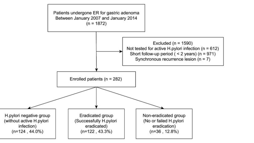
Between January 2007 and January 2014, 1872 patients were diagnosed with low- and high-grade gastric dysplasia and underwent ER at Severance Hospital, Seoul, Korea. Of these
patients, we excluded 612 patients who did not undergo tests for activeH.pyloriinfection
including a urea breath test, a rapid urease test, or histopathological examination. We also
excluded 971 patients with only a short-term follow-up period (<2 years) and 7 patients who
had a recurrence in a previous ER site. Ultimately we enrolled a total of 282 patients for analy-sis in this study. The ER procedures were performed by expert endoscopists who had a previ-ous experience of over 1000 gastric endoscopy cases per year. The patients were divided into
three groups according to presence of activeH.pyloriinfection and successful eradication. The
patient selection and grouping flow diagram is shown inFig 1: (1) those without activeH.
pyloriinfection at the time of ER (theH.pylori-negative group, n = 124), (2) those with a
suc-cessfully treatedH.pyloriinfection (theH.pylori-eradicated group, n = 122), (3) those who
failed treatment ofH.pyloriinfection or were untreated (theH.pylori-persistent group,
n = 36). A time interval of at least 6 months was observed betweenH.pyloritreatment and the
development of metachronous gastric neoplasms to accurately assess the effect ofH.pylori
drinking history were divided into yes or no. The clinicopathological characteristics were ana-lyzed retrospectively from patient medical records. The Institutional Review Board (IRB) of Severance Hospital approved this study. We received a consent exemption from the IRB. Patients records and information was anonymized.
Evaluation of
H
.
pylori
infection status and treatment
To determine theH.pyloriinfection status rapid urease and urea breath tests and histological
assessment were used. Patients who showed negative results for all three examinations were
assigned to theH.pylori-negative group. The patients who had a positive result for at least one
test among these were assigned to the activeH.pyloriinfection group. The activeH.pylori
infection group was subdivided into two groups depending on their infection treatment
out-come and assigned to either the eradicated or persistent groups.H.pylorieradication therapy
was carried out in accordance with the guideline for treatment ofH.pyloriinfection in Korea
[17]. Of the total 282 patients, 122 patients assigned to the eradicated group received triple
therapy of a standard dose of a proton pump inhibitor (PPI), 500 mg of clarithromycin, and 1000 mg of amoxicillin twice a day for 7 days. Twelve patients in the eradicated group who ini-tially failed eradication after conventional triple therapy underwent a secondary regimen of bis-muth-based quadruple therapy.
Follow-up schedule after the endoscopic resection
Follow-up endoscopic examinations with endoscopy and biopsy were conducted at regular intervals after endoscopic resection (3 and 12 months and annually after the first year of Fig 1. A flow chart of patient enrollment.
follow-up). A biopsy was performed at the previous ER site and any other sites suspected to represent a recurrence of metachronous neoplasm.
Histopathological evaluation of the gastric lesion
Biopsy or resection specimens were examined by two expert pathologists. The histology of gas-tric cancers were reviewed according to the new Japanese classification for gasgas-tric carcinomas
[18] and the grade of gastric dysplasia was categorized according to the Vienna classification
[19]. A metachronous neoplasm was defined as evident dysplasia or carcinoma that developed
subsequently (i.e., more than 6 months after ER of the primary gastric dysplasia).
Statistical analysis
The chi-square test and Fisher’s exact test were used to compare the clinicopathological factors
between the groups according to the presence/absence of activeH.pyloriinfection and failed/
successful eradication. The Student’st-test was used for non-categorical variables in the
inter-group comparisons of clinicopathological characteristics. The threshold for statistical
signifi-cance was set atp<0.05. The incidence of metachronous gastric neoplasms was calculated by
the Kaplan-Meier method, and compared among the three groups by the log-rank test. A Cox proportional hazards model and multivariate analyses were used for risk assessment. Statistical analyses were performed using the Statistical Package for Social Sciences Version 18.0 (SPSS Inc., Chicago, IL, USA).
Results
Baseline characteristics of the study population
The baseline characteristics of the three groups are summarized inTable 1. The enrolled
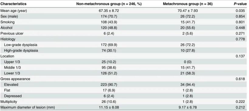
patients included 200 (70.9%) males and 82 (29.1%) females and the median age was 68 years old. There were no significant differences among the three groups in the sex ratio, smoking and alcohol history, previous ulcer history and histology, or the location and size of the tumor. The clinicopathological characteristics of patients according to the development of metachronous
neoplasms are shown inTable 2. Among the 282 patients, metachronous recurrence developed
in 36 (14.6%) patients after ER of gastric dysplasia. There were no significant differences between the two groups in the sex ratio, smoking and alcohol consumption history, initial his-tology, lesion size, or location of the tumor. The metachronous group was significantly older
than the non-metachronous group (p= 0.035).
Metachronous recurrence
During the follow-up period, 36 metachronous recurrences were found on endoscopy and con-firmed by biopsy. Metachronous recurrence was diagnosed in 19 of 124 patients (15.3%) in the H.pylori-negative group, in 10 of 122 patients (8.2%) in the eradicated group, and in 7 of 36 patients (19.4%) in the persistent group. The median time until metachronous recurrence was
36 months (range, 6–85 months). The cumulative incidence of metachronous recurrence was
significantly lower in theH.pylori-eradicated group in comparison with either of theH.
pylori-persistent (non-eradicated or failed) groups (p= 0.039,Fig 2A). Moreover, this result was
dis-tinct by 36 months after ER. Similarly, the incidence of metachronous recurrence was
signifi-cantly lower in theH.pylori-eradicated group compared with theH.pylori-negative group
(p= 0.041,Fig 2B). Univariate analysis using Cox’s proportional hazards model showed that
the persistent group had a higher risk of developing metachronous gastric neoplasms than the
Discussion
According to the current Korean guidelines for treatment ofH.pyloriinfection, eradication
therapy is recommended after ER of EGC.H.pyloriinfection plays an important role in the
development of gastric cancer. Huanget al. show by a meta-analysis that the odds ratio for the
development of gastric cancer inH.pylori-infected patients is 1.92, and younger patients with
H.pyloriinfection have a higher relative risk to develop gastric cancer than older patients [20].
Therefore, there is some evidence thatH.pylorieradication could prevent metachronous
recur-rence after ER of EGC; however, data are still lacking whether there are prophylactic benefits of H.pylorieradication to prevent the development of metachronous lesions after ER of pre-malignant lesions such as gastric dysplasia.
Rapid urease test, histology, and urea breath tests are the recommended diagnostic tests for H.pyloriinfection. The urea breath test had high sensitivity and specificity (95%). However, false negative rates greater than 30% have been reported when antibiotics or PPI were used. For
the rapid urease test, test sensitivity ranges from 85–98% and specificity ranges from 89–100%.
Hematoxylin and eosin staining has a sensitivity of 69–93% and a specificity of 87–90% [17].
Table 1. Baseline characteristics of enrolled patients.
Characteristic H.pylorinegative group (n = 124, %)
Eradicated group (n = 122, %)
Persistent group (n = 36, %)
P -value
Mean age (year)a 68.89±8.78 66.64±8.55 67.61±8.49 0.126
Sex 0.621
Male 91 (73.4) 83 (68.0) 26 (72.2)
Female 33 (26.6) 39 (32.0) 10 (27.8)
Smoking 47 (37.9) 62 (50.8) 14 (38.9) 0.347
Alcohol 58 (46.8) 62 (50.8) 20 (55.6) 0.324
Family history of Gastric cancer 19 (15.3) 26 (21.3) 6 (16.7) 0.462
Previous ulcer 5 (4.0) 1 (0.8) 2 (5.6) 0.793
Histology 0.597
Low-grade dysplasia 85 (68.8) 87 (71.3) 26 (72.2)
High-grade dysplasia 39 (31.5) 35 (28.7) 10 (27.8)
Location 0.418
Upper 1/3 11 (8.9) 13 (10.7) 1 (2.8)
Middle 1/3 53 (42.7) 40 (32.8) 17 (47.2)
Lower 1/3 60 (48.4) 69 (56.6) 18 (50.0)
Gross appearance 0.504
Elevated 117 (94.4) 106 (86.9) 20 (55.6)
Flat 4 (3.2) 13 (10.7) 1 (2.8)
Depressed 3 (2.4) 3 (2.5) 1 (2.8)
Multiplicity 14 (11.3) 12 (9.8) 1 (2.8) 0.178
Maximum diameter of lesion (mm)a 10.84±7.56 11.08±8.87 10.55±6.22 0.960
Metachronous recurrenceb 19 (15.3) 10 (8.2) 7 (19.4) 0.095
Dysplasia 15 (79.0) 6 (60.0) 4 (57.2)
Cancer 4 (21.0) 4 (40.0) 3 (42.9)
Follow up duration (months, median, range)
53.0 (26.3–85.7) 58.3 (24.3–85.9) 57.2 (28.1–85.2) 0.026
Values are presented as mean±SD or n (%).H.pylori,Helicobacter pylori. aStatistical signi
ficance were tested by oneway analysis of variances among groups bStatistical signi
ficance were tested by Fisher’s exact test between three groups.
Table 2. Comparison of clinicopathologic characteristics between patients with and without metachronous recurrence.
Characteristics Non-metachronous group (n = 246, %) Metachronous group (n = 36) P-value
Mean age (year) 67.35±8.72 70.47±7.93 0.035
Sex (male) 174 (70.7) 26 (72.2) 0.854
Smoking 108 (43.9) 15 (41.7) 0.801
Alcohol 120 (48.8) 20 (55.6) 0.448
Previous ulcer 6 (2.4) 2 (5.6) 0.271
Histology 0.778
Low-grade dysplasia 172 (69.9) 26 (72.2)
High-grade dysplasia 74 (30.1) 10 (27.8)
Location 0.137
Upper 1/3 25 (10.2) 0 (0)
Middle 1/3 95 (38.6) 15 (41.7)
Lower 1/3 126 (51.2) 21 (58.3)
Gross appearance 0.618
Elevated 223 (90.7) 34 (94.4)
Flat 17 (6.9) 1 (2.8)
Depressed 6 (2.4) 1 (2.8)
Multiplicity 26 (10.6) 1 (2.8) 0.222
Maximum diameter of lesion (mm) 11.15±8.08 9.17±6.78 0.212
Values are presented as mean±SD or n (%).H.pylori,Helicobacter pylori.
doi:10.1371/journal.pone.0143257.t002
Fig 2. (A) The cumulative incidence of metachronous recurrence was significantly lower in theH.pylori-eradicated group in comparison with theH. pylori-persistent (non-eradicated or failed) groups (p= 0.039). (B) The cumulative incidence of metachronous recurrence was significantly lower in theH. pylori-eradicated group in comparison with theH.pylorinegative group (p= 0.041).
Recently, ER of gastric dysplasia has become accepted as a reasonable treatment option. Although the ER of low-grade dysplasia diagnosed by forceps biopsy is controversial, tumors
with a size>1 cm and/or with surface redness or nodularity are believed to have significant
risk for high-grade dysplasia or EGC [21]. Therefore, in Korea, ER is commonly performed for
patients with both low- and high-grade dysplasia.
Since a risk of metachronous recurrence still exists in the remnant stomach following ER of gastric dysplasia, surveillance for metachronous gastric neoplasms is believed important during
the follow-up period. Gutierrez-Gonzalezet al. show that identical genetic alterations are
pres-ent in dysplasia and the surrounding intestinal metaplasia (IM) [22]. They also demonstrate
that genetic changes in the IM are likely to lead to dysplasia, and further genetic alterations can
cause the progression of dysplasia to gastric cancer referred to as“cancer in adenoma”[22,23].
Several causative genetic and epigenetic changes in the IM or dysplasia are reversed byH.pylori
eradication. In this study, successful eradication ofH.pyloriinfection significantly lowers
metachronous recurrence; therefore, the eradication ofH.pyloriinfection may prevent the
emergence of metachronous gastric neoplasms after ER of gastric dysplasia.
There are some reports thatH.pylorieradication has a prophylactic effect to prevent
meta-chronous gastric neoplasms after ER of EGC. One large, randomized controlled trial shows a reduced incidence of metachronous gastric cancer (odds ratio [OR] 0.353, 95% confidence
interval [CI] 0.161–0.775,p= 0.009), and recommends that prophylactic eradication should be
performed after ER [12]. However, this trial doesn’t address whether there is a similar benefit
to eradicateH.pyloriinfection after ER of gastric dysplasia. Unlike previous studies, we focused
the role ofH.pylorieradication in the development of metachronous lesions after ER of gastric
dysplasia which was a precancerous lesion. And, in our study,H.pylorieradication could also
prevent metachronous lesions after ER of gastric dysplasia, similar EGC.
H.pyloriinfection both initiates and promotes the gastric carcinogenesis; therefore, eradica-tion should both inhibit newly developed gastric cancers and reduce the growth rate of existing
gastric cancers [24]. After a median follow-up period of 36 months, metachronous recurrence
developed in 36 patients. Although a 3-year follow-up period is short, eradication ofH.pylori
after ER of dysplasia might inhibit the occurrence of new gastric neoplasms and delay the
growth rate of gastric cancer. Our study has some limitations. TheH.pylori-negative group
showed a lower metachronous recurrence rate than non-eradicated group, but a higher
recur-rence rate than the eradicated group. This unexpected result may be due to false negativeH.
pyloriinfection status. Additional potential causes for the false negative result might be due to antibiotic therapy administered to treat other infections, the masking effect of proton pump
inhibitors (PPIs), inadequate sampling, or suboptimal staining technique [25]. We didn’t
ana-lyze the background mucosa for atrophy and intestinal metaplasia, which might also affect the
rate of metachronous recurrence in theH.pylori-negative group. We didn’t consider the
re-infection ofH.pylori. However, five (4.1%) patients were re-infected withH.pyloriin
eradi-cated group. Therefore, the annual re-infection rate was quite low.
In conclusion, this study demonstrates thatH.pylorieradication after ER in patients with
gastric dysplasia may reduce the development of subsequent metachronous gastric neoplasms.
Therefore,H.pylorieradication is recommended after ER of gastric dysplasia.
Author Contributions
References
1. Schistosomes, liver flukes and Helicobacter pylori. IARC Working Group on the Evaluation of Carcino-genic Risks to Humans. Lyon, 7–14 June 1994. IARC Monogr Eval Carcinog Risks Hum. 1994; 61: 1–
241.
2. Correa P. A human model of gastric carcinogenesis. Cancer Res. 1988; 48: 3554–3560. PMID:
3288329
3. de Vries AC, Haringsma J, Kuipers EJ. The detection, surveillance and treatment of premalignant gas-tric lesions related to Helicobacter pylori infection. Helicobacter. 2007; 12: 1–15.
4. Schlansky B, Sonnenberg A. Epidemiology of noncardia gastric adenocarcinoma in the United States. Am J Gastroenterol. 2011; 106: 1978–1985. doi:10.1038/ajg.2011.213PMID:22008896
5. Yim JY, Kim N, Choi SH, Kim YS, Cho KR, Kim SS, et al. Seroprevalence of Helicobacter pylori in South Korea. Helicobacter. 2007; 12: 333–340. PMID:17669107
6. Woodward M, Morrison C, McColl K. An investigation into factors associated with Helicobacter pylori infection. Journal of Clinical Epidemiology. 2000; 53: 175–181. PMID:10729690
7. Everhart JE, Kruszon-Moran D, Perez-Perez GI, Tralka TS, McQuillan G. Seroprevalence and ethnic differences in Helicobacter pylori infection among adults in the United States. Journal of Infectious Dis-eases. 2000; 181: 1359–1363. PMID:10762567
8. Kim YJ, Park JC, Kim JH, Shin SK, Lee SK, Lee YC, et al. Histologic diagnosis based on forceps biopsy is not adequate for determining endoscopic treatment of gastric adenomatous lesions. Endoscopy. 2010; 42: 620–626. doi:10.1055/s-0030-1255524PMID:20623445
9. Kang KJ, Lee JH. Characteristics of Gastric Cancer in Korea—with an Emphasis on the Increase of the Early Gastric Cancer (EGC). Journal of the Korean Medical Association. 2010; 53: 283–289.
10. Nasu J, Doi T, Endo H, Nishina T, Hirasaki S, Hyodo I. Characteristics of metachronous multiple early gastric cancers after endoscopic mucosal resection. Endoscopy. 2005; 37: 990–993. PMID:16189772
11. Isomoto H, Shikuwa S, Yamaguchi N, Fukuda E, Ikeda K, Nishiyama H, et al. Endoscopic submucosal dissection for early gastric cancer: a large-scale feasibility study. Gut. 2009; 58: 331–336. doi:10.1136/ gut.2008.165381PMID:19001058
12. Fukase K, Kato M, Kikuchi S, Inoue K, Uemura N, Okamoto S, et al. Effect of eradication of Helicobac-ter pylori on incidence of metachronous gastric carcinoma afHelicobac-ter endoscopic resection of early gastric cancer: an open-label, randomised controlled trial. Lancet. 2008; 372: 392–397. doi: 10.1016/S0140-6736(08)61159-9PMID:18675689
13. Bae SE, Jung HY, Kang J, Park YS, Baek S, Jung JH, et al. Effect of Helicobacter pylori eradication on metachronous recurrence after endoscopic resection of gastric neoplasm. Am J Gastroenterol. 2014; 109: 60–67. doi:10.1038/ajg.2013.404PMID:24343545
14. Kim YI, Choi IJ, Kook MC, Cho SJ, Lee JY, Kim CG, et al. The association between Helicobacter pylori status and incidence of metachronous gastric cancer after endoscopic resection of early gastric cancer. Helicobacter. 2014; 19: 194–201. doi:10.1111/hel.12116PMID:24612125
15. Maehata Y, Nakamura S, Fujisawa K, Esaki M, Moriyama T, Asano K, et al. Long-term effect of Helico-bacter pylori eradication on the development of metachronous gastric cancer after endoscopic resec-tion of early gastric cancer. Gastrointest Endosc. 2012; 75: 39–46. doi:10.1016/j.gie.2011.08.030
PMID:22018552
16. Choi J, Kim SG, Yoon H, Im JP, Kim JS, Kim WH, et al. Eradication of Helicobacter pylori after endo-scopic resection of gastric tumors does not reduce incidence of metachronous gastric carcinoma. Clin Gastroenterol Hepatol. 2014; 12: 793–800.e791. doi:10.1016/j.cgh.2013.09.057PMID:24100112
17. Kim SG, Jung HK, Lee HL, Jang JY, Lee H, Kim CG, et al. Guidelines for the diagnosis and treatment of Helicobacter pylori infection in Korea, 2013 revised edition. J Gastroenterol Hepatol. 2014. 2014/04/25. doi:10.1111/jgh.12607
18. Japanese classification of gastric carcinoma: 3rd English edition. Gastric Cancer. 2011; 14: 101–112. doi:10.1007/s10120-011-0041-5PMID:21573743
19. Stolte M. The new Vienna classification of epithelial neoplasia of the gastrointestinal tract: advantages and disadvantages. Virchows Arch. 2003; 442: 99–106. PMID:12596058
20. Huang JQ, Sridhar S, Chen Y, Hunt RH. Meta-analysis of the relationship between Helicobacter pylori seropositivity and gastric cancer. Gastroenterology. 1998; 114: 1169–1179. PMID:9609753
22. Gutierrez-Gonzalez L, Graham TA, Rodriguez-Justo M, Leedham SJ, Novelli MR, Gay LJ, et al. The clonal origins of dysplasia from intestinal metaplasia in the human stomach. Gastroenterology. 2011; 140: 1251–1260 e1251–1256. doi:10.1053/j.gastro.2010.12.051PMID:21223968
23. Sugano K. Premalignant conditions of gastric cancer. Journal of Gastroenterology and Hepatology. 2013; 28: 906–911. doi:10.1111/jgh.12209PMID:23560829
24. Kato M, Asaka M, Ono S, Nakagawa M, Nakagawa S, Shimizu Y, et al. Eradication of Helicobacter pylori for primary gastric cancer and secondary gastric cancer after endoscopic mucosal resection. Journal of Gastroenterology. 2007; 42: 16–20.