Geane Moreira
Migração de células T regulatórias em carcinoma de células
escamosas de cavidade bucal e de lábio: fatores de prognóstico
clínico e microscópico
.
Belo Horizonte
Geane Moreira
Migração de células T regulatórias em carcinoma de células
escamosas de cavidade bucal e de lábio: fatores de prognóstico
clínico e microscópico.
Dissertação apresentada ao Programa de Pós-Graduação da Faculdade de Odontologia da Universidade Federal de Minas Gerais, como requisito parcial para obtenção do título de mestre em Odontologia. Área de concentração: Patologia bucal
Orientador (a): Prof (a): Dra. Tarcília Aparecida da Silva
Belo Horizonte
Ao meu bom Deus, por me proporcionar mais uma conquista.
Aos meus pais, que vivem como se fossem seus os meus sonhos, dificuldades e vitórias.
Ao meu namorado pelo incentivo, amor e paciência.
Agradecimentos
À professora Tarcília Aparecida da Silva, por ter me acolhido como orientanda concedendo-me subsídios para a realização deste trabalho.
Aos professores Maria Cássia Ferreira de Aguiar, Maria Auxiliadora Vieira do Carmo, Ricardo Santiago Gomez, Ricardo Alves de Mesquita pela competência e estímulo.
Ao professor Alfredo Maurício Batista de Paula, exemplo de dedicação e amizade por ter sido o primeiro incentivador pela vida acadêmica.
Aos professores do departamento de patologia bucal da Faculdade de Odontologia da Universidade Federal de Goiás (UFG) e Hospital Araújo Jorge, Associação de combate ao câncer de Goiás, pela contribuição na seleção dos casos e na coleta de dados dos pacientes.
Aos amigos da patologia, em especial Bruna, Aline, Adriana, Inês, Heloísa, Patrícia, Tânia, Jeane e Daniela pela amizade e inestimável ajuda.
Aos alunos de iniciação científica Fernanda e Lívia pela amizade e grande contribuição na execução deste trabalho.
Sumário
Lista de Abreviaturas e Siglas---06
Resumo---07
Abstract---08
Síntese Bibliográfica---09
Artigo---13
Considerações Finais---38
Conclusões---41
Referências Bibliográficas---42
Anexo A---49
Anexo B---50
Lista de Abreviaturas e Siglas
CEC: Carcinoma de células escamosas
CCEB: Carcinoma de células escamosas de boca CCEL: Carcinoma de células escamosas de lábio Treg: Células T regulatórias
CD4: Cluster of differentiation 4 CD25: Cluster of differentiation 25
CTLA-4: Cytotoxic T lymphocyte antigen-4 Foxp3: Forkhead transcription factor
INCA: Instituto Nacional do Câncer NK: Células natural killer
MHC: Major histocompatibility complex IL-2: Interleucina-2
IL-10: Interleucina-10
RESUMO
ABSTRACT
T Regulatory (Treg) cells represent a T CD4+ lymphocytes subpopulation that displays important roles in the regulation and suppression of immune responses. We investigated the expression of Treg cell markers CD4, CD25, CTLA-4 and FoxP3 by immunohistochemistry in samples of oral cavity squamous cell carcinoma (OCSCC) and lip squamous cell carcinoma (LSCC). The relationship of Treg markers with survival data was also evaluated. We observed a higher percentual of CD4 (P=0.019) and FoxP3 (P=0.040) positive cells in OCSCC samples when compared with LSCC. OCSCC showed lower percentual of CTLA-4+ cells than LSCC (P<0.0001). Moreover, CD4/FoxP3, CD4/CD25 and CD4/CTLA-4 ratio was significantly greater in OCSCC, indicating higher numbers of Treg cell phenotype in OCSCC. A log-rank test showed that patients with high counts of CD4; CD4/FoxP3, CD4/CD25 and CD4/CTLA-4 showed a decrease of survival in relation to patients with low cell counts (P<0.05). In line with this findings, samples with high numbers of CD4 (P=0.015) and FoxP3 (P=0.018) exhibited greater proliferative index. Our findings suggest an association of Treg cells phenotype with poor prognosis; this might result from suppression of anti-tumor immune responses by Treg cells in OSCC.
Síntese Bibliográfica
O carcinoma de células escamosas (CEC) é o câncer de boca mais comum no Brasil. O Instituto Nacional do Câncer (INCA) estima uma incidência de 14160 novos casos de neoplasias malignas de cavidade bucal no Brasil em 2008, sendo 10380 para o sexo masculino e 3780 para o sexo feminino [1].
O Carcinoma de células escamosas de cavidade bucal (CECB) acomete principalmente indivíduos entre a quinta e oitava décadas de vida, do sexo masculino e de raça branca [2]. Os principais locais de aparecimento das lesões incluem região posterior da língua e o soalho bucal, seguido por palato mole, gengiva, mucosa jugal e palato duro [3]. Os principais fatores ambientais de risco para o CECB são os consumos de tabaco e álcool. No entanto, essa condição é conhecida por também se desenvolver na ausência desses fatores, o que sugere a importância de eventos relacionados ao hospedeiro [4].
O Carcinoma de células escamosas de lábio (CCEL) ocorre principalmente em homens, da raça branca entre a quinta e sétima décadas de vida [5]. A mucosa do lábio inferior é a localização anatômica mais comum do carcinoma epidermóide de lábio [6]. A exposição crônica à radiação solar é apontada como o principal fator de risco para o CCEL apesar de se admitir uma etiopatogenia multifatorial para esta condição [5].
[7]. Neste sentido, as imunidades inata e adaptativa desempenham um importante papel na vigilância imunológica e destruição tumoral. A imunidade inata é a primeira linha de defesa do hospedeiro contra patógenos e células tumorais. Os tipos celulares da imunidade inata incluem células natural-killer (NK), macrófagos e neutrófilos que desempenham um papel crítico na proteção do hospedeiro contra o câncer. Já a imunidade adaptativa está envolvida na eliminação de patógenos e na defesa do hospedeiro em fases mais tardias do crescimento tumoral. O linfócito é o tipo celular mais predominante na imunidade adquirida e pode também ocorrer produção de anticorpos. A imunidade adaptativa também é a responsável por uma resposta mais específica e pela memória imunológica [10].
A presença de um infiltrado imune/inflamatório em contato com as células neoplásicas pode ser um sinal indicativo de uma resposta imunológica favorável por parte do hospedeiro, contra o câncer [7]. Entretanto, os diferentes tipos celulares da resposta inata e adaptativa podem apresentar efeitos que favorecem ou antagonizam o crescimento tumoral [7, 8].
Existem muitos fatores que concorrem para a falha do sistema imune do hospedeiro no controle do crescimento tumoral. Algumas dessas razões incluem o desenvolvimento de variantes tumorais que escapam do reconhecimento imunológico ou uma menor regulação da classe de moléculas MHC (major
histocompatibility complex), supressão imune mediada pelas células T regulatórias (Treg) e outras células supressoras da imunidade inata [9-13].
caracterizadas pela expressão da proteína transmembrana CD25 que é a cadeia α do receptor da interleucina-2 (IL-2); CTLA-4 (cytotoxic T lymphocyte antigen-4) e Foxp3 (forkhead transcription factor) [9-15].
Estudos prévios revelaram que as células T regulatórias são potentes inibidores da resposta imune anti-tumoral e estão associadas com prognóstico desfavorável em diferentes tipos de câncer [12, 16-29]. Estes trabalhos demonstraram que a depleção de células Treg CD4+ CD25+ resultou em uma menor taxa de crescimento do tumor [19]. Além disso, uma alta prevalência de células T regulatórias tem sido observada no estroma do adenocarcinoma pancreático ductal e estas células estão fortemente correlacionadas com diversos fatores de malignidade como: presença de metástases à distância, grau de proliferação celular avançado e estágio precoce de metástase linfonodal [24]. Alguns estudos também têm observado crescente número de células Treg no sangue periférico de pacientes portadores de CEC de cabeça e pescoço [18, 30, 31].
anti-tumoral resultando em um acionamento da resposta imune contra o tumor [16, 17, 20, 22, 26, 27].
O Foxp3 é considerado o marcador mais específico das células Treg. É um membro da família forkhead de fatores transcripcionais que está criticamente envolvido no desenvolvimento e função das células Treg CD25+ [13, 15, 32, 33]. No câncer humano, a expressão do Foxp3 está sendo usualmente correlacionada com um curso desfavorável da doença. Dessa forma, talvez, essa expressão possa representar, no futuro, uma variável prognóstica independente em termos de sobrevida geral e sobrevida livre da doença [21, 24, 29, 23].
A importância dos marcadores das células Treg no carcinoma de células escamosas de cavidade bucal e lábio ainda não tem sido determinada. Assim, o objetivo deste estudo foi investigar a expressão do CD4, CD25, CTLA-4 e Foxp3 no CECB e CCEL e sua implicação com a agressividade tumoral e prognóstico da doença.
Original article, submitted May 2008, to Oral Oncology
T regulatory cell markers in oral squamous cell carcinoma: relationship with survival and tumor agressiveness
Short title: T regulatory cell markers in squamous cell carcinoma
Geane Moreiraa
Fernanda Oliveira Ferreiraa Lívia Bonfim Fulgêncioa
Rita de Cássia Gonçalves Alencarb Elismauro Francisco de Mendonçac Cláudio Rodrigues Lelesc
Aline Carvalho Batistac Tarcília Aparecida da Silvaa*
a
Department of Oral Surgery and Pathology, Dental School, Federal University of Minas Gerais, Belo Horizonte, Brazil;
b
Anatomopathology and Cytopathology Division of Araújo Jorge Hospital, Association of Cancer Combat of Goiás, Goiânia, Brazil;
c
Department of Oral Medicine (Oral Pathology), Dental School, Federal University of Goiás, Goiânia, Brazil;
*Corresponding author: Tarcília Aparecida da Silva Mailing address: Departamento de Clínica,
Original article, submitted May 2008, to Oral Oncology
Abstract
Background: T Regulatory (Treg) cells represent a T CD4+ lymphocytes subpopulation that displays important roles in the regulation and suppression of immune responses.
Aims/Methods: We investigated the expression of Treg cell markers CD4, CD25, CTLA-4 and FoxP3 by immunohistochemistry in samples of oral cavity squamous cell carcinoma (OCSCC) and lip squamous cell carcinoma (LSCC). The relationship of Treg markers with survival data was also evaluated.
Original article, submitted May 2008, to Oral Oncology
Introduction
It is becoming accepted that tumor-infiltrating immune cells may have a dual function: inhibiting or promoting tumor growth and progression1, 2. There are many reasons that account for the failure of host immune systems to control tumor growth such as development of tumor variants that escape immune recognition or downregulation of MHC (major histocompatibility complex) class molecules; immune suppression mediated by T regulatory (Treg) cells and other supressor cells of innate immune cells3-7.
Treg cells comprise 5-10% of the total population of CD4+ T cells in mice and men and were primarily thought to be critically involved in the repression of autoimmune disorders 3-7. Treg which are characterized by the constitutive expression of a transmembrane protein (CD25) that is alpha chain of the receptor for interleukin-2 (IL-2); CTLA-4 (cytotoxic T lymphocyte antigen-4) and forkhead transcription factor (FoxP3)3-9.
Original article, submitted May 2008, to Oral Oncology
Cytotoxic T lymphocyte antigen-4 (CTLA-4) is a member of immunoglobulin superfamily and binds to the B7.1 and B7.2 coestimulatory molecules3-8. The CTLA-4 gene encodes a receptor transiently expressed on activated T-cells that plays a pivotal role in immune regulation by providing a negative feedback signal to the T cell once an immune response has been initiated and completed8. Cytotoxic T lymphocyte antigen-4 (CTLA-4) plays roles in the supressive activity of CD4 CD25 Treg against CD4 or CD8 T cells6, 8. Specific antibodies that block CTLA-4 have been used as an anti-tumor agent resulting in enhancement of anti-tumor immune response10, 11, 14, 16, 20, 21.
FoxP3 expression has been thought to be the most specific marker of Treg cells. It is a member of the forkhead family of transcription factors critically involved in the development and function of CD25+ regulatory T cells7, 9, 26, 27. In human cancer, FoxP3 expression has usually been correlated to an unfavorable course of disease and may even represent an independent prognostic variable in terms of overall survival and progression-free survival15, 18, 23, 17.
Original article, submitted May 2008, to Oral Oncology
Materials and Methods
Patient population
Surgically-excised specimens of primary OSCC were obtained from the files of the Anatomopathology and Cytopathology Division of Araujo Jorge Hospital, Association of Cancer Combat of Goias, Goiania, Brazil. This study has been approved by Ethics Committee of Universidade Federal de Minas Gerais (UFMG) and Araujo Jorge Hospital.
Original article, submitted May 2008, to Oral Oncology
Immunohistochemistry
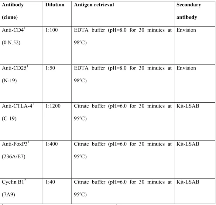
Secctions of 4 μm from routinely processed paraffin embedded blocks were desparaffinized and dehydrated. The sections were deparaffinized by immersion in xylene, and this was followed by immersion in alcohol and then incubation with 3% hydrogen peroxide diluted in Tris-buffered saline (TBS) (pH 7.4) for 40 minutes. Antigen retrieval was obtained as described in Table 1. Endogenous peroxidase activity was blocked using 0.3% hydrogen peroxide. The slides were then incubated with the primary antibodies, all from Santa Cruz Biotecnology (Santa Cruz, CA) (Table 1) 18 hours at 4ºC. After washing in TBS, the sections were treated with the EnVision® + Dual Link System-HRP (Dako Corporation, Carpinteria, CA) or using LSAB®+system, HRP Peroxidase Kit (Dako). The sections were then incubated in 3,3’-Diaminobenzidine (DAB) (Dako) for 2 to 5 minutes. Finally, the sections were stained with Mayer´s hematoxylin and were covered. Negative controls were obtained by the omission of primary antibodies, which were substituted by 1% PBS-BSA and by non-immune rabbit (X0902, Dako) or mouse (X501-1, Dako) serum.
Cell counting and statistical analysis
(4740680000000-Netzmikrometer 12.5x, Carl Zeiss, Göttingen, Germany). The percentage of positive cells in the stroma was calculated as the proportion of the total of immune/inflammatory cells.
Original article, submitted May 2008, to Oral Oncology
The cell densities/proportions were expressed as density per mm2 and percentages (mean ± SD). A P value of less than 0.05 was considered to be statistically significant. The comparative analyses between experimental groups were performed using the non-parametric Kuskall Wallis, followed by Dunn test, and/or Mann-Whitney.
Original article, submitted May 2008, to Oral Oncology
Results
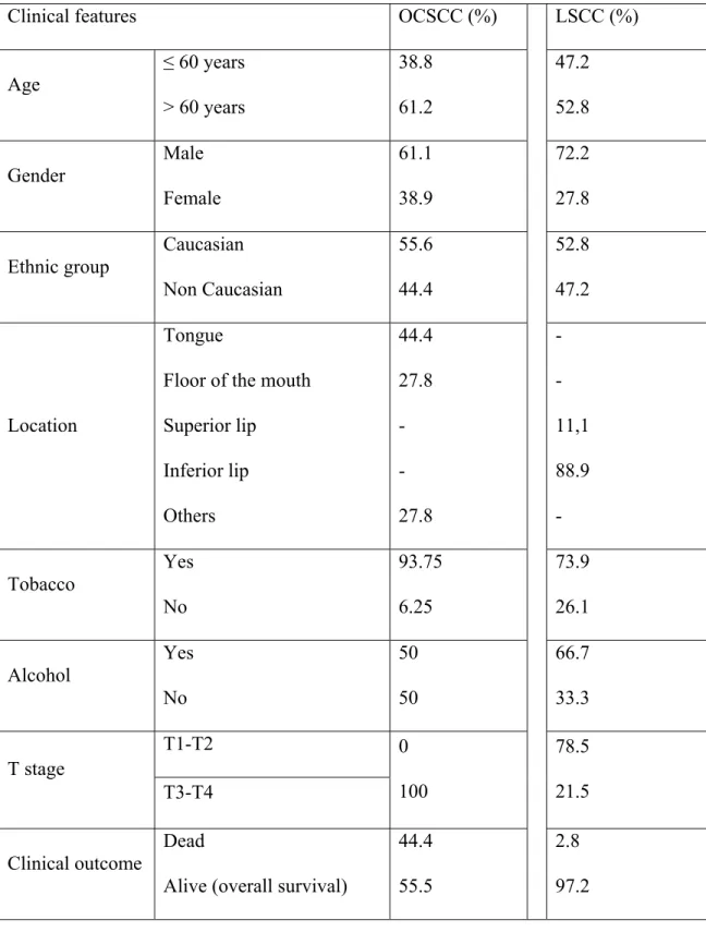
The main clinical features of our series of 18 patients with oral OCSCC and 36 patients with LSCC are summarized in Table 2.
In OCSCC and LSCC, CD4, CD25, CTLA-4 and FoxP3 positive cells were distributed throughout the tumoral stroma. All stained cells had a mononuclear appearance in both groups (Fig. 1A-1D).
We observed higher percentual of CD4 (P=0.019) and FoxP3 (P=0.040) positive cells in OCSCC samples when compared with LSCC (Fig. 2A and 2D). On the other hand, lower percentual of CTLA-4+ cells were observed in OCSCC group (P<0.0001) (Fig. 2C). Similar numbers of CD25+ cells were observed in both groups (Fig. 2B). When OCSCC samples were dicotomized in metastatic and non-metastatic groups, no statistical significance was observed for all cells markers comparing these two groups. Moreover, positive correlations were observed when analyzing CD4 and FoxP3 cells population (P=0.027); CD25 and CD4 (P=0.099) and CD25 with FoxP3 (P=0.089) (Pearson Chi-square test).
Original article, submitted May 2008, to Oral Oncology
To analyze the relationship of T regulatory cell markers and proliferative index of tumoral cells, the values were dicotomized in high and low groups by using the median. We obtained that samples with high counts of CD4 (12.23 ± 2.01) (P=0.015) and FoxP3 (12.50 ± 1.99) (P=0.018) exhibited greater proliferative index than samples with low counts (6.39 ± 1.03; 7.27 ± 1.04; respectively for CD4 and FoxP3). No significant differences in the proliferative index were observed when high and low CD25 and CTLA-4 groups were compared.
Original article, submitted May 2008, to Oral Oncology
showed significant increase of survival (144 ± 6 and 31 ± 4 months, respectively for high and low groups; P=0.006).
Original article, submitted May 2008, to Oral Oncology
Discussion
Original article, submitted May 2008, to Oral Oncology
FoxP3 expression was detected in the melanoma cells28 and various types of tumor cells29. However, we did not verified FoxP3 expression in neoplastic epithelial cells. It is also important to consider that the evaluation of the number of positive cells does not necessarily reflect the level of expression of these molecules at each cell. Indeed, FoxP3 expression can be influenced by different cytokines such as TGF-β, IL-10 or IL-27, 9, 27
. We have observed a slightly increase in the IL-10 concomitant with FoxP3 expression in OSCC samples (data not shown).
CTLA-4. It is under known that LSCC patients usually have a good prognosis and a low rate of regional lymph node metastasis and mortality when compared with oral cavity
Original article, submitted May 2008, to Oral Oncology
SCC31, 32. These results could be in part explained by recently demonstrated the function of CTLA-4 on destruction of tumor cells in vivo via interaction with B733. Moreover, the CTLA-4 expression seems not to be exclusive of Treg cells3, 6, 8. On the hand, we consider the double positive CD4/CTLA-4 population; we observed a poor prognosis in patients with high counts of these cells, corroborating the potential role of CTLA-4 blockade in the cancer immunotherapy10, 11, 14, 16, 20, 21.
We obtained that patients with high counts of CD4 and FoxP3 exhibited greater proliferative index. Consistent with these results patients with low counts of CD4 showed a significant increase of survival in relation to patients with high CD4 counts. Furthermore, we observed a tendency of groups with high counts of FoxP3 and CD25 to have lower survival compared with groups with low counts of these cells. When evaluating the double positive CD4/CD25 and CD4/FoxP3 cells we obtained that patients with high counts showed significant decrease of survival. Our results are corroborated by previous data showing an association of Tregs with inhibition of anti-tumoral immunity and consequently poor prognosis10-23.
Original article, submitted May 2008, to Oral Oncology
Acknowledgments
Original article, submitted May 2008, to Oral Oncology
References
1. Hanahan D, Lanzavecchia A, Mihich E. Fourteenth Annual Pezcoller Symposium: the novel dichotomy of immune interactions with tumors. Cancer Res 2003; 63(11): 3005-3008.
2. Oliveira-Neto HH, Leite AF, Costa NL, Alencar RC, Lara VS, Silva TA et al. Decrease in mast cells in oral squamous cell carcinoma: possible failure in the migration of these cells. Oral Oncol 2007; 43(5):484-490.
3. Akbar AN, Taams LS, Salmon M, Vukmanovic-Stejic M. The peripheral generation of CD4+ CD25+ regulatory T cells. Immunology 2003;109(3):319-25.
4. Wang HY, Lee DA, Peng G, Guo Z, Li Y, Kiniwa Y et al. Tumor-specific human CD4+ regulatory T cells and their ligands: implications for immunotherapy. Immunity 2004;20(1):107-118.
5. Wei WZ, Morris GP, Kong YC. Anti-tumor immunity and autoimmunity: a balancing act of regulatory T cells. Cancer Immunol Immunother 2004;53(2):73-78.
6. Wang RF. Regulatory T cells and innate immune regulation in tumor immunity. Springer Semin Immunopathol 2006;28(1):17-23.
7. Yamaguchi T, Sakaguchi S. Regulatory T cells in immune surveillance and treatment of cancer. Semin Cancer Biol 2006;16(2):115-23.
9. Chen W, Jen W, Hardegen N, Lei K, Marinos N, McGrady G et al. Conversion of peripheral CD4+ CD25 naive T cells to CD4+ CD25+ regulatory T cells by TGF-β induction of transcription factor FoxP3. J Exp Med 2003;198:1875-1886.
10.Hurwitz AA, Yu TF, Leach DR, Allison JP. CTLA-4 blockade synergizes with tumor-derived granulocyte-macrophage colony-stimulating factor for treatment of an experimental mammary carcinoma. Proc Natl Acad Sci U S A 1998;95(17):10067-10071.
11.Hurwitz AA, Foster BA, Kwon ED, Truong T, Choi EM, Greenberg NM et al. Combination immunotherapy of primary prostate cancer in a transgenic mouse model using CTLA-4 blockade. Cancer Res 2000;60(9):2444-2448.
12.Tartour E, Mosseri V, Jouffroy T, Deneux L, Jaulerry C, Brunin F et al. Serum soluble interleukin-2 receptor concentrations as an independent prognostic marker in head and neck cancer. Lancet 2001;357(9264):1263-1264.
13.Jones E, Dahm-Vicker M, Simon AK, Green A, Powrie F, Cerundolo V et al. Depletion of CD25+ regulatory cells results in suppression of melanoma growth and induction of autoreactivity in mice. Cancer Immun 2002;2:1-12.
15.Curiel TJ, Coukos G, Zou L, Alvarez X, Cheng P, Mottram P et al. Specific recruitment of regulatory T cells in ovarian carcinoma fosters immune privilege and predicts reduced survival. Nat Med 2004; 10(9):942-949.
16.Ghaderi A, Yeganeh F, Kalantari T, Talei AR, Pezeshki AM, Doroudchi M et al. Cytotoxic T lymphocyte antigen-4 gene in breast cancer. Breast Cancer Res Treat 2004;86(1):1-7.
17.Wolf D, Wolf AM, Rumpold H, Fiegl H, Zeimet AG, Muller-Holzner E, Deibl M, Gastl G, Gunsilius E, Marth C. The expression of the regulatory T cell-specific forkhead box transcription factor FoxP3 is associated with poor prognosis in ovarian cancer. Clin Cancer Res 2005;11(23):8326-8331.
18.Hiraoka N, Onozato K, Kosuge T, Hirohashi S. Prevalence of FOXP3+ regulatory T cells increases during the progression of pancreatic ductal adenocarcinoma and its premalignant lesions. Clin Cancer Res 2006;12(18):5423-5434.
19.Larkin J, Tangney M, Collins C, Casey G, O'Brien MG, Soden D et al. Oral immune tolerance mediated by suppressor T cells may be responsible for the poorer prognosis of foregut cancers. Med Hypotheses 2006;66(3):541-544.
20.Downey SG, Klapper JA, Smith FO, Yang JC, Sherry RM, Royal RE et al. Prognostic factors related to clinical response in patients with metastatic melanoma treated by CTL-associated antigen-4 blockade. Clin Cancer Res 2007;13(22):6681-6688.
compartment without affecting regulatory T-cell function. Clin Cancer Res 2007; 13(7):2158-2167.
22.Fu J, Xu D, Liu Z, Shi M, Zhao P, Fu B et al. Increased regulatory T cells correlate with CD8 T-cell impairment and poor survival in hepatocellular carcinoma patients. Gastroenterology 2007; 132(7): 2328-2339.
23.Miracco C, Mourmouras V, Biagioli M, Rubegni P, Mannucci S, Monciatti I et al. Utility of tumour-infiltrating CD25+FOXP3+ regulatory T cell evaluation in predicting local recurrence in vertical growth phase cutaneous melanoma. Oncol Rep 2007;18(5):1115-1122.
24.Strauss L, Bergmann C, Whiteside TL. Functional and phenotypic characteristics of D4+CD25highFoxp3+ Treg clones obtained from peripheral blood of patients with cancer. Int J Cancer 2007;121(11):2473-2483.
25.Chikamatsu K, Sakakura K, Whiteside TL, Furuya N. Relationships between regulatory T cells and CD8+ effector populations in patients with squamous cell carcinoma of the head and neck. Head Neck 2007;29(2):120-127.
26.Wang J, Ioan-Facsinay A, van der Voort EI, Huizinga TW, Toes RE. Transient expression of FOXP3 in human activated nonregulatory CD4+ T cells. Eur J Immunol 2007;37:129–138.
27.Zheng Y, Rudensky AY. Foxp3 in control of the regulatory T cell lineage. Nat Immunol. 2007;8(5):457-462.
29.Karanikas V, Speletas M, Zamanakou M, Kalala F, Loules G, Kerenidi T et al. Foxp3 expression in human cancer cells. J Transl Med 2008;6(1):19.
30.Wong YK, Chang KW, Cheng CY, Liu CJ. Association of CTLA-4 gene polymorphism with oral squamous cell carcinoma. J Oral Pathol Med 2006;35(1):51-54.
31.Antunes JLF, Biazevic MGH, Araujo ME, Tomita NE, Chinellato LEM, Narvai PC. Trends and spatial distribution of oral cancer mortality in São Paulo, Brazil, 1980-1998. Oral Oncol 2001;37(4):345-350.
32.Vartanian JG, Carvalho AL, Filho MJA, Júnior MH, Magrin J, Kowalski LP. Predictive factors and distribution of lymph node metastasis in lip cancer patients and their implications on the treatment of the neck. Oral Oncol 2004; 40(2): 223-227.
[Original article, submitted May 2008, to Oral Oncology
Table 1: Antibodies and protocol of immunohistochemical reaction
Antibody (clone)
Dilution Antigen retrieval Secondary
antibody Anti-CD41
(0.N.52)
1:100 EDTA buffer (pH=8.0 for 30 minutes at 98ºC)
Envision
Anti-CD251 (N-19)
1:50 EDTA buffer (pH=8.0 for 30 minutes at 98ºC)
Envision
Anti-CTLA-41 (C-19)
1:1200 Citrate buffer (pH=6.0 for 30 minutes at 95ºC)
Kit-LSAB
Anti-FoxP31 (236A/E7)
1:400 Citrate buffer (pH=6.0 for 30 minutes at 95ºC)
Kit-LSAB
Cyclin B12 (7A9)
1:40 Citrate buffer (pH=6.0 for 30 minutes at 95ºC)
Kit-LSAB
1
Table 2 Main clinical findings of patients with OSCC (oral cavity and lip): Clinical features OCSCC (%) LSCC (%)
Age
≤ 60 years > 60 years
38.8 61.2 47.2 52.8 Gender Male Female 61.1 38.9 72.2 27.8 Ethnic group Caucasian Non Caucasian 55.6 44.4 52.8 47.2 Location Tongue
Floor of the mouth Superior lip Inferior lip Others 44.4 27.8 - - 27.8 - - 11,1 88.9 - Tobacco Yes No 93.75 6.25 73.9 26.1 Alcohol Yes No 50 50 66.7 33.3 T1-T2 T stage T3-T4 0 100 78.5 21.5 Clinical outcome Dead
Alive (overall survival)
44.4 55.5
Survival time ≥ 48 months < 48 months
0 100
Original article, submitted May 2008, to Oral Oncology
Legends of Figures
Figure 1 Representative immunostaning for CD4 (A), CD25 (B), CTLA-4 (C) and Foxp3 (D) in oral squamous cell carcinoma of OSCC. Positive cells for CD4 (100X), CD25 (100X), CTLA-4 (100X) and Foxp3 (X400) distributed throughout the tumoral stroma.
A B
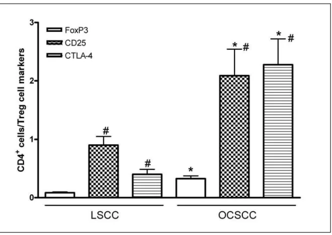
Figure 3 CD4/Treg markers ratio in primary squamous cell carcinoma of lip (LSCC) and oral cavity (OCSCC). The total number of CD4 positive cells (mm2) was divided by number of CD25, CTLA-4 and FoxP3 positive cells (mm2) to obtain CD4/Treg markers ratio in each respective group.
Considerações Finais
A imunidade inata e adaptativa desempenha um papel essencial na vigilância imunológica e destruição tumoral. Ambos os efeitos das respostas imunológicas são regulados por diferentes tipos celulares, dentre os quais se incluem as células T regulatórias (Treg) [9-13]. Em tumores humanos, as células Treg estão relacionadas à supressão da resposta imunológica podendo então contribuir para o crescimento tumoral [12, 16-29].
No presente estudo foi encontrado um elevado percentual de células positivas para CD4 e Foxp3 em amostras de CECB quando comparado ao índice de marcação no CCEL. Além disso, a relação CD4/Foxp3, CD4/CD25, e CD4/CTLA-4 foi significativamente maior no CECB o que indica uma maior expressão fenotípica desta célula nesta lesão. Resultados semelhantes têm sido encontrados revelando aumento da população de células Treg em outros tipos de câncer, tais como pâncreas [24], tumores ovarianos [23], melanoma metastático [29] e neoplasias malignas de cabeça e pescoço [18, 30,31]. Nossos dados, também indicam que a maior população celular dentro das células CD4 positivas são as células CD25 para CCEL e igualmente CD25 e CTLA-4 para o CECB sugerindo um diferente perfil celular nestas lesões.
CD4/Foxp3 sugere que outros tipos celulares poderiam contribuir para a expressão do Foxp3 em ambas as lesões. De fato, a expressão do Foxp3 foi transitoriamente induzida em células humanas através da ativação do receptor T celular [32,33]. Recentemente, a expressão do Foxp3 foi atribuída estar restrita a linhagem de células T. No entanto, alguns trabalhos observaram a expressão do Foxp3 em células neoplásicas do melanoma [34] e vários outros tipos de células tumorais [35]. Entretanto, no presente trabalho, não foi observado a expressão deste marcador em células epiteliais neoplásicas. Também é importante considerar que a avaliação do número de células positivas não reflete necessariamente o nível de expressão das moléculas de cada célula. Soma-se a isso, o fato da expressão do Foxp3 poder ser influenciada por diferentes citocinas tais como TGF-Β, IL-10 ou IL-2 [13, 15, 33].
O CTLA-4 inibe a ativação da célula T e também finaliza a resposta da célula T pelo bloqueio de sinais estimuladores via CD28. Em geral, o CTLA-4 não está expresso em células T CD4+ CD25- mas a sua estimulação sobre as células T pode ser induzida por diferentes mecanismos [11, 13 14]. Muitas evidências apontam para a importância do bloqueio do CTLA-4 para a prevenção de malignidades e invasão metastática [16, 17, 20, 22, 26, 27]. Além disso, no CECB, o polimorfismo A/A do gene CTLA-4, que resulta em alto fenótipo produtor, foi associado a uma menor sobrevida [36].
apresentam bom prognóstico e baixo índice de mortalidade e de metástases em linfonodos regionais quando comparado com CECB [37,38]. Estes resultados podem em parte ser explicados pela recente demonstração da função do CTLA-4 sobre a destruição das células tumorais in vivo via interação com B7. Além disso, a expressão do CTLA-4 não é exclusiva de células Treg [9, 12, 14]. Neste sentido, foi considerado que em pacientes com elevadas contagens de CD4/CTLA-4 observou-se um prognóstico ruim, o que vai ao encontro do potencial papel do bloqueio do CTLA-4 na imunoterapia para o câncer [16, 17, 20, 22, 26, 27].
Os pacientes com elevadas contagens de CD4 e Foxp3 exibiram um maior índice de proliferatividade. Em acordo com estes resultados, pacientes com baixas contagens de CD4 revelaram um aumento significativo da sobrevida em relação aos pacientes com altas contagens. Além disso, observou-se uma tendência dos grupos com elevadas contagens de Foxp3 e CD25 apresentarem menor sobrevida quando comparados com grupos com baixas contagens destas células. Quando foi avaliada a dupla positividade para CD4/CD25 e CD4/Foxp3 observou-se que pacientes com maiores contagens mostraram redução significativa da sobrevida. Tais achados são semelhantes a estudos prévios que descreveram uma associação de células Treg com a inibição da imunidade anti-tumoral e conseqüente prognóstico desfavorável [16-29].
Conclusões
Referências Bibliográficas
1-Instituto Nacional do Câncer [Base de dados na Internet]. Brasil: Ministério da Saúde [acesso em 2008 abril 02] Estimativa 2008 - Incidência de Câncer no Brasil. Disponível em: http://www.inca.gov.br/estimativa/2008.
2-Chen JK, Eisenberg E, Krutchkoff DJ, Katz RV. Changing trends in oral câncer in the United States, 1935 to 1985: a Connecticut study. J Oral Maxillofac Surg 1991; 49 (11): 1152-1158.
3-Jordan RC, Daley T. Oral squamous cell carcinoma: new insights. J Canad Dent Ass 1997; 63 (7): 517-518.
4-Scully C, Field JK, Tanzawa H. Genetic aberrations in oral or head and neck squamous cell carcinoma 2: chromosomal aberrations. Oral Oncol. 2000; 36 (4): 311-27.
5-Moore SR, Johnson NW, Pierce AM, Wilson DF. The epidemiology of lip cancer: a review of global incidence and etiology. Oral diseases 1999; 5 (3): 185-195.
7-Hanahan D, Lanzavecchia A, Mihich E. Fourteenth Annual Pezcoller Symposium: the novel dichotomy of immune interactions with tumors. Cancer Res 2003; 63(11): 3005-3008.
8-Oliveira-Neto HH, Leite AF, Costa NL, Alencar RC, Lara VS, Silva TA et al. Decrease in mast cells in oral squamous cell carcinoma: possible failure in the migration of these cells. Oral Oncol 2007; 43(5):484-490.
9-Akbar AN, Taams LS, Salmon M, Vukmanovic-Stejic M. The peripheral generation of CD4+ CD25+ regulatory T cells. Immunology 2003;109(3):319-25.
10-Wang HY, Lee DA, Peng G, Guo Z, Li Y, Kiniwa Y et al. Tumor-specific human CD4+ regulatory T cells and their ligands: implications for immunotherapy. Immunity 2004;20(1):107-118.
11-Wei WZ, Morris GP, Kong YC. Anti-tumor immunity and autoimmunity: a balancing act of regulatory T cells. Cancer Immunol Immunother 2004;53(2):73-78.
12-Wang RF. Regulatory T cells and innate immune regulation in tumor immunity. Springer Semin Immunopathol 2006;28(1):17-23.
14-Chen L. Co-inhibitory molecules of the B7-CD28 family in the control of T-cell immunity. Nat Rev Immunol 2004;4(5):336-347.
15-Chen W, Jen W, Hardegen N, Lei K, Marinos N, McGrady G et al. Conversion of peripheral CD4+ CD25 naive T cells to CD4+ CD25+ regulatory T cells by TGF-β induction of transcription factor FoxP3. J Exp Med 2003;198:1875-1886.
16-Hurwitz AA, Yu TF, Leach DR, Allison JP. CTLA-4 blockade synergizes with tumor-derived granulocyte-macrophage colony-stimulating factor for treatment of an experimental mammary carcinoma. Proc Natl Acad Sci U S A 1998;95(17):10067-10071.
17-Hurwitz AA, Foster BA, Kwon ED, Truong T, Choi EM, Greenberg NM et al. Combination immunotherapy of primary prostate cancer in a transgenic mouse model using CTLA-4 blockade. Cancer Res 2000;60(9):2444-2448.
18-Tartour E, Mosseri V, Jouffroy T, Deneux L, Jaulerry C, Brunin F et al. Serum soluble interleukin-2 receptor concentrations as an independent prognostic marker in head and neck cancer. Lancet 2001;357(9264):1263-1264.
20-Phan GQ, Yang JC, Sherry RM, Hwu P, Topalian SL, Schwartzentruber DJ et al. Cancer regression and autoimmunity induced by cytotoxic T lymphocyte-associated antigen 4 blockade in patients with metastatic melanoma. Proc Natl Acad Sci U S A 2003;100(14):8372-8377.
21-Curiel TJ, Coukos G, Zou L, Alvarez X, Cheng P, Mottram P et al. Specific recruitment of regulatory T cells in ovarian carcinoma fosters immune privilege and predicts reduced survival. Nat Med 2004; 10(9):942-949.
22-Ghaderi A, Yeganeh F, Kalantari T, Talei AR, Pezeshki AM, Doroudchi M et al. Cytotoxic T lymphocyte antigen-4 gene in breast cancer. Breast Cancer Res Treat 2004;86(1):1-7.
23-Wolf D, Wolf AM, Rumpold H, Fiegl H, Zeimet AG, Muller-Holzner E, Deibl M, Gastl G, Gunsilius E, Marth C. The expression of the regulatory T cell-specific forkhead box transcription factor FoxP3 is associated with poor prognosis in ovarian cancer. Clin Cancer Res 2005;11(23):8326-8331.
25-Larkin J, Tangney M, Collins C, Casey G, O'Brien MG, Soden D et al. Oral immune tolerance mediated by suppressor T cells may be responsible for the poorer prognosis of foregut cancers. Med Hypotheses 2006;66(3):541-544.
26-Downey SG, Klapper JA, Smith FO, Yang JC, Sherry RM, Royal RE et al. Prognostic factors related to clinical response in patients with metastatic melanoma treated by CTL-associated antigen-4 blockade. Clin Cancer Res 2007;13(22):6681-6688.
27-Fecci PE, Ochiai H, Mitchell DA, Grossi PM, Sweeney AE, Archer GE et al. Systemic CTLA-4 blockade ameliorates glioma-induced changes to the CD4+ T cell compartment without affecting regulatory T-cell function. Clin Cancer Res 2007; 13(7):2158-2167.
28-Fu J, Xu D, Liu Z, Shi M, Zhao P, Fu B et al. Increased regulatory T cells correlate with CD8 T-cell impairment and poor survival in hepatocellular carcinoma patients. Gastroenterology 2007;132(7):2328-2339.
30-Strauss L, Bergmann C, Whiteside TL. Functional and phenotypic characteristics of D4+CD25highFoxp3+ Treg clones obtained from peripheral blood of patients with cancer. Int J Cancer 2007;121(11):2473-2483.
31-Chikamatsu K, Sakakura K, Whiteside TL, Furuya N. Relationships between regulatory T cells and CD8+ effector populations in patients with squamous cell carcinoma of the head and neck. Head Neck 2007;29(2):120-127.
32-Wang J, Ioan-Facsinay A, van der Voort EI, Huizinga TW, Toes RE. Transient expression of FOXP3 in human activated nonregulatory CD4+ T cells. Eur J Immunol 2007;37:129–138.
33-Zheng Y, Rudensky AY. Foxp3 in control of the regulatory T cell lineage. Nat Immunol. 2007;8(5):457-462.
34-Ebert LM, Tan BS, Browning J, Svobodova S, Russell SE, Kirkpatrick N et al. The regulatory T cell-associated transcription factor FoxP3 is expressed by tumor cells. Cancer Res 2008;68(8): 3001-3009.
36-Wong YK, Chang KW, Cheng CY, Liu CJ. Association of CTLA-4 gene polymorphism with oral squamous cell carcinoma. J Oral Pathol Med 2006;35(1):51-54.
37-Antunes JLF, Biazevic MGH, Araujo ME, Tomita NE, Chinellato LEM, Narvai PC. Trends and spatial distribution of oral cancer mortality in São Paulo, Brazil, 1980-1998. Oral Oncol 2001;37(4):345-350.
38-Vartanian JG, Carvalho AL, Filho MJA, Júnior MH, Magrin J, Kowalski LP. Predictive factors and distribution of lymph node metastasis in lip cancer patients and their implications on the treatment of the neck. Oral Oncol 2004;40(2):223-227.


