Abstract
Background
Congenital heart disease (CHD) is the most prevalent type of birth defect in human, with high morbidity in infant. Several genes essential for heart development have been identified. GATA4 is a pivotal transcription factor that can regulate the cardiac development. Many GATA4 mutations have been identified in patients with different types of CHD.
Aims
In this study, the NKX2-5, HAND1 and GATA4 coding regions were sequenced in a family spanning three generations in which seven patients had CHD. Disease-causing potential variation in this family was evaluated by bioinformatics programs and the transcriptional activity of mutant protein was analyzed by the dual luciferase reporter assay.
Results
A novel GATA4 mutation, c.C931T (p.R311W), was identified and co-segregated with the affected patients in this family. The bioinformatics programs predicted this heterozygous mutation to be deleterious and the cross-species alignment of GATA4 sequences showed that the mutation occurred within a highly conserved amino acid. Even though it resided in the nuclear localization signal domain, the mutant protein didn’t alter its intracellular distribu-tion. Nevertheless, further luciferase reporter assay demonstrated that the p.R311W muta-tion reduced the ability of GATA4 to activate its downstream target gene.
Conclusions
Our study identified a novel mutation in GATA4 that likely contributed to the CHD in this fam-ily. This finding expanded the spectrum of GATA4 mutations and underscored the patho-genic correlation between GATA4 mutations and CHD.
OPEN ACCESS
Citation:Zhang X, Wang J, Wang B, Chen S, Fu Q, Sun K (2016) A Novel Missense Mutation of GATA4 in a Chinese Family with Congenital Heart Disease. PLoS ONE 11(7): e0158904. doi:10.1371/journal. pone.0158904
Editor:Yong-Bin Yan, Tsinghua University, CHINA
Received:March 15, 2016
Accepted:June 23, 2016
Published:July 8, 2016
Copyright:© 2016 Zhang et al. This is an open access article distributed under the terms of the
Creative Commons Attribution License, which permits unrestricted use, distribution, and reproduction in any medium, provided the original author and source are credited.
Data Availability Statement:All relevant data are within the paper and its Supporting Information files.
Funding:This work was supported by the National Natural Science Foundation of China (No.81371893 to QF), the Innovation Program of Shanghai Municipal Education Commission (No.13ZZ086 to QF), the research fund of Shanghai Municipality Health Bureau (No.20114y072 to JW). The funders had no role in study design, data collection and analysis, decision to publish, or preparation of the manuscript.
Introduction
Congenital heart disease (CHD) is the most common developmental abnormality, with a post-natal incidence of 1% [1]. During the development of heart, multiple molecular pathways are involved. Emerging evidences have indicated the crucial role of genetic defects in the occur-rence of CHD. Pathogenic gene mutations, chromosomal aberrations and microRNA lesion can all result in CHD [2]. Tetralogy of Fallot (TOF) is the most common type of cyanotic CHD [3]. Several transcription factors essential for cardiac development have been identified in caus-ing TOF, includcaus-ing NKX2-5, HAND1 and GATA4 [4–7].
GATA4 belongs to a family of DNA binding proteins. They share highly conserved zinc fin-ger domains and target the consensus DNA sequence“GATA”element [8]. In vertebrates, six members of the GATA family have been identified. GATA1, GATA2 and GATA3 are mainly expressed in the hematopoietic cells, whereas GATA4, GATA5 and GATA6 are predominantly expressed in tissues such as the heart, liver and gonads [9]. During the development, GATA4 is highly expressed in the embryonic heart and adult cardiomyocytes. By cooperating with target genes, GATA4 can regulate multiple processes essential for normal cardiac morphogenesis [10].
Small changes of GATA4 can dramatically influence cardiac development and embryonic survival. In mice, GATA4 homozygous knockout embryos die in early embryonic stage because of failed heart tube formation and abnormal ventral folding [11]. Mouse embryos expressing reduced GATA4 protein display congenital cardiac defects including common atrioventricular canal, double outlets of the right ventricle and myocardial hypoplasia [12]. In Xenopus embryos, GATA4 is sufficient to induce functional cardiomyocyte differentiation in embryonic ectoderm [13]. Functional experiments in chick, fly and fish also demonstrate the importance of GATA4 in their heart development [14]. In human, GATA4 mutations can lead to many kinds of congenital heart diseases [15]. Till now, more than 90 missense mutations have been reported in CHD patients (S1 Table).
In our study, a novel heterozygous GATA4 mutation, c.C931T (p.R311W), was identified in a family spanning three generations and seven individuals in this family had CHD. All family members were negative for mutation screening in NKX2-5 and HAND1. The proband of this family was affected with TOF. Detailed clinical and genetic evaluations were reviewed for all family members, and displayed an autosomal dominant pattern of inheritance. The p.R311W mutation was functionally analyzed, and in vitro assays showed that the mutation compro-mised the ability of GATA4 to transactivate its downstream target gene. This finding expanded the spectrum of GATA4 mutations and supported the essential role of GATA4 in cardiac development.
Materials and Methods
Study Subjects
our study, a signed informed consent was obtained from their guardians. This research was approved by the Ethics Committee of Shanghai Children's Medical Center.
Genetic studies
Peripheral blood samples of all cases were obtained and genomic DNA was extracted using QIAmp DNA Blood Mini Kit (QIAGEN, Hilden, Germany) by standard protocols. DNA was quality tested by optical density (OD) 260/280 nm ratios, quantified by UV spectrophotometry (NanoDrop Technologies, Wilmington, DE, USA), and stored at -20°C until use. All exon regions and flanking intronic sequences of NKX2-5, HAND1and GATA4 were amplified by polymerase chain reaction (PCR) to screen gene mutations.
Multiple sequence alignments of GATA4
To evaluate the evolutionary conservation of the mutation, protein sequences of GATA4 from 7 animal species including human (Homo sapiens: NP_001295022), pig (Sus scrofa:
NP_999458), cattle (Bos taurus: NP_001179806), rat (Rattus norvegicus: NP_653331), mouse (Mus musculus: NP_001297539), rabbit (Oryctolagus cuniculus: XP_008247110), dog (Canis lupus: NP_001041577) were aligned using the BioEdit program.
Disease-causing potential prediction of the GATA4 mutation
To predict the disease-causing potential of GATA4 mutation in our report, several bioinfor-matic programs as MutationTaster (http://www.mutationtaster.org), PolyPhen-2 (http:// genetics.bwh.harvard.edu/pph2) and SIFT (http://sift.jcvi.org/) were used. Swiss PdbViewer software version 3.7 was applied to analyze the three-dimensional conformation of GATA4. We selected the neighboring residues of amino 311 for H-bond computation to assess the influ-ence of p.R311W mutation.
Plasmid Constructs and Site Directed Mutagenesis
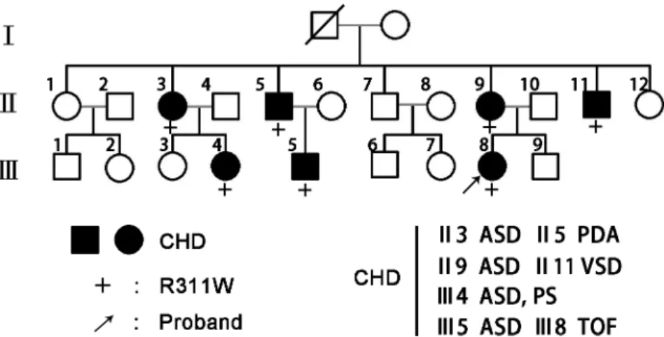
Human GATA4 cDNA (ENST00000532059) was PCR amplified and cloned into pcDNA3.1 (+) expression vector. The identified mutation was introduced using the QuikChange II Site-Fig 1. GATA4 mutation segregates with familial CHD.Family members in the pedigree chart are indicated by generations (I-III) and numbers. Males are symbolized by squares while females are circles and the symbols with a slash indicate the deceased members. Patients affected with CHD are designated with filled symbols and the plus signs (+) represent carriers of the heterozygous R311W mutation. The proband of the family is indicated by arrow.
Directed Mutagenesis Kit (Stratagene, La Jolla, CA) following manufacturer’s instructions. Both wild-type and mutant inserts were sequenced in sense and antisense directions to confirm the desired sequence and exclude any further base exchanges.
Cell Culture and Transfection
Hela cell line was purchased from the Type Culture Collection of the Chinese Academy of Sciences (Shanghai, China) and cultured in the DMEM medium supplemented with 10% FBS, penicillin (100 units/ml) and streptomycin (100μg/ml). Cells were incubated at 37°C in a humid atmosphere
with 5% CO2, 95% air. Plasmids were transfected into Hela cells with Lipofectamine 3000 reagent (Invitrogen, Carlsbad, CA) using the methodology recommended by the manufacturer.
Western Blot and Immunofluorescence
For overexpression experiments, Hela cells were transfected with pcDNA3.1-GATA4-wt or mutant plasmid DNA in 24-well plates. To confirm that GATA4 protein was expressed in tran-siently transfected cells, western blot analysis was carried out. Cells were harvested 48hr after transfection and lysed in 1×SDS buffer. Cell lysates were analyzed by sodium dodecyl sulfate-polyacrylamide gel electrophoresis and transferred onto PVDF membranes (Amersham Biosci-ences, Piscataway, USA). Blocking of the membrane was done with 5% BSA in Tris-buffered saline containing 0.1% Tween-20. Then we incubated the membrane with anti-human GATA4 antibody (Bioworld, Nanjing, China) and anti-GAPDH antibody (Bioworld) at a dilution of 1:1000. The antigen-antibody complex was then incubated with horseradish peroxidase-conju-gated secondary antibodies (Bioworld) and visualized with an ImageQuant LAS 4000 (GE Healthcare life science, USA). For immunofluorescence, Hela cells which have been transfected with GATA4 wide-type or mutant plasmid were fixed using 4% paraformaldehyde/PBS for 20min, permeabilized with 1% Triton X-100/PBS for 10min, and blocked using 1% BSA/PBS solution for 30min. Treated cells were incubated with anti-human GATA4 antibody at a dilu-tion of 1:1000 and then with fluorescein isothiocyanate conjugated secondary antibody for 1hr. The cells were observed using a Laser Scanning Confocal Microscope (Leica, Germany).
Luciferase Assay
The human ANF promoter from -644 to +35 was PCR amplified and inserted into the pGL4.10 vector (Promega, Madison, USA) to produce ANF-Luc [4]. To analyze the transcrip-tional activity of GATA4 protein, Hela cells were transfected with the human ANF-Luc plas-mid, GATA4-pcDNA3.1 plasmid (wide-type, R311W mutant) as well as an internal control reporter plasmid pRL-TK (Promega). Briefly,*1.0 × 10⁵cells were seeded per well in 24-well
plates and incubated at 37°C in a CO2incubator. At*80% confluence, the cells were
cotrans-fected with 200 ng of pGL4.10-ANF luciferase reporter, 200 ng of pcDNA3.1-GATA4 plasmids (wild-type or R311W mutant) and 5 ng of pRL-TK. Thirty six hours after transfection, the luciferase activity of cells was measured with Dual-Glo luciferase assay system (Promega) according to the manufacturer’s instructions. Triple transfection experiments were performed three times for wild-type and GATA4 mutant. The student’s t-test was used for statistical anal-ysis, and a P-value<0.05 was considered to be statistically significant.
Results
Identification of GATA4 mutation
same mutation was present in all affected family members, but absent in unaffected individuals (Fig 1). In addition, we screened the NKX2-5 and HAND1 genes in this family; no mutations were identified. The multiple sequence alignments of GATA4 protein showed that the muta-tion occurred within a highly conserved amino acid, as presented inFig 2, suggesting its critical function.
Disease-causing potential of the GATA4 mutation
The c.C931T mutation of GATA4 was predicted by SIFT to be damaging with a SIFT score of 0 which suggested a high‘security’of the prediction. Meanwhile, the pathogenicity of the muta-tion was also supported by Polyphen-2 and Mutamuta-tiontaster with scores of nearly 1, predicting the mutation to be probably damaging and disease-causing, respectively (Fig 3A). High patho-genicity scores calculated by different prediction software suggested the deleterious effect of the mutation.
To further explore the functional effect of p.R311W, we used Swiss PdbViewer to predict the three-dimensional structure of GATA4 protein. Results showed that Arg311 was inclined to form multiple hydrogen bonding (H-bonds) and could make direct H-bond with Met310 (Fig 3B). Meanwhile, arginine was polar and positively charged which was ideal for binding negatively charged groups, e.g., nucleic acids. A model of GATA4-DNA binding domain was predicted by using the temple of highly conserved zinc finger domains of GATA3 (PDB code 3DFX with 80% sequence identity,Fig 3C). As shown inFig 3C and 3D, p.R311 located in the DNA-binding domain. When arginine was mutated to the nonpolar hydrophobic amino acid tryptophan, the charge of the residue altered and might reduce GATA4 protein-DNA binding activity.
Nuclear localization of GATA4 mutant protein
GATA4 is a nucleic transcription factor and localizes completely in the nucleus [16]. The p. R311W mutation falls within the highly conserved nuclear localization signal domain, a region that is of great importance to protein location. We hypothesized that as a consequence of this mutation, the protein may have altered intracellular distribution, which would prevent GATA4 Fig 2. Distribution of the identified GATA4 mutation and multiple sequence alignment of GATA4 protein.(A) Sequencing results of the GATA4 mutation. The arrow indicates the heterozygous nucleotides of C/T. (B) A schematic diagram of GATA4 shows the location of the p.R311W mutation. (C) Multiple sequence alignment of the GATA4 indicates that residue 311 is highly evolutionarily conserved in mammals.
from properly functioning. To test this hypothesis, we performed immunofluorescence stain-ing. However, the result showed that the wild-type and the mutant GATA4 proteins translo-cated completely into the nucleus with normal distribution (Fig 4).
Decreased transcriptional activity of GATA4 mutant protein
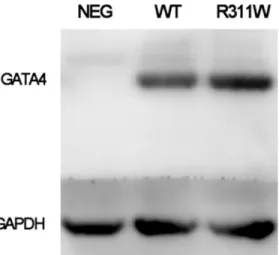
The expression of GATA4 plasmid in Hela cells was quantified by western blot and there was no significant difference between the wide-type and mutant protein (Fig 5,S1 Fig). It has been Fig 3. Disease-causing potential of the GATA4 mutation.(A) Functional significance of mutation prediction done by SIFT, PolyPhen-2 and MutationTaster. (B) Three-dimensional structure of GATA4 protein predicted by Swiss-PdbViewer. Upper panel is the reference structure; the lower panel is with arginine in position 311 mutated to tryptophan. (C) Double GATA4 bound to a tandem GATA4 binding site. Amino acid in position 311 is highlighted as yellow cube. (D) A schematic diagram of GATA4 protein.
doi:10.1371/journal.pone.0158904.g003
Fig 4. Normal subcellular distribution of GATA4 mutant protein.The immunofluorescence staining of the transfected HeLa cells shows that the GATA4 p.R311W mutant protein doesn’t change its nuclear
distribution.
previously demonstrated that ANF is one of the direct cardiac downstream target genes of GATA4 [17]. To determine whether p.R311W affected the functional activity of GATA4, ANF promoter luciferase reporter together with either wide-type or mutant GATA4 plasmid were co-transfected to HeLa cells. As shown inFig 6, the p.R311W plasmid present a significantly reduced transactivation of the ANF promoter (P<0.05), demonstrating a diminished target gene activation of the mutant protein.
Discussion
In this study, we identified a novel heterozygous GATA4 mutation p.R311W. Multiple sequence alignment of GATA4 protein showed that the altered amino acid was highly con-served in mammals. Meanwhile, this mutation was predicted to be damaging or disease causing by several in silico bioinformatic tools (MutationTaster, polyphen2 and SIFT), suggesting that this site p.R311 was vital in the function of GATA4.
Fig 5. Expression levels of GATA4 wide-type and p.R311W mutant protein in Hela cells.Western blot exhibits equal amount of GATA4 p.R311W protein as compared to the wild-type. Protein loading is normalized by the level of GAPDH.
doi:10.1371/journal.pone.0158904.g005
Fig 6. Decreased transcriptional activity of GATA4 mutant protein.Relative luciferase activation of ANF-Luc in Hela cells shows significantly reduced transcriptional activity of the p.R311W mutant protein. (*, P<0.05).
As a crucial modulator, GATA4 is required for proper regulation of many genes expressed during heart development, includingα-myosin heavy chain,β-myosin heavy chain, brain natriuretic peptide and atrial natriuretic factor (ANF) [9,18]. GATA4 contains regulatory ele-ments of these genes and can potently activate their promoters. In our research, Dual-Glo lucif-erase assay was carried out to evaluate the effect of p.R311W on the transactivational function of GATA4 and the result revealed a significantly reduced transcriptional activity on the down-stream gene.
Human GATA4 maps to a region of chromosome 8 and consists of 7 exons encoding 442 amino acids. The functional GATA4 protein comprises of two independent transcriptional activation domains (TAD) required for transcriptional activity, two highly conserved zinc fin-ger domains (ZnF) essential for DNA sequence identification and protein-DNA binding, and one nuclear localization signal (NLS) region associated with the subcellular trafficking and dis-tribution of GATA4 [19]. The GATA4 mutation, p.R311W, found in our study is identified in NLS region and may disrupt the normal subcellular localization of GATA4. We performed immunofluorescence staining to determine whether the p.R311 was necessary for the nuclear import of GATA4. The result showed that the mutant GATA4 protein translocated normally into the nucleus. Several mutations in NLS region had been identified in CHD patients (S1 Table) and some of the mutant proteins still accumulated to the nuclei (S2 Fig) [20]. All these findings suggested that not all the mutations in NLS region were sufficient to inhibit GATA4 nuclear targeting. This was consistent with the results revealed by Philips, et al. 2007 [21] that four amino acids Arg283, Arg284, Arg318, Arg320 were essential for GATA4 nuclear localiza-tion and proteins with individual mutated site in this region all showed normal nuclear distribution.
Besides the NLS region, p.R311 also locates in the GATA4 DNA-binding domain and can have a dual function in both nuclear import and DNA binding. Even though individual muta-tion is not sufficient to inhibit the nuclear import, it may impair GATA4 protein-DNA binding [21]. In our study, the amino acid at position 311 of GATA4 changes from arginine to trypto-phan. Arginine is ideal for binding negatively charged groups and is inclined to form multiple H-bonds. The opposite charge attraction, the length and flexibility of the side chain, and the ability to produce excellent hydrogen-bonding geometries with nucleobases or phosphate groups make arginine a vital residue for protein-DNA binding [22]. But tryptophan belongs to hydrophobic amino acid carrying aromatic ring. The substitution of strongly basic arginine by neutral tryptophan alters the structure and charge of the residue. The p.R311W mutation may reduce GATA4 protein-DNA binding activity and this is the genetic susceptible basis for the diminished transactivation of downstream target genes.
gene-because testing for p.R311W in new family members can be translated into effective preven-tion, accurate diagnosis and efficient treatment [29].
Conclusions
In this study, we reported a novel GATA4 mutation (p.R311W) in a three generation family with congenital heart disease. The present identification expands the mutation spectrum of GATA4 and provides new clues implicated in the mechanism of CHD.
Supporting Information
S1 Fig. Original western blot result for data inFig 5.M310V and Q316E are two of the GATA4 mutations located in NLS region which have been identified in CHD patients in other research. Western blot displays equal amount of GATA4 R311W/M310V/Q316E protein as compared to wild-type.
(TIF)
S2 Fig. Nuclear Localization of the GATA4 Mutants.The immunofluorescence of the trans-fected HeLa cells shows that the GATA4 R311W/M310V/Q316E mutant proteins don’t change their nuclear localization.
(TIF)
S1 Table. Missense mutations of GATA4 reported in patients with CHD.
(PDF)
Acknowledgments
We thank all the patients for their participating in this study. This work has been presented on the 2015 national conference of Chinese society of Laboratory Medicine.
Author Contributions
Conceived and designed the experiments: QF KS. Performed the experiments: XZ JW. Ana-lyzed the data: SC. Contributed reagents/materials/analysis tools: XZ BW. Wrote the paper: XZ JW XZ BW.
References
2. Bruneau BG. The developmental genetics of congenital heart disease. Nature. 2008; 451(7181):943– 8. doi:10.1038/nature06801PMID:18288184
3. Shah GS, Singh MK, Pandey TR, Kalakheti BK, Bhandari GP. Incidence of congenital heart disease in tertiary care hospital. Kathmandu Univ Med J 2008; 6(1):33–6.
4. Yang YQ, Gharibeh L, Li RG, Xin YF, Wang J, Liu ZM, et al. GATA4 Loss-of-Function Mutations Under-lie Familial Tetralogy of Fallot. Hum Mutat. 2013; 34(12):1662–71. doi:10.1002/humu.22434PMID:
24000169
5. Wang J, Lu Y, Chen H, Yin M, Yu T, Fu Q. Investigation of somatic NKX2-5, GATA4 and HAND1 muta-tions in patients with tetralogy of Fallot. Pathology. 2011; 43(4):322–6. doi:10.1097/PAT.
0b013e32834635a9PMID:21519287
6. Goldmuntz E, Geiger E, Benson DW. NKX2.5 mutations in patients with tetralogy of Fallot. Circulation. 2001; 104(21):2565–8. PMID:11714651
7. Lu CX, Gong HR, Liu XY, Wang J, Zhao CM, Huang RT, et al. A novel HAND2 loss-of-function mutation responsible for tetralogy of Fallot. International journal of molecular medicine. 2016; 37(2):445–51. doi:
10.3892/ijmm.2015.2436PMID:26676105
8. Molkentin JD, Kalvakolanu DV, Markham BE. Transcription factor GATA-4 regulates cardiac muscle-specific expression of the alpha-myosin heavy-chain gene. Mol Cell Biol. 1994; 14(7):4947–57. PMID:
8007990
9. Pikkarainen S, Tokola H, Kerkelä R, Heikki Ruskoaho. GATA transcription factors in the developing and adult heart. Cardiovascular research. 2004; 63(2):196–207. PMID:15249177
10. Agnihotri S, Wolf A, Munoz DM, Smith CJ, Gajadhar A, Restrepo A, et al. A GATA4-regulated tumor suppressor network represses formation of malignant human astrocytomas. J Exp Med. 2011; 208 (4):689–702. doi:10.1084/jem.20102099PMID:21464220
11. Chen MW, Sheng PY, Guo Y, Pan JH, Liu BL, Shen J, et al. GATA4 mutations in Chinese patients with congenital cardiac septal defects. Pediatr Cardiol. 2010; 31(1):85–9. doi:10.1007/s00246-009-9576-1
PMID:19915893
12. Pu WT, Ishiwata T, Juraszek AL, Ma Q, Izumo S. GATA4 is a dosage-sensitive regulator of cardiac morphogenesis. Dev Biol. 2004; 275(1):235–44. PMID:15464586
13. LatinkićBV, Kotecha S, Mohun TJ. Induction of cardiomyocytes by GATA4 in Xenopus ectodermal explants. Development. 2003; 130(16):3865–76. PMID:12835401
14. Epstein JA, Parmacek MS. Recent advances in cardiac development with therapeutic implications for adult cardiovascular disease. Circulation. 2005; 112(4):592–7. PMID:16043659
15. Yang YQ, Li L, Wang J, Liu XY, Chen XZ, Zhang W, et al. A Novel GATA4 Loss-of-Function Mutation Associated With Congenital Ventricular Septal Defect. Pediatr Cardiol. 2012; 33(4):539–46. doi:10. 1007/s00246-011-0146-yPMID:22101736
16. Wang E, Sun S, Qiao B, Duan W, Huang G, An Y, et al. Identification of Functional Mutations in GATA4 in Patients with Congenital Heart Disease. PLoS One. 2013; 8(4):e62138. doi:10.1371/journal.pone. 0062138PMID:23626780
17. Garg V, Kathiriya IS, Barnes R, Schluterman MK, King IN, Butler CA, et al. GATA4 mutations cause human congenital heart defects and reveal an interaction with TBX5. Nature. 2003; 424(6947):443–7. PMID:12845333
18. Heineke J, Auger-Messier M, Xu J, Oka T, Sargent MA, York A, et al. Cardiomyocyte GATA4 functions as a stress-responsive regulator of angiogenesis in the murine heart. J Clin Invest. 2007; 117 (11):3198–210. PMID:17975667
19. Liu XY, Wang J, Zheng JH, Bai K, Liu ZM, Wang XZ, et al. Involvement of a novel GATA4 mutation in atrial septal defects. Int J Mol Med. 2011; 28(1):17–23. doi:10.3892/ijmm.2011.638PMID:21373748
20. Takaya T, Kawamura T, Morimoto T, Ono K, Kita T, Shimatsu A, et al. Identification of p300-targeted acetylated residues in GATA4 during hypertrophic responses in cardiac myocytes. J Biol Chem. 2008; 283(15):9828–35. doi:10.1074/jbc.M707391200PMID:18252717
21. Philips AS, Kwok JC, Chong BH. Analysis of the Signals and Mechanisms Mediating Nuclear Traffick-ing of GATA-4. J Biol Chem. 2007; 282(34):24915–27. PMID:17548362
22. Fuhrmann J, Clancy KW, Thompson PR. Chemical biology of protein arginine modifications in epige-netic regulation. Chem Rev. 2015; 115(11):5413–61. doi:10.1021/acs.chemrev.5b00003PMID:
25970731