CASE REPORTS
UDC: 616-006.442:616.311
Non-Hodgkin Lymphomas of Oral Cavity
Mario Vicente-Barrero*, Ines Garcia-Castro", MilanKnezevic",Jose Juan Castellano-Reyes*, Francisco Garcia-Jimenez*, Maria del Carmen
Camacho-Garcia", Beatriz Baez-Acosta", SlobodanLoncarevics
Insular University Hospital, *Oral and Maxillofacial Surgery Department, "Oncclogy Department, *Pathological Anatomy Department, Las Palmas de Gran Canaria,
Spain
Non-Hodgkin lymphomas (NHL) often show up in an extranodal pattern, especially in the head and neck. Intraoral locations are much less frequent, particularly when they are single. This, in turn, can lead to a prolonged diagnosis and even to inadequate treatment. Different patients with initial extranodal location of NHL which were not previously diagnosed and in which it was manifested only intraoraly are presented in this paper. These cases are presented together with the additional examinations used for the early diagnosis and with the corresponding clinical pictures, as well as with the
overview of other cases from the available literature.
Key words: mouth neoplasms; lymphoma, non-Hodgkin; diagnosis.
Introduction
Malignant lymphomas form a heterogeneous group of neoplasms of the lymphoid tissue with different clinical courses, depending on the treatment and the prognosis (1). Non-Hodgkin lymphomas (NHL) often show up in extran-odal sites of the head and neck (2), but intraoral locations are much less frequent, especially when they are the cnly manifestations of this disease(3). This is why the diagnosis is frequently postponed and the treatment improper (2).
Various cases of NHL that had initial extranodal intra-oral presentations, not previously diagnosed, are presented in this paper. In some cases this type was the only presenta-tion of NHL. Such patients were sent to the Dental, Oral and Maxillofacial Surgery Department of the Insular Uni-versity Hospital of Las Palmas de Gran Canaria for review-ing and studreview-ing.
Case I
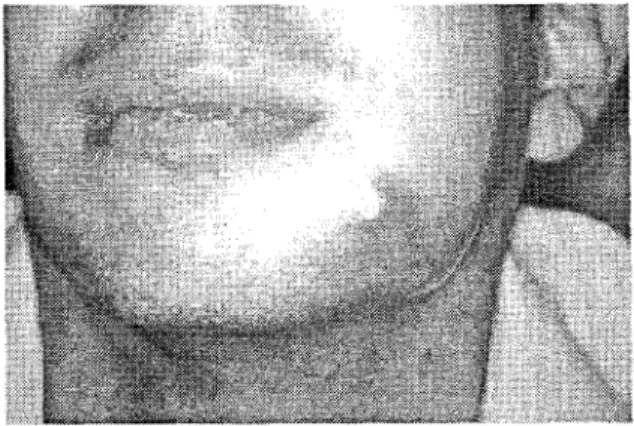
A 27-year-old male, with no medical history of imme-diate interest, sought treatment for the left perimandibular swelling that had developed over several weeks. He had previously been treated with antibiotics, with no positive re-sponse. Upon the exploration a hyperemic mass that fluctu-ated slightly at touch was found.Itwas painful and was in-tra-orally adhered to the external mandibular cortex.It was
near the fistulisation, and could also be felt intra-orally (Fig 1). Ultrasonographic examination hinted at the existence of an adenoma or an abscess. Since there was no defined den-tal cause, it was decided to perform a fine needle biopsy which confirmed the existence of a malignant tumor. A re-moval/biopsy of the lesion was programmed.
Fig. 1 - In Case 1 there occures a hyperemic mass with light fluctuation on palpation, painful and adhered on the
external mandible cortex with the aspect of imminent fistulisation.
A diffuse infiltration of polygonal amphophilic cyto-plasm cells was detected by histological analysis as well as
the large nuclei, irregular in places, multicolored, with one or several nucleoli (Fig 2), diagnosed as anaplastic large-cell Ki1 (+) NHL. Computed tomography revealed a tumor in the left mandibular region 4.1×2.6 cm thick; there was blurriness in the surrounding fat (Fig 3). The extension study was negative, thereby classifying it as a stage I E. The patient received 6 cycles of CHOP (cyclophosphamide, doxorubicin, vincristine, prednisone) chemotherapy and ra-diotherapy to the affected area. After 9 months of follow-up, there was no evidence of relapse.
Fig. 2 −Neoplastic cells have an anaplastic occurence with ampholytic cytoplasm (HE×40)
Fig. 3 −CAT scan reveals a tumor in the left side of the mandible of 4.1×2.6 cm with enlargement and disorder of
adjacent fat tissue.
Case 2
A 41-year-old male patient with no medical back-ground of immediate interest complained of unspecific dis-comfort in the right perimandibular area. The x-rays did not reveal any characteristic signs, but since he was sensitive to slight discomfort upon digital pressure on the area (with no dental cause that would justify it), a CAT scan was re-quired. The scan images revealed an expansive lesion in the right horizontal mandibular ramus, which produced the thinning and the break of the cortex with the internal areas of sclerosis, which led to the assumption of malignancy, thus a biopsy was carried out.
Histologically, the tumor cells diffusely infiltrated the soft tissue areas; they were made of small lymphocytes with hyperchromatic nuclei, slightly irregular, with extensively isolated figures of mitosis. Immunohistochemically, neo-plastic cells were monoclonal and they expressed lympho-cyte B markers, which led to the diagnosis of small NHL cell of low degree malignity. The rest of the stage study re-sults were strictly normal. The patient received 8 cycles of CHOP chemotherapy. He remained in remission during 30 months of follow-up.
Case 3
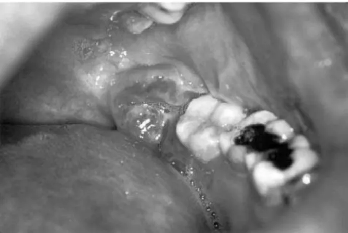
A 40-year-old male patient entered the Emergency Unit due to an episode of retromolar pericoronitis of the lower left wisdom tooth (Fig 4), that was accompanied by intense pain and fever of 38.5 – 40°C, not responding to the antibiotic/anti-inflammatory ambulatory treatment. HIV+ and Hepatitis B, diagnosed six years earlier, were in his pertinent medical history. The patient was not receiv-ing antiviral treatment at a time. He had left peripheral fa-cial paralysis for months earlier, treated with vitamin complexes. The analyses detected a CD 4 count of 37. Be-cause of his medical history a biopsy was ordered. His-tologically, there was a diffuse infiltration of poorly de-fined eosinophile cytoplasm cells, though clear in some areas. The nuclei were vesiculous and of the irregular morphology, with nucleoli. There were many mitotic fig-ures. Immunohistochemically the tumor cells were lym-phocytes B, which led to the diagnosis of diffuse large cell immunophenotype B NHL. The patient was sent to his country of origin to undergo treatment.
Fig. 4 −Behind the third molar there is an ulcerous lesion, which may be confused as a stage of pericoronitis.
Case 4
re-ported that he had been on sick leave due to the intense lower back pain that radiated to his lower limbs. Upon the exploration, multiple painful bilateral lymphadenopaties were found, the largest one being located in the right sub-mandibular area. Histologically a diffuse infiltration was identified, made of medium-sized cells mixed with macro-phages that gave it a starry sky appearance. Tumor cells had regular nuclei and hardly any cytoplasm, with one or sev-eral unimpressive nucleoli. Abundant mitotic figures were identified. Immunohistochemically the tumor cells were lymphocytes B, which lead to the diagnosis of diffuse small cells NHL, immunophenotype B, Burkitt type. The CAT scan revealed an adenopathic mass located in the area of the mandibular angle, along with multiple cervical lymphade-nopathy. The bone marrow biopsy of the iliac crest revealed severe lymphoblastic leukemia of B cells (Burkitt type), morphology L3 of the FAB, stage IV E. In the treatment the CODOX-M protocol was used and the leukemia entered in complete remission, consolidated with IVA C. After that the patient was without recurrence.
Discussion
Although the predominant sites of lymphoma oc-curence are lymph nodes, where it shows progressive en-largement, it also occurs in other organs, as an exclusive manifestation (1). NHL in 24% were of extra lymph node localization and 25% of these occured in the head and neck region (4). In his study of 1 467 cases of extra lymph node NHL, Freeman et al (5) found that 28% affected the head and neck area and 2% were located in the oral cavity. Taka-hashi et al (6) obtained the results demonstrating that 8% of extra lymph node NHL was in the oral cavity. This could be considered a low incidence of NHL oral manifestation, even more so if focused only on its intraosseous occurrence. In this paper four Caucasian males 22, 27, 40 and 41 years of age were presented, in which NHL was diagnosed only through its oral symptoms, although one of them was with a complete clinical picture in the final two months and the other was HIV positive. Primary lymphoma located in the mandible was not frequent. Current literature described about 100 cases of mandible NHL (7). In addition, there are various publications on isolated NHL occurence (4, 8−12). Some authors reported two cases of NHL in the same article (14, 15). Larson et al (2) in their review of 100 patients with extra lymph node lymphoma of the head and neck reported no mandible localization, while in the study of Conley et al (16) there was only one case with the oral manifestation. Econopoulos´ review (17) of 116 cases of head and neck NHL treated between 1977 and 1997 reported 9 cases (7.8%) with mandible and/or gingival involvement. Eisnbound et al (18) analyzed 31 case of oral cavity NHL and found 14 patients with bone invasion, 5 of them in the mandible. In their retrospective analysis of 31 cases of Bur-kitt´s lymphoma in young adolescents between 12 and 17 years of age, Anavi et al (19) reported 2 cases of mandible
involvement. In this paper 3 cases of primary localization and diagnostic of NHL of the mandible are presented (one of the cases not considered as primary NHL of mandible); one was located in the left retromolar triangle and two oth-ers were on the right side of mandible body. Two of the pa-tients were classified as being in stage IE, while the third patient was in stage IV. Eisenbud et al (18) presented 31 case in four stages of NHL, respectively. Evaluating only the cases with mandible involvement, the patients were pressented as 1 at stage I, 2 at stage III and 1 at stage IV.
but there were indications that other types of lymphoma or lymphoproliferative disease could be incorporated into the current definition of AIDS. In the study of Ioachim et al (25) 111 cases of AIDS-associated lymphomas were evalu-ated, only 3 of them localized in the oral cavity. Stolarski et al (26) published one case of NHL where the patient was not aware of being HIV positive. In the third case of this article it was known that the patient was HIV positive, but until that moment there was not sufficient proof to consider it as AIDS.
Conclusion
NHL of the head and neck region, particularly of the mandible, are rare and may be confused with other neo-plasms or periapical or periodontal inflammatory processes. There is usually a mass or swelling, showing rapid growth and enlargement, but this is not necessarily the case. Some-times there is only numbing or teeth motility. Considering this, it is supposed that in every doubtful case a biopsy and CAT scan are indicated.
R E F E R E N C E S
1. Bunch C, Gatter KC. The lymphomas. In: Weatherall
DJ, editor. Oxford Text Book of Medicine. Oxford: Oxford Medical Publications; 1996. p. 3568−87.
2. Larson DL, Robbins KT, Butler JJ. Lymphoma of the
head and neck. A diagnostic dilemma. Am J Surg 1984; 148(4): 433−7.
3. Eisenbud L, Sciubba J, Mir R, Sachs SA. Oral
presen-tations in non-Hodgkin's lymphoma: a review of thirty-one cases. Part II. Fourteen cases arising in bone. Oral Surg Oral Med Oral Pathol 1984; 57(3): 272−80.
4. Piatelli A, Croce A, Tete S, Artese L. Primary
non-Hodgkin's lymphoma of the mandible: a case report. J Oral Maxillofac Surg 1997; 55(10): 1162−6.
5. Freeman C, Berg JW, Cutler SJ. Occurrence and
prog-nosis of extranodal lymphomas. Cancer 1972; 29(1): : 252−60.
6. Takahashi H, Fujita S, Okabe H, Tsuda N, Tezuka F.
Immunophenotypic analysis of extranodal non-Hodgkin´s lymphomas in the oral cavity. Pathol Res Pract 1993; 189(3): 300−11.
7. Bachaud JM, Coppin D, Douchez J, Boutault F, Paty
E, Saboye J, et al. Primary malignant lymphoma of the mandible. A Study of 3 cases and a review of the lit-erature. Rev Stomatol Chir Maxillofac 1992; 93(6): : 372−6.
8. Bertolotto M, Cecchini G, Martinoli C, Perrone R,
Garlaschi G. Primary lymphoma of the mandible with diffuse widening of the mandibular canal: report of a case. Eur Radiol 1996; 6(5): 637−9.
9. Gusenbauer AW, Katsikeris NF, Brown A. Primary
lymphoma of the mandible: report of a case. J Oral Maxillofac Surg 1990; 48(4): 409−15.
10. Heng CK, Heng J. Implications of malignant
lym-phoma on a periapical mandibular lesion. Gen-Dent 1995; 43(5): 454−8.
11. Kawasaki G, Nakai M, Mizuno A, Nakamura T, Okabe
H. Malignant lymphoma of the mandïble: report of a case. Oral Surg Oral Med Oral Pathol Oral Radiol En-dod 1997; 83(3): 345−9.
12. Moretti A, Croce A, D'Agostino L, Bianchedi M,
Lat-tanzio G. Primary non-Hodgkin's lymphoma of the
mandible: a description of a clinical case and review of literature. Acta Otorhinolaryngol Ital 1997; 17(2): : 140−5.
13. Ugar DA, Turker M, Memis L. Primary lymphoma of
the mandible: report of a case. J Oral Maxillofac Surg 1995; 53(7): 827−9.
14. Macintyre DR. Lymphomas of the mandible presenting
as acute alveolar swellings. Br Dent J 1986; 161(7): : 253−4.
15. Rios Martin JJ, Villar Rodriguez JL, Vazquez Ramirez FJ, Illanes Moreno M, Parra Martin JA, Gonzalez Campora R, et al. Mandibular lymphomas with sclero-sis. Oral Surg Oral Med Oral Pathol Oral Radiol Endod 1996; 81(3): 321−7.
16. Conley SF, Staszak C, Clamon GH, Maves MD.
Non-Hodgkin's lymphoma of the head and neck: the Univer-sity of Iowa experience. Laryngoscope 1987; 97(3Pt1): : 291−300.
17. Economopoulos T, Fountzilas G, Kostourou A,
Daniilidis J, Pavlidis N, Andreopoulou H, et al. Pri-mary extranodal non-Hodgkin's lymphoma of the head and neck in adults: a clinicopathological comparison between tonsillar and non tonsillar lymphomas. (Hel-lenic co-Operative Oncology Group). Anticancer Res 1998; 18(6B): 4655−60.
18. Eisenbud L, Sciubba J, Mir R, Sachs SA. Oral presen-tations in non-Hodgkin's lymphoma: a review of thirty-one cases. Part I. Data analysis. Oral Surg Oral Med Oral Pathol 1983; 56(2): 151−6.
19. Anavi Y, Kaplinsky C, Calderon S, Zaizov R. Head,
neck, and maxillofacial childhood Burkitt´s lymphoma: a retrospective analysis of 31 patients. J Oral Maxilofac Surg 1990; 48(7): 708−13.
20. Yoshida H, Yusa H, Suzuki K, Dierks EJ. Painless
mandibular gingival mass. J Oral Maxillofac Surg 1995; 53(11): 1341−6.
21. Wright JM, Radman WP. Intrabony lymphoma
simu-lating periradicular inflammatory disease. J Am Dent Assoc 1995; 126(1): 101−5.
22. Ribera JM. Linfomas en pacientes con infección por
JM, Mallolas J, editors. Guía práctica del SIDA. Clín-ica, diagnóstico y tratamiento. Barcelona: Editorial Masson S.A.; 1998. p. 376−91.
23. CDC 1992 revises classification system for human im-munodeficiency virus infection and expanded AIDS surveillance case definition for adolescents and adults. (Draft, 15 November, 1991. CDC. Public Health Serv-ice). Atlanta: US Department of Health and Human Services; 1991.
24. CDC Revision of the CDC surveillance definition for acquired immunodeficiency syndrome [editorial].
MMWR Morb Mortal Wkly Rep 1987; 36(1S Suppl): : 3S−15S.
25. Ioachim HL, Dorsett B, Cronin W, Maya M, Wahl S. Ac-quired immunodeficiency syndrome-associated lympho-mas: clinical, pathologic, immunologic and viral charac-teristics of 111 cases. Hum Pathol 1991; 22(7): 659−73.
26. Stolarski CR, Boguslaw BI, Hoffman CH, Gates PE.
Small-cell noncleaved non-Hodgkin's lymphoma of the mandible in previously unrecognized human immuno-deficiency virus infection: report of a case. J Oral Maxillofac Surg 1997 55(8): 853−6.
Rad je primljen 4 VI 2002. god.
A p s t r a k t
Vicente-Barrero M, García-Castro I, Kne`evi} M, Castellano-Reyes JJ, Garcí a--Jimenez F, Camacho-García MC, Baez-Acosta B, Lon~arevi} S. Vojnosanit Pregl
2002; 59(6): 669−673.
NON-HODŽKINOVI LIMFOMI USNE DUPLJE
Non-Hod`kinovi limfomi se ~esto javljaju ekstranodalno, naro~ito na glavi i vratu. Intraoralna lokalizacija nije ~esta, naro~ito kad je jedina. To mo`e da dovede do prolongirane dijagnoze i ~ak do neadekvatnog le~enja. U radu se prikazuju razli~iti slu~ajevi s inicijalnim ekstranodalnim lokalizacijama koji nisu prethodno dijagnos-tikovani, od kojih je nekima NHL manifestovan samo intraoralno. Prikazani su uz dodatna ispitivanja kori{}ena za ranu dijagnostiku i odgovaraju}e klini~ke slike i pregled drugih slu~ajeva iz raspolo`ive literature.