Health Surveillance of Culture Negative Bacterial
Meningitis in Sa˜o Paulo, Brazil
Claudio T. Sacchi1*, Lucila O. Fukasawa1, Maria G. Gonc¸alves1, Maristela M. Salgado1, Kathleen A. Shutt2, Telma R. Carvalhanas3, Ana F. Ribeiro3, Brigina Kemp4, Maria C. O. Gorla5, Ricardo K. Albernaz3, Eneida G. L. Marques6, Angela Cruciano7, Eliseu A. Waldman8, M. Cristina C Brandileone5, Lee H. Harrison2, Sa˜o Paulo RT-PCR Surveillance Project Team"
1Division of Medical Biology, Department of Immunology, Instituto Adolfo Lutz, Sa˜o Paulo, Brazil,2Infectious Diseases Epidemiology Research Unit, University of Pittsburgh Graduate School of Public Health and School of Medicine, Pittsburgh, Pennsylvania, United States of America,3Center for Epidemiologic Surveillance, Sa˜o Paulo, Brazil,4Center for Epidemiologic Surveillance, Campinas, Brazil,5Division of Medical Biology, Department of Bacteriology, Instituto Adolfo Lutz, Sa˜o Paulo, Brazil, 6Bacteriology Area, Department of Medical Biology, Instituto Adolfo Lutz Regional Laboratory of Campinas, Campinas, Brazil,7Department of International Health, Johns Hopkins Bloomberg School of Public Health, Baltimore, Maryland, United States of America,8Faculdade de Sau´de Pu´blica, Universidade de Sa˜o Paulo, Sa˜o Paulo, Brazil
Abstract
Real-time (RT)-PCR increases diagnostic yield for bacterial meningitis and is ideal for incorporation into routine surveillance in a developing country. We validated a multiplex RT-PCR assay forStreptococcus pneumoniae, Neisseria meningitidis, and Haemophilus influenzaein Brazil. Risk factors for being culture-negative, RT-PCR positive were determined. The sensitivity of RT-PCR in cerebrospinal fluid (CSF) was 100% (95% confidence limits, 96.0%–100%) forN. meningitidis, 97.8% (85.5%–99.9%) forS. pneumoniae, and 66.7% (9.4%–99.2%) forH. influenzae. Specificity ranged from 98.9% to 100%. Addition of RT-PCR to routine microbiologic methods increased the yield for detection ofS. pneumoniae,N. meningitidis, andH. influenzaecases by 52%, 85%, and 20%, respectively. The main risk factor for being culture negative and RT-PCR positive was presence of antibiotic in CSF (odds ratio 12.2, 95% CI 5.9-25.0). RT-PCR using CSF was highly sensitive and specific and substantially added to measures of meningitis disease burden when incorporated into routine public health surveillance in Brazil.
Citation:Sacchi CT, Fukasawa LO, Gonc¸alves MG, Salgado MM, Shutt KA, et al. (2011) Incorporation of Real-Time PCR into Routine Public Health Surveillance of Culture Negative Bacterial Meningitis in Sa˜o Paulo, Brazil. PLoS ONE 6(6): e20675. doi:10.1371/journal.pone.0020675
Editor:Baochuan Lin, Naval Research Laboratory, United States of America
ReceivedDecember 6, 2010;AcceptedMay 9, 2011;PublishedJune 22, 2011
Copyright:ß2011 Sacchi et al. This is an open-access article distributed under the terms of the Creative Commons Attribution License, which permits unrestricted use, distribution, and reproduction in any medium, provided the original author and source are credited.
Funding:This work was supported by the Secretary of Health of Sa˜o Paulo State, Brazil; the Brazilian Council for Science and Technology Development (CNPq grant 301340/2007-2, to Dr. Brandileone); a career development award to Dr. Harrison, National Institute of Allergy and Infectious Diseases (K24 AI52788); and by a Fogarty International Center Global Infectious Diseases Research Training Program grant, National Institutes of Health, to the University of Pittsburgh (D43TW006592). The funders had no role in study design, data collection and analysis, decision to publish, or preparation of the manuscript.
Competing Interests:The authors have declared that no competing interests exist.
* E-mail: ctsacchi@gmail.com
"Membership of the Sa˜o Paulo RT-PCR Surveillance Project Team is provided in the Acknowledgments.
Introduction
Bacterial meningitis is a serious and often fatal infection.
Neisseria meningitidis, Streptococcus pneumoniae, and Haemophilus influenzaetype b (Hib) account for the vast majority of bacterial meningitis cases outside the neonatal period. Understanding the burden of bacterial meningitis is important because of recent advances in vaccines for these infections. Hib conjugate vaccines have led to the near disappearance of invasive Hib disease in Brazil and elsewhere [1]. Pneumococcal conjugate vaccine has had a huge impact on the incidence of invasive pneumococcal disease in the United States and is being incorporated into the routine pediatric immunization schedule in Brazil, as is serogroup C meningococcal conjugate vaccine [2,3].
In many developing countries, surveillance for bacterial meningitis is hampered by limited use of bacterial culture and a high frequency of negative cultures [4]. Availability of over-the-counter antibiotics, administration of antibiotics before
performance of lumbar puncture, lack of microbiology resources for bacterial culture, and variable quality of microbiology services are among the reasons for culture negativity. This problem leads to an underestimate of disease burden and assessments of the potential impact of vaccination. Non-culture methods, such as real-time (RT)-PCR, can increase the diagnostic yield for bacterial meningitis in both developed and developing countries [4,5,6,7,8,9,10].
identify factors associated with being culture negative, RT-PCR positive in Sa˜o Paulo.
Methods
Institutional review board (IRB) approvals
The study was approved by the Instituto Adolfo Lutz (IAL) and University of Pittsburgh IRBs, as well as the Comissa˜o Nacional de E´ tica em Pesquisa, the Brazilian national IRB.
Data sources
We included nine hospitals in the city of Sa˜o Paulo (Instituto de Infectologia Emı´lio Ribas, Casa de Sau´de Santa Marcelina, Hospital Estadual do Grajau´, Hospital da Santa Casa de Miserico´rdia de Sa˜o Paulo, Hospital Municipal Infantil Menino Jesus, Hospital Sa˜o Paulo, Hospital Estadual Regional Sul, Conjunto Hospitalar do Mandaqui, and Hospital Municipal do Tatuape´) and three in nearby Campinas (Hospital das Clı´nicas-Unicamp, Hospital Mario Gatti, and Hospital Celso Pierro), selected because they are centers for treatment of bacterial meningitis, report through SINAN, and send specimens for performance of counter immunoelectrophoresis forN. meningitidis
and Hib at IAL, a Sa˜o Paulo State reference laboratory, or to IAL Regional Laboratory of Campinas. IAL serves as the Brazilian National Reference Laboratory for Bacterial Meningitis. Eleven of the 12 hospitals conduct active surveillance for meningitis.
SINAN was queried for patients from these facilities for May 4, 2007 through March 31, 2009. Two non-mutually-exclusive groups of patients were included: those with blood or cerebrospi-nal fluid (CSF) culture positive for one of the three study organisms and patients whose CSF had$100 leukocytes/mm3and
$60% neutrophils, regardless of culture results. Patients in the culture positive group were used in the analyses of the sensitivity of RT-PCR, regardless of whether they met the CSF criteria. Patients who were culture negative but met the CSF criteria were used to determine the additional yield of RT-PCR. Other patients with suspected meningitis who did not meet these criteria but who had CSF submitted to IAL were used for the survival analyses described in the data analysis section.
Information gathered from SINAN included results of blood and CSF culture, demographic variables, and clinical and laboratory variables. Medical record reviews were conducted to collect information that was missing from SINAN. CSF and serum specimens were sent to IAL for RT-PCR testing.
Culture results were obtained from SINAN, which were confirmed during site visits to each of the hospital laboratories. Some isolates were also sent to IAL for confirmation; those results were added to the database if missing from SINAN.
Laboratory assays
A multiplex RT-PCR assay was used to identify patients with bacterial meningitis caused by the three study organisms. The assay was developed and performed at IAL and composed of three sets of primers and a probe targeting 1), meningococcal capsular transport genectrA[11], 2) pneumococcal autolysin gene
lytA[12], and 3) H. influenzaepolysaccharide capsular expression genebexA, which detectsH. influenzaeserotypes a, b, c, and d, but not e and f. [4].
The laboratory sensitivity of the assay was assessed with 55 isolates ofN. meningitidis, 54H. influenzae, and 66S. pneumoniae. For laboratory specificity, 125 bacterial and fungal isolates representing other species were used to validate the three individual RT-PCR assays (ctrA, lytAandbexA), and also the multiplex RT-PCR assay:
Acinetobacter baumannii, Cryptococcus sp, Coccidioides immitis, Chlamydia
pneumoniae, Chlamydia trachomatis, Corynebacterium ulcerans, Corynebacte-rium diphtheria, Haemophilus parainfluenzae, Haemophilus influenzae
biogroup aegyptius,Enterobacter cloaceae, Enterococcus faecalis, Histoplas-ma capsulatum, Klebsiella pneumoniae, Legionella sp, Leptospira interrogans, Moraxella catarrhalis, Moraxella genitalium, Mycobacterium tuberculosis, Nocardia asteroides, Pseudomonas aeruginosa, Paracoccidioides brasiliensis, Pneumocystis carinii, Streptococcus agalactiae, Streptococcus mitis, Streptococcus pyogenes, Neisseria lactamica, Neisseria sicca, Neisseria subflava, Salmonella sp, Streptococcus viridians,andListeria monocytogenes.
The lower limit of detection (LLD) was determined using extracted DNA from one isolate each ofN. meningitidis, H. influenzae
andS. pneumoniae. DNA concentration was adjusted to 100 ng/mL from which 10 fold serial dilutions (1021to 1029) were made in PCR-grade water. The LLD for the individual and multiplex assays were determined to be the dilution that yielded a Ct value#39.
DNA extraction was performed using QIAGEN QIAamp DNA Blood mini kit (QIAGEN, Valencia, CA) as described by the manufacturer except that ideally an aliquot of 500mL of CSF or
200mL of serum was processed and the DNA was eluted in 50mL
or 100 mL of TE buffer, respectively. However, specimens with
smaller available volumes, representing around 40% of specimens, were also included. 200mL of PCR-grade water was included as an extraction-negative control.
RT-PCR reactions were prepared in a separate area in which no DNA or bacteria were handled and in a biosafety cabinet cleaned with 1% sodium hypochlorite and exposed to UV light for at least 20 minutes. Final reactions were prepared in 96 well plates that included three separate positive controls, one for each
of the target organisms: strain N.24/75 serogroup A N.
meningitidis, strain ST.782/06 serotype 3 S. pneumoniae, and H.18/06 Hib. These controls were prepared in 0.01 M PBS pH 7.2 with 0.02% sodium azide, heated to 56uC for 30 minutes,
and maintained at 220uC. The extracted water and four
reactions without any DNA template were also included in each plate as negative controls.
The assays were carried out in a final 25ml reaction volume and were performed using TaqMan Universal Master Mix (Applied Biosystems, Foster City, CA), with 5ml of sample extracted DNA. Forward primer, reverse primer, and probe for each gene target were used in concentrations of, respectively, 300, 900, and 100 nM forctrA; 300, 600, and 100 nM forlytA; and 300, 300, and 100 nM forbexA. The ctrA,lytA, andbexAprobes were labeled at the 59end with FAM, VIC, and NED, respectively.
All reactions were run in duplicate and the assays were performed using an Applied Biosystems 7300 Real-Time PCR System (Applied Biosystems) using the following cycling param-eters: 50uC for 2 min, 95uC for 10 min, followed by 45 cycles of 95uC for 15 s and 60uC for 1 min. Extension of 55uC for 1 min was used for single RT-PCR genogrouping assay for serogroups B, W135, and Y. A positive result was defined as a cycle threshold (Ct) value below 39, an inconclusive result as a Ct value of 40 or 41, and a negative result as a Ct value of zero or .41. All inconclusive results and inconsistent replicates were repeated. Other than Ct values, the positive reactions were confirmed for each probe by the increase in fluorescence during the RT-PCR reaction. To determine the influence of RT-PCR Ct cut-off value on assay sensitivity, analysis using Ct values #35 as a second definition for positivity was used for all culture positive samples.
For further evaluation of culture-negative, RT-PCR-positive specimens, confirmation using a second gene was performed using RT-PCR assays targeting group-specific genes of N. meningitidis
CSF specimens with sufficient volume were tested for the presence of antibiotic [6,14]. A 5 mm blank filter paper disc was
soaked with 15mL of CSF and placed on Mueller Hinton agar
plate with a lawn of the pansensitive bacteriumKocuria rhizophila
(ATCC 9341). The plate was incubated overnight at 37uC and zone of inhibition around the disc was measured at 24 hours. Any zone was considered positive for presence of antibiotic. A disc containing 0.015 U of penicillin was used as a positive control and a blank disk was used as a negative control.
Data analysis
The sensitivity of RT-PCR for each pathogen was calculated as the proportion of culture (blood and/or CSF) positive specimens that had a positive result by RT- PCR for the same pathogen. Specificity was calculated using two approaches. In the first, as the proportion of all patients who were culture positive for one of the other two pathogens that were RT-PCR negative for the pathogen in question (specificity method 1). Patients who were culture positive for organisms other thanN. meningitidis, S. pneumoniaeorH. influenzae
(e.g., Escherichia coli) were excluded. The presumption was that specimens that are, for example, culture positive forN. meningitidisor
S. pneumoniaeshould be negative by RT-PCR forH. influenzae. In the second, culture-negative specimens that met the CSF criteria of
$100 leukocytes/mm3and$60% neutrophils were also included (specificity method 2, unadjusted. To determine whether RT-PCR positive, culture-negative specimens represented true RT-PCR positives (because of false negative culture results), these specimens were tested by PCR for a second gene target as indicated above. Specimens that were positive for the second target were interpreted as being true positives and therefore removed from the specificity calculation (specificity method 2, adjusted).
For the analyses of percent of patients with positive RT-PCR assay by CSF WBC count and CSF percent neutrophils, patients who were culture negative and who met the CSF criteria were included. CSF specimens that were culture negative or culture unknown that were submitted for routine testing for suspected bacterial meningitis, regardless of CSF profile, were also included. Survival analyses were performed to determine the proportion of RT-PCR positive samples that would be detected and the proportion of samples that would be tested at any specific WBC value.
To investigate factors associated with being culture-negative, cases were defined as patients who met the CSF criteria above and were culture negative and RT-PCR positive. Controls were patients who had positive CSF and/or blood cultures. Univariate logistic regression was done in SAS (version 9.2, SAS Institute) to identify risk factors. Transformations were performed on contin-uous factors that were not approximately linear to facilitate inclusion in the model. Factors with a p-value,0.1 were eligible for inclusion in multivariable logistic regression analysis. A backward logistic regression model with a stay criterion of 0.05 was run to identify independent risk factors for being culture negative. Factors remaining in the final model were checked for colinearity.
Results
Laboratory validation of RT-PCR assay
All N. meningitidis, H. influenzae and S. pneumoniae used in the evaluation of sensitivity of the RT-PCR assays were positive in the species-specific assay targeting the ctrA, bexA and lytA genes, respectively (laboratory sensitivity of 100%). DNA from strains representing other species gave negative results in all assays
(laboratory specificity of 100%). The LLD for the individual and multiplex RT-PCR assays was 20 fg of genomic DNA.
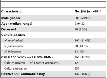
Validation of RT-PCR assay on clinical specimens There were 162 patients who were culture positive for one of the three target organisms and 460 who met the CSF criteria (Table 1). Of the 460 patients, 123 were culture positive for one of the three target organisms and 337 were culture negative. The median age was 9 years, 60.9% were male, and 9.6% were recorded as having died. Around a third of patients had the presence of antibiotic in CSF. Among 11 of the hospitals, the proportion of patients with presence of antibiotic in CSF ranged from 0.0% to 50.0% and in the one remaining hospital, a large referral facility, the proportion was 88.1%. For some analyses (Figures 1, 2, 3), an additional 188 specimens from patients with unknown culture results were included. Among 428 CSF speci-mens, 173, 70, and 3 were RT-PCR positive forN. meningitidis, S. pneumoniae,orH. influenzae,respectively. For 232 serum specimens tested, 48, 17, and 0 were RT-PCR positive for the same organisms, respectively.
The sensitivity of RT-PCR in CSF for diagnosis of meningitis was 100% (95% confidence limits, 96.0%–100%) forN. meningitidis, 97.8% (88.5%–99.9%) forS. pneumoniae, and 66.7% (9.4%–99.2%) for H. influenzae (Table 2). Specificity for the three organisms ranged from 98.9% to 100% (specific methods 1 and 2, adjusted), with narrow confidence limits. Positive and negative predictive values (PPV and NPV, respectively) of RT-PCR in CSF ranged from 98.3%–100% and 98.9%–100%, respectively (data not shown). For RT-PCR in serum, the sensitivities were lower and specificities were 94.1%–100% (Table 2).
Molecular genogrouping ofN. meningitidisin CSF Among 90 meningococcal culture-positive patients, the sero-group distribution as determined by molecular genosero-grouping was 10 (11.1%) group B, 67 (74.4%) group C, 11 (12.2%) group W-135, 1 (1.1%) group Y, and 1 (1.1%) non-groupable. For 83 culture negative, RT-PCR positive cases, the distribution was 10 (12.0%) group B, 64 (77.1%) group C, 4 (4.8) group W-135, 1 group Y (1.2%), and 4 (4.8%) non-groupable.
Table 1.Characteristics of patients included in the study.
Characteristic No. (%) (n = 499)*
Male gender 301 (60.9%)
Age (median, range) 9 (0–82)
Deceased 40 (9.6%)
Culture-positive
N. meningitidis 107 (21.4%)
S. pneumoniae 50 (10.0%)
H. influenzae 5 (1.0%)
CSF$100 WBCs and$60% PMNs 460 (93.7%)
Culture positive, 1 of 3 target organisms 123
Culture negative 337
Positive CSF antibiotic assay 142 (34.6%)
Of the 499 patients, 263 had CSF sample only, 67 had serum sample only, and 169 had both CSF and serum samples.
*Denominators for some characteristics,499 because of missing data for some patients
Increased yield of RT-PCR over and above culture-based results
A total of 26 culture-negative, RT-PCR-positive patients with pneumococcal meningitis were identified, for additional yield of 52.0% of RT-PCR over the 50 culture-based results; of the 26, 23 were identified by testing CSF only, 2 by both CSF and serum, and 1 by serum only. ForN. meningitidis, there were 91 culture negative patients with positive PCR results for an additional yield of RT-PCR over the 107 culture-positive patients, an 85.0% increased yield; of the 91, 67 were identified by testing CSF only, 13 by both CSF and serum and 11 by serum only. The increased yield was 20% forH. influenzae, with 5 culture positive cases and 1 additional RT-PCR, culture negative case, which was identified by testing CSF.
Relationship between CSF characteristics and RT-PCR positivity
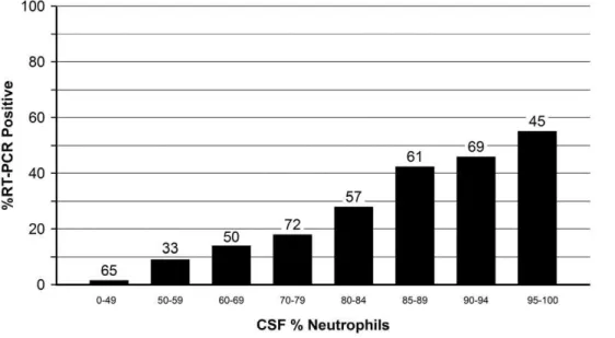
Among culture-negative and culture-unknown specimens, there was a strong relationship between the WBC and percent neutrophils in CSF and the percent of CSFs that were RT-PCR positive (Figures 1 and 2). For example, 4.9% of CSF specimens with 0–499 WBCs were positive, as compared to 73.2% for CSFs with at least 2,000 WBCs (Figure 1). Similarly, the proportion of specimens that were RT-PCR positive increased with increasing CSF percent neutrophils (Figure 2).
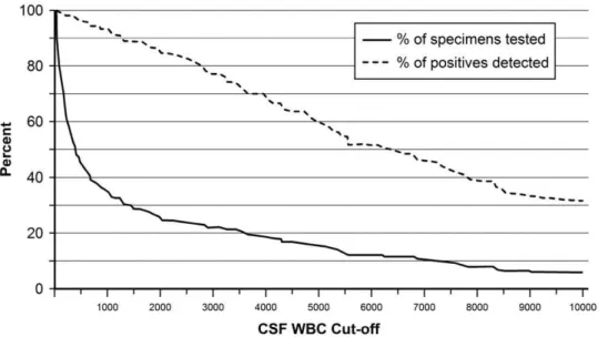
An analysis of all culture negative and culture unknown CSF specimens that were tested was conducted to determine optimal CSF cut-offs (Figure 3). For example, if we had used a cut-off of at least
Figure 2. Proportion of culture-negative and culture-unknown 452 CSF specimens that were RT-PCR positive, by cerebrospinal
fluid (CSF) percent neutrophils.Numbers above bars represent the number of specimens tested. Includes 188 patients with unknown culture
results.
doi:10.1371/journal.pone.0020675.g002
Figure 1. Proportion of culture-negative and culture-unknown 481 CSF specimens that were RT-PCR positive, by cerebrospinal
fluid (CSF) white blood cell (WBC) count (leukocytes/mm3).Numbers above bars represent the number of specimens tested. Includes 188
500 CSF leukocytes/mm3 to determine which specimens should undergo RT-PCR testing, we would have tested only 44% of sub-mitted specimens yet identified 95% of specimens that were RT-PCR positive. Alternatively, a cut-off of at least 1,000 CSF leukocytes/ mm3would have required the testing of only 34% of specimens, which would have resulted in detection of 92% of positives.
Risk factors for being culture negative and RT-PCR positive
In univariate analysis of risk factors for being a culture negative, RT-PCR positive case (n = 118), using culture positive patients as controls (n = 162), presence of antibiotic in CSF (odds ratio 15.5, 95% CL 8.3-29.1, p,0.0001), age$18 (OR 2.3, 95% CL 1.4, 3.7, p,0.0012), higher CSF WBC (median 4,400 among cases, 1,280
among controls, p,0.0001), and being from one of three hospitals (hospital numbers 3, 6, or 11) (OR 6.8, 95% CL 4.0-11.6) were all associated with being a case (Table 3).
In multivariable analysis, being from one of the three hospitals, presence of antibiotic in CSF, age $18, and infection withN. meningitidiswere independent risk factors (Table 4). Hospitals 3, 6, and 11 had the highest proportion of cases with antibiotic in CSF (35.7%, 41.7%, and 88.1%, respectively).
Discussion
We found that RT-PCR, when incorporated into routine public health surveillance performed well and added substantially to estimates of public health burden for N. meningitidis and S. pneumoniae. This indicates that RT-PCR could be used as an Figure 3. Relationship between minimum CSF WBC count used as a cut-off to determine what specimens are tested by RT-PCR and 1) the proportion of the total RT-PCR positives (n = 122) that are identified (dashed line) and 2) the proportion of all specimens
(n = 481) that would be tested (solid line).For example, if only those specimens with a CSF WBC of at least 1000 had been tested, about 34% of
the 481 CSF specimens would have been tested, which would have detected about 92% of the 122 specimens that were RT-PCR positive. Data were censored at a CSF WBC cut-off of 10,000. Data are for culture-negative and culture unknown specimens. Includes 188 patients with unknown culture results.
doi:10.1371/journal.pone.0020675.g003
Table 2.Determination of sensitivity and specificity of RT-PCR assay forN. meningitidis(ctrA),S. pneumoniae(lytA), and H. influenzae(bexA) in cerebrospinal fluid and serum specimens (see text for details).
Pathogen No.* Sensitivity 95% CIs
Specificity-Method 1** No.* 95% CIs
Specificity-Method
2, unadjusted*** No. 95% CIs
Specificity-Method
2, adjusted**** No.* 95% CIs
CEREBROSPINAL FLUID
N. meningitidis 90 100% 96.0–100 100% 51 93.0–100 75.7% 342 70.8–80.2 98.9% 262 96.7–99.8
S. pneumoniae 46 97.8% 88.5–99.9 100% 94 96.2–100 93.5% 386 90.6–95.8 100% 361 99.0–100
H. influenzae 3 66.7% 9.4–99.2 100% 139 97.4–100 99.8% 433 98.7–100 100% 432 99.2–100
SERUM
N. meningitidis 21 57.1% 34.0–78.2 100% 12 73.5–100 83.1% 223 77.4–87.9 94.1% 188 89.8–97.0
S. pneumoniae 10 80.0% 44.4–97.5 100% 25 86.3–100 96.2% 235 92.9–98.2 100% 226 98.4–100
H. influenzae 2 0% 0–84.2 100% 33 89.4–100 100% 243 98.5–100 100% 243 98.5–100
*Number of specimens contributing to the calculation of sensitivity or specificity **Specificity determined using specimens positive for other organisms
***Specificity determined using specimens positive for other organisms or culture negative
adjunct for surveillance for bacterial meningitis in developing countries. Although Brazil is a middle-income country, RT-PCR has been demonstrated to be useful in all types of settings, from low income to highly developed countries [4,6,7]. RT-PCR has the added advantage of providing results more rapidly than culture and has also been shown to be an excellent tool for epidemics [15]. We performed RT-PCR in a public health reference laboratory to increase the sensitivity of bacterial meningitis surveillance, not for clinical diagnosis. However, when used in the clinical setting, timeliness is a major advantage of RT-PCR because it can generally be completed on the same day as opposed to bacterial culture, which generally requires 2–3 days.
The RT-PCR assay we used had a high sensitivity and specificity in CSF when compared to bacterial culture. A high sensitivity (around 90%) has been previously described for RT-PCR in CSF forN. meningitidis and S. pneumoniae[4]. We found that the RT-PCR assay we used, although quite specific, was not as sensitive in serum, which is in line with a recent systematic
review and meta-analysis [16]. Serum is known to contain PCR inhibitors and the DNA polymerase we used is sensitive to inhibition [17,18]. In addition, collection of blood in ethylene-diaminetetraacetic acid (EDTA) is generally preferred over serum for PCR because of EDTA inhibition of DNA degradation. Despite the relative lack of sensitivity, RT-PCR in serum still managed to detect a substantial number of culture-negative cases. However, given the high sensitivity of the assay in CSF and the modest increased yield from testing serum alone, one could argue that serum should be tested only if a CSF specimen were not available.
Lack of specificity of PCR for detection of S. pneumoniae in serum usingplyas the gene target in children with high rates of pneumococcal carriage has been reported [19]. However, the specificity of the pneumococcal component of our assay in serum was very high, despite inclusion of sera from 31 children,2 years old (data not shown) in the specificity calculation; this may be due to the higher specificity of lytA as a primary gene target as
Table 3.Univariate analysis of risk factors for being a RT-PCR positive, culture-negative case-patient, using culture positive patients as controls.
Risk Factor Cases, No. (%) Controls, No. (%) OR 95% CI p-value
Male gender 69 (58.5%) 102 (63.4%) 0.8 0.5-1.3 0.41
Age$18 years 59 (50.0%) 49 (30.6%) 2.3 1.4–3.7 0.0012
Hospitalized 113 (99.1%) 160 (99.4%) 0.7 0.04–11.4 0.81
Race - White 61 (67.8%) 66 (55.9%) Baseline 0.22
White vs. Black 4 (4.4%) 7 (5.9%) 0.6 0.2–2.2
White vs. Brown 25 (27.8%) 45 (38.1%) 0.6 0.3–1.1
Antibiotic in CSF 79 (76.7%) 25 (17.5%) 15.5 8.3–29.1 ,0.0001
CSF MIC (mm) 21.0 (7–53) 21.0 (9–40) 0.59
CSF glucose 16 (0–101) 10 (0–610) 0.06
CSF WBC count (cell/mm3) 4,400 (100–29600) 1,280 (1–95200)
,0.0001
CSF % neutrophils 89 (60–99) 86 (0–100) 0.11
CSF protein (xx) 286.5 (22–4854) 230 (11–2789) 0.14
Hospital
Hospital 10 1 (0.9%) 22 (13.6%) Baseline ,0.0001
Hospital 1 vs. 10 10 (8.5%) 22 (13.6%) 10.0 1.2–84.9
Hospital 2 vs. 10 8 (6.8%) 21 (13.0%) 8.4 0.96–72.9
Hospital 3 vs. 10 19 (16.1%) 26 (16.1%) 16.1 2.0–129.9
Hospital 4 vs. 10 1 (0.9%) 2 (1.2%) 11.0 0.5–250.9
Hospital 5 vs. 10 5 (4.2%) 22 (13.6%) 5.0 0.5–46.4
Hospital 6 vs. 10 8 (6.8%) 4 (2.5%) 44.0 4.3–454.9
Hospital 7 vs. 10 2 (1.7%) 5 (3.1%) 8.8 0.7–117.2
Hospital 8 vs. 10 2 (1.7%) 14 (8.6%) 3.1 0.3–38.0
Hospital 9 vs. 10 4 (3.4%) 9 (5.6%) 9.8 0.96–99.9
Hospital 11 vs. 10 57 (48.3%) 13 (8.0%) 96.5 11.9–781.9
Hospital 12 vs. 10 1 (0.9%) 2 (1.2%) 11.0 0.5–250.9
Hospital 3, 6, 11 vs. others 84 (71.2%) 43 (26.5%) 6.8 4.0–11.6 ,0.0001
Bacterial species
N. meningitidis 91 (77.1%) 108 (66.7%) 1.7 0.98–2.9 0.06
S. pneumoniae 26 (22.0%) 50 (30.9%) 0.6 0.4–1.1 0.10
H. influenzae 1 (0.9%) 5 (3.1%) 0.3 0.03–2.3 0.23
There were a total of 118 case-patients and 162 controls. For some calculations, denominators differ because of missing data for some patients.
OR, odds ratio; CI, confidence interval; CSF, cerebrospinal fluid; MIC, minimum inhibitory concentration of CSF against strain ofKocuria rhizophilaamong specimens with detectable antibiotic; WBC, white blood cell.
compared toply[20]. The small number ofH. influenzaecases was due to the decline in Hib disease following incorporation of Hib conjugate vaccines into the routine pediatric immunization schedule in Brazil, which limited our ability at assess performance of theH influenzaecomponent of our assay.
This study points out the difficulty in evaluating a diagnostic test that performs better than the ‘‘gold standard’’, bacterial culture in this case, to which it is compared. Culture of normally sterile body fluids is generally considered to be very specific but can suffer from lack of sensitivity, particularly in the presence of antibiotics. This phenomenon can make the diagnostic test appear to be less specific than it truly is when culture negative specimens (some of which may have the presence of the organism despite the negative culture) are included. We attempted to circumvent this problem by measuring specificity first by using only specimens that were culture positive for another pathogen and second after correcting for what appeared to be false positive RT-PCR results by testing these ‘‘false positives’’ using a second gene target. In this study, the calculated specificities were very high using both approaches, which makes us confident that we have accurately assessed the true specificity of the assay.
The results of this study suggest that surveillance programs that use RT-PCR for diagnosis of bacterial meningitis can establish WBC count cut-offs that optimize both detection of cases and utilization of laboratory resources. For example, a large number of samples could be excluded without missing a substantial proportion of bacterial meningitis cases using WBC count cut-offs of, for example, 500 or 1000. Programs with patient characteristics that differ from those in our study should consider similar analyses to determine the optimal cut-offs for their setting.
We were able to determine the meningococcal group among culture-negative cases using a PCR-based assay; a similar approach is available for H. influenzae (10, 12). We are also exploring non-culture approaches for determining pneumococcal serotype [21]. These serotype and serogroup data are important for monitoring the impact of the conjugate vaccines that are currently being used in Brazil.
We found that the most important risk factor for being culture negative/RT-PCR positive was presence of antibiotic in CSF, which has been previously described [6]. This finding is not surprising because antibiotics are widely available over-the-counter in Brazil [22]. In addition, it is likely that some patients were given antibiotics by healthcare workers before CSF and/or blood were obtained for culture. We suspect that the finding that being from one of three hospitals was a risk factor for being culture negative, RT-PCR positive was due to residual confounding because these hospitals had the highest rates of CSF specimens with the presence of antibiotic. However, other hospital-specific
factors cannot be excluded, such as how specimens were collected, stored, and prepared for submission at the hospitals. The finding that infection withN. meningitidisis a risk factor for being culture-negative, RT-PCR positive may reflect the fact that this organism tends to be sensitive to most antibiotics, whereas drug resistance is common amongS. pneumoniaeisolates submitted to IAL [23,24]. Why age $18 years was a risk factor is unclear but residual confounding may be a factor.
Public health surveillance in Brazil has been changed substantially as a result of this study. RT-PCR is now performed routinely on all serum and CSF specimens from patients with suspected bacterial meningitis submitted to IAL. In addition, IAL is acquiring two additional RT-PCR systems for its regional laboratories to expand availability of the assay in Sa˜o Paulo State. Furthermore, the Brazilian Ministry of Health is asking all state reference laboratories to introduce RT-PCR into their routine.
In conclusion, RT-PCR for diagnosis of bacterial meningitis was successfully incorporated into public health surveillance for bacterial meningitis in Sa˜o Paulo. Future studies will involve use of novel molecular approaches that could supplant our current assay and provide diagnosis for a broader array of pathogens [25,26]. Despite success of this program in increasing the proportion of laboratory diagnosed bacterial meningitis cases, we still have a substantial number of pyogenic meningitis cases without an etiologic diagnosis. Given the public health impact and potential vaccine preventability of this disease, further investiga-tion is needed.
Acknowledgments
We thank Leonard Mayer and Xin Wang for their technical advice on RT-PCR and Daise Cristina Carvalho Becare for her assistance with surveillance.
Other members of the Sa˜o Paulo RT-PCR Surveillance Project Team:Division of Medical Biology, Immunology Department, Adolfo Lutz Institute, Sa˜o Paulo, Brazil: Anna Vera Custo´dio and Terezinha P. Arau´jo; Division of Medical Biology, Bacteriology Depart-ment, Adolfo Lutz Institute, Sa˜o Paulo, Brazil: Rosemeire Cobo Zanella, Ana Paula Silva de Lemos, Maria Luiza L S Guerra, Conceic¸a˜o Martins da Costa Zanelato, Marta Galhardo; Center for Epidemiologic Surveillance, Sa˜o Paulo, Brazil: Vera Malheiro; Centro de Controle e Prevenc¸a˜o de Doenc¸as, COVISA, Secretaria Municipal de Sau´de de Sa˜o Paulo: Rosa M.D. Nakazaki, Rachel Fernandes; Instituto da Crianc¸a - Faculdade de Medicina, Universidade de Sa˜o Paulo: Sonia R. T. da Silva Ramos; Instituto de Infectologia Emı´lio Ribas: Ce´lia E. Guarnieri, Simone A. de Souza; Casa de Sau´de Santa Marcelina: Marcelo Maki, Fabio Valdetaro; Hospital da Santa Casa de Miserico´rdia de Sa˜o Paulo: Maria J. P. Rujula, Elza L. U. Vicentine; Hospital Estadual Regional Sul: Sueli Rosenfeld, Elaine Magalha˜es; Hospital Estadual do Mandaqui: Fabio Rossi, Marilia V. Nogueira; Hospital Sa˜o Paulo da Universidade Federal de Sa˜o Paulo: Katsumi Osiro, Eliete A. M. Frigatto; Hospital Municipal Carmino Caricchio: Juang H. Jyh, Regina M. Loda; Hospital Municipal Infantil Menino Jesus: Walter B. G. Amorin; Hospital Estadual do Grajau´: Marcia C. de Arau´jo, Rossini Modesto; Hospital das Clı´nicas da Universidade de Campinas: Veronica M. Sincok, Carlos E. Levy; Hospital Celso Pierro da Pontifı´cia Universidade Cato´lica de Campinas: Elizabete A. Russi, Nereide A. B. Badur; Hospital Municipal Mario Gatti: Ricardo A. Cocolisce, Roge´rio A. Kuboyama.
Author Contributions
Conceived and designed the experiments: CTS LOF RKA EAW MCCB LHH. Performed the experiments: CTS LOF MGG MMS TRC AFR BK MCOG RKA EGLM AC MCCB. Analyzed the data: CTS KAS LHH. Contributed reagents/materials/analysis tools: CTS KAS LHH. Wrote the paper: CTS LHH. Reviewed the manuscript: CTS LOF MGG MMS KAS TRC AFR BK MCOG RKA EGLM AC EAW MCCB LHH. Table 4.Multivariable analysis of risk factors for being a
RT-PCR positive, culture-negative case-patient, using culture positive patients as controls.
Risk Factor OR 95% CI p-value
Hospital 3, 6, or 11 4.3 2.1–8.6 ,0.0001
Antibiotic in CSF 12.2 5.9–25.0 ,0.0001
Age$18 years 2.8 1.3–5.8 0.006
N. meningitidis 3.3 1.5–7.7 0.005
References
1. Adams WG, Deaver KA, Cochi SL, Plikaytis BD, Zell ER, et al. (1993) Decline of childhoodHaemophilus influenzaetype b (Hib) disease in the Hib vaccine era. JAMA 269: 221–226.
2. Whitney CG, Farley MM, Hadler J, Harrison LH, Bennett NM, et al. (2003) Decline in invasive pneumococcal disease after the introduction of protein-polysaccharide conjugate vaccine. N Engl J Med 348: 1737–1746.
3. Hsu HE, Shutt KA, Moore MR, Beall BW, Bennett NM, et al. (2009) Effect of pneumococcal conjugate vaccine on pneumococcal meningitis. N Engl J Med 360: 244–256.
4. Corless CE, Guiver M, Borrow R, Edwards-Jones V, Fox AJ, et al. (2001) Simultaneous detection of Neisseria meningitidis, Haemophilus influenzae, and
Streptococcus pneumoniaein suspected cases of meningitis and septicemia using real-time PCR. J Clin Microbiol 39: 1553–1558.
5. Kilpatrick ME, Mikhail IA, Girgis NI (1987) Negative cultures of cerebrospinal fluid in partially treated bacterial meningitis. Trop Geogr Med 39: 345–349. 6. Saha SK, Darmstadt GL, Yamanaka N, Billal DS, Nasreen T, et al. (2005)
Rapid diagnosis of pneumococcal meningitis: implications for treatment and measuring disease burden. Pediatr Infect Dis J 24: 1093–1098.
7. Chanteau S, Sidikou F, Djibo S, Moussa A, Mindadou H, et al. (2006) Scaling up of PCR-based surveillance of bacterial meningitis in the African meningitis belt: indisputable benefits of multiplex PCR assay in Niger. Trans R Soc Trop Med Hyg 100: 677–680.
8. Afifi S, Wasfy MO, Azab MA, Youssef FG, Pimentel G, et al. (2007) Laboratory-based surveillance of patients with bacterial meningitis in Egypt (1998-2004). Eur J Clin Microbiol Infect Dis 26: 331–340.
9. Pedro LG, Boente RF, Madureira DJ, Matos JA, Rebelo CM, et al. (2007) Diagnosis of meningococcal meningitis in Brazil by use of PCR. Scand J Infect Dis 39: 28–32.
10. Wang X, Mair R, Hatcher C, Theodore MJ, Edmond K, et al. (2011) Detection of bacterial pathogens in Mongolia meningitis surveillance with a new real-time PCR assay to detectHaemophilus influenzae. Int J Med Microbiol.
11. Mothershed EA, Sacchi CT, Whitney AM, Barnett GA, Ajello GW, et al. (2004) Use of real-time PCR to resolve slide agglutination discrepancies in serogroup identification ofNeisseria meningitidis. J Clin Microbiol 42: 320–328.
12. Carvalho MD, Tondella ML, McCaustland K, Weidlich L, McGee L, et al. (2007) Evaluation and improvement of real-time PCR detection assays tolytA,
ply, andpsaAgenes for detection of pneumococcal DNA. J Clin Microbiol. 13. Maaroufi Y, De Bruyne JM, Heymans C, Crokaert F (2007) Real-time PCR for
determining capsular serotypes ofHaemophilus influenzae. J Clin Microbiol 45: 2305–2308.
14. Markowitz M, Gordis L (1968) A mail-in technique for detecting penicillin in urine: application to the study of maintenance of prophylaxis in rheumatic fever patients. Pediatrics 41: 151–153.
15. Nathan N, Rose AM, Legros D, Tiendrebeogo SR, Bachy C, et al. (2007) Meningitis serogroup W135 outbreak, Burkina Faso, 2002. Emerg Infect Dis 13: 920–923.
16. Avni T, Mansur N, Leibovici L, Paul M (2010) PCR using blood for diagnosis of invasive pneumococcal disease: systematic review and meta-analysis. J Clin Microbiol 48: 489–496.
17. Radstrom P, Knutsson R, Wolffs P, Lovenklev M, Lofstrom C (2004) Pre-PCR processing: strategies to generate PCR-compatible samples. Mol Biotechnol 26: 133–146.
18. Abu Al-Soud W, Radstrom P (1998) Capacity of nine thermostable DNA polymerases To mediate DNA amplification in the presence of PCR-inhibiting samples. Appl Environ Microbiol 64: 3748–3753.
19. Dagan R, Shriker O, Hazan I, Leibovitz E, Greenberg D, et al. (1998) Prospective study to determine clinical relevance of detection of pneumococcal DNA in sera of children by PCR. J Clin Microbiol 36: 669–673.
20. Abdeldaim G, Herrmann B, Molling P, Holmberg H, Blomberg J, et al. (2010) Usefulness of real-time PCR forlytA,ply, andSpn9802on plasma samples for the diagnosis of pneumococcal pneumonia. Clin Microbiol Infect 16: 1135–1141. 21. Findlow H, Laher G, Balmer P, Broughton C, Carrol ED, et al. (2009)
Competitive inhibition flow analysis assay for the non-culture-based detection and serotyping of pneumococcal capsular polysaccharide. Clin Vaccine Immunol 16: 222–229.
22. Silva LR, Vieira EM (2004) [Pharmacists’ knowledge of sanitary legislation and professional regulations]. Rev Sau´de Pu´blica 38: 429–437.
23. Mantese OC, de Paula A, Almeida VV, de Aguiar PA, Wolkers PC, et al. (2009) Prevalence of serotypes and antimicrobial resistance of invasive strains of pneumococcus in children: analysis of 9 years. J Pediatr (Rio J) 85.
24. Brandileone MC, Casagrande ST, Guerra ML, Zanella RC, de Andrade AL, et al. (2005) Increase in penicillin resistance of invasiveStreptococcus pneumoniaein Brazil after 1999. J Antimicrob Chemother 56: 437–439.
25. Boving MK, Pedersen LN, Moller JK (2009) Eight-plex PCR and liquid-array detection of bacterial and viral pathogens in cerebrospinal fluid from patients with suspected meningitis. J Clin Microbiol 47: 908–913.