Sulcus vocalis: evidence for autosomal
dominant inheritance
R.H.G. Martins1, T.M. Gonçalves1, D.S. Neves1, T.A. Fracalossi1, E.L.M. Tavares1 and D. Moretti-Ferreira2
1Departamento de Otorrinolaringologia, Faculdade de Medicina de Botucatu,
Universidade Estadual de São Paulo “Júlio de Mesquita Filho”, Botucatu, SP, Brasil
2Departamento de Genética, Instituto de Biociências,
Universidade Estadual de São Paulo “Júlio de Mesquita Filho”, Botucatu, SP, Brasil
Corresponding author:D. Moretti-Ferreira E-mail: sag@fmb.unesp.br
Genet. Mol. Res. 10 (4): 3163-3168 (2011) Received May 5, 2011
Accepted September 5, 2011 Published December 19, 2011
DOI http://dx.doi.org/10.4238/2011.December.19.5
ABSTRACT. We found evidence of autosomal dominant hereditary transmission of sulcus vocalis. Four dysphonic patients from three generations of the same family were submitted to videolaryngoscopic examination (three patients) and to direct laryngoscopy (one patient) to diagnose the hoarseness. Sulcus vocalis was diagnosed in all four patients.
The inding of four affected individuals in three generations, with vertical
transmission affecting man and women, is more consistent with autosomal dominant inheritance pattern; it is an etiological model that we propose for the sulcus vocalis in this pedigree.
INTRODUCTION
Sulcus vocalis is a rare lesion of the true vocal folds, in which there is a linear groove on the mucosal lining, extending along its vibratory surface (Bouchayer et al., 1985; Pontes et al., 1994). Its depth varies, and it can involve one or both vocal folds. Its diagnosis is not
always straightforward, especially in children, and in many cases it is conirmed only during
surgery. Its etiology is a controversial matter among different authors. It occurs secondary to
chronic laryngeal inlammatory processes, i.e., gastroesophageal relux and chronic laryngitis
and iatrogenic complications during vocal fold handling in microsurgeries, or it may have a congenital origin (Bouchayer et al., 1985; Pontes et al., 1994; Ford et al., 1996). In the present study, we report its diagnosis in four individuals from the same family, suggesting a congenital and genetic origin for this disorder.
MATERIAL AND METHODS
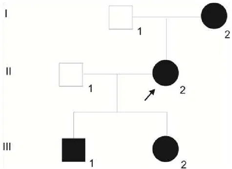
This study was approved by our Ethics Committee (protocol No. 3321-2009). Four dysphonic individuals from the same family (Figure 1) were seen in the Voice Disorders Ward of the Otolaryngology Department of the Medical School of Botucatu, Sao Paulo State Uni-versity (UNESP), and they were submitted to videostroboscopy through a multifunctional im-age capture system - XE-30, Eco X-TFT/USB (Germany) coupled to a rigid 8-mm, 70-degree (Asap) scope, and the images were recorded with a DVD. They were also submitted to quali-tative voice analysis using GRBASI and acoustic voice analysis using a Multi-Dimensional Voice Program - MDVP (model 5105, version 3.1.4, software, Multi-Speech 3700 - Kaypen-tax, USA), where the results are described below.
Case 1
MLM, female, 54 years old (I-2; Figure 1), with lifelong hoarseness that has gotten worse in the past four years. This patient did not report smoking, drinking or voice abuse, did not have na-sal or esophageal symptoms or undergo any type of surgery. Videostroboscopy showed a sulcus vo-calis on the right vocal fold (Figure 2). Vocal assessment was: G1R1B1A0S1I1, and acoustic analysis with the following values: Fo = 197.627; % shimmer = 2.777; % jitter = 0.955, and NHR = 0.148.
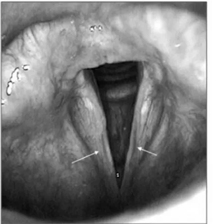
Figure 2. Sulcus vocalis in right vocal fold (case 1, arrow). Videolaryngoscopic examination.
Case 2
AM, female, 30 years old (II-2; Figure 1, arrow), hoarseness since childhood. This patient did not report smoking, drinking, vocal abuse, nasal or esophageal symptoms, or sur-geries. Videostroboscopy showed a bilateral sulcus vocalis (Figure 3). Vocal assessment was: G2R2B2A0S2I1, and acoustic analysis with the following values: Fo = 190.807; % shimmer = 2.484; % jitter = 0.852, and NHR = 0.112.
Case 3
LM, female, 12 years old (III-2; Figure 1), hoarseness since childhood. This patient reported vocal abuse, but denied nasal or esophageal symptoms or surgeries. Videostrobos-copy showed bilateral sulcus vocalis (Figure 4). Vocal assessment was: G2R2B2A0S2I1,and the acoustic analysis showed the following values: Fo = 217.035; % shimmer = 5.041; % jitter = 0.737, and NHR = 0.121.
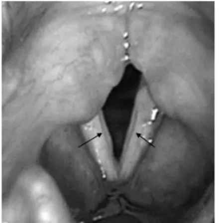
Figure 4. Bilateral sulcus vocalis (case 3, arrows). Videolaryngoscopic examination.
Case 4
DTM, male, 4 years old (III-1; Figure 1), lifelong hoarseness, without respiratory, nasal or esophageal symptoms, or surgery. This patient reported sporadic vocal abuse. The child did not collaborate during the videostroboscopy examination, and under direct laryngoscopy we found sulcus vocalis on the right-side vocal fold and bilateral vocal nodules (arrows, Figure 5). Vocal assessment was: G2R1B2A0S2I0, and the following values were recorded on the acoustic analysis: Fo = 313.412; % shimmer = 3.514; % jitter = 1.838, and NHR = 0.093 (Figure 5).
DISCUSSION
Ford et al. (1996) morphologically classiied sulcus vocalis as follows. Type I, or phys -iological, is found in cases of atrophy, presbyphonia and vocal fold paralysis. This type is seen upon endoscopic examinations during maximum vocal fold abduction. Type II, or true sulcus vocalis, corresponds to the stria major (vergeture) or stria minor types. Most times, this type of sulcus is adhered to the vocal ligament, thus considerably impairing muco-undulatory
move-ment. Type III, or pouch-shaped coniguration, is where the epithelial change is restricted to a
given area of the vocal fold.
When we discuss the origin of sulcus vocalis, our major concern is not with type 1 sulcus, which has very little effect on vocal quality, but rather with types II and III, in which dysphonia is marked, as seen in the patients of the present study, especially in bilateral le-sions, responsible for glottic incompetence and reduction of the vibratory wave. Sulcus voca-lis etiology is controversial according to different authors. For some, it is acquired and arises
from inlammatory processes on the vocal folds, or iatrogenic sequelae or even vocal fold
atrophy (Bouchayer et al., 1985; Pontes et al., 1994; Ford et al., 1996). Others advocate a ge-netic origin, in which sulcus vocalis stems from embryonic defects in laryngeal development
(Bouchayer et al., 1985). The latter has support from the indings of these lesions in monozy -gotic twins (Cakir et al., 2010) and in many members of the same family (Martins et al., 2007), found by our group in another family unrelated to the one presented here, and also from its association with other congenital laryngeal lesions, i.e., cysts, mucosal bridges and microwebs
(Bouchayer et al., 1985; Pontes et al., 1994; Villagomez and Rosen, 2000).
In the present study, sulcus vocalis was detected in four individuals and both gen-ders, in three generations of the same family. All individuals had had a lifelong hoarseness, thus strengthening the hypothesis of a congenial origin for this lesion, which is further
rein-forced by the indings of sulcus vocalis in the four-year-old patient (case 4), a rare diagnosis
at this age. The very laryngeal presentation of the lesion in the members of this family, which recurred in three generations and in both genders, seems to suggest autosomal dominant in-heritance. In a previous paper, we presented four siblings with sulcus vocalis (1 female and 3 males), and we suggested a likely genetic transmission (Martins et al., 2007); however, it was not possible to establish a genetic transmission mode for this disorder.
CONCLUSIONS
In the present case report, we describe the genetic etiology of sulcus vocalis, and we
suggest the need for investigating vocal disorders in irst-degree relatives (parents, children and siblings), as well as doing a detailed videoendoscopic examination for conirmation of di -agnosis. Complementary molecular studies in a larger group of individuals with sulcus vocalis may pinpoint the gene involved in this disorder.
REFERENCES
Bouchayer M, Cornut G, Witzig E, Loire R, et al. (1985). Epidermoid cysts, sulci, and mucosal bridges of the true vocal
cord: a report of 157 cases. Laryngoscope 95: 1087-1094.
Ford CN, Inagi K, Khidr A, Bless DM, et al. (1996). Sulcus vocalis: a rational analytical approach to diagnosis and management. Ann. Otol. Rhinol. Laryngol. 105: 189-200.
Martins RH, Silva R, Ferreira DM and Dias NH (2007). Sulcus vocalis: probable genetic etiology. Report of four cases in close relatives. Braz. J. Otorhinolaryngol. 73: 573.
Pontes PL, Behlau M and Gonçalves MI (1994). Alterações estruturais mínimas da laringe: considerações básicas. Acta Awho 13: 2-6.