ABSTRACT: Dental bleaching is a simple and conservative procedure for aesthetic restoration of vital and non-vital discolored teeth. Nevertheless, a number of studies have demonstrated the risk of tissue damage from the contact of these agents with the oral mucosa. In the current study, the genotoxic potential associated with exposure to den-tal bleaching agents was assessed by the single cell gel (comet) assay in vitro. Chinese hamster ovary (CHO) cells in vitro were exposed to six commercial dental bleaching agents (Clarigel Gold – Dentsply; Whitespeed – Discus Dental; Nite White – Discus Dental; Magic Bleaching – Vigodent; Whiteness HP – FGM and Lase Peroxide - DMC). The results pointed out that all dental bleaching agents tested contributed to DNA damage as depicted by the mean tail moment, being the strongest effect observed with the highest dose of hydrogen peroxide (Whiteness HP and Lase Peroxide, at a 35% concentration). On the other hand, Magic Bleaching (Vigodent) induced the lowest level of DNA breakage. Negative and positive controls displayed absence and presence of DNA-damaging, respectively. Taken together, these results suggest that dental bleaching agents may be a factor that increases the level of DNA damage. A higher concentration of hydrogen peroxide produced higher noxious activities in the genome as detected by single cell gel (comet) assay.
DESCRIPTORS: CHO cells; Comet assay; Tooth bleaching; Mutagenicity tests.
RESUMO: Clareamento dental é um procedimento simples e conservador para restaurar esteticamente a cor de dentes vitais e não-vitais. Entretanto, alguns estudos têm demonstrado o risco de dano tecidual a partir do con-tato desses agentes com a mucosa bucal. Neste presente estudo, o potencial genotóxico associado à exposição aos agentes clareadores dentais foi avaliado pelo teste de células individualizadas em gel (teste do cometa) in vitro. Células de ovário de hamster chinês (CHO) in vitro foram expostas a seis agentes clareadores dentais comercial-mente disponíveis (Clarigel Gold – Dentsply; Whitespeed – Discus Dental; Nite White – Discus Dental; Magic Ble-aching – Vigodent; Whiteness HP – FGM e Lase Peroxide – DMC). Os resultados mostraram que todos os agentes clareadores testados contribuíram para os danos no DNA, como demonstrado pela média do momento da cauda, sendo o efeito mais forte observado na mais alta dose de peróxido de hidrogênio (Whiteness HP e Lase Peroxide, na concentração de 35%). Por outro lado, Magic Bleaching (Vigodent) induziu o menor nível de quebras no DNA. Os controles negativo e positivo apresentaram ausência e presença de danos no DNA, respectivamente. Em suma, esses resultados sugerem que os agentes clareadores dentais podem ser um fator que aumenta o nível de danos no DNA. Uma concentração de peróxido de hidrogênio mais elevada produziu atividades nocivas mais severas no genoma como detectado pelo teste do cometa.
DESCRITORES: Células CHO; Ensaio em cometa; Clareamento de dente; Testes de mutagenicidade.
INTRODUCTION
Dental bleaching is a simple and conserva-tive procedure for aesthetic restoration of vital and non-vital discolored teeth. There are many bleaching agents commercially available with vari-ous constituents, such as hydrogen peroxide and carbamide peroxide. Carbamide peroxide decom-poses to produce hydrogen peroxide29, which may be considered as the active ingredient of choice for bleaching because of its low molecular weight
and its ability to denature proteins. Nevertheless, a number of studies have demonstrated the risk of tissue damage from the contact of these agents with the oral mucosa8,13,17. Hydrogen peroxide is able to interact both directly with DNA and through highly reactive oxygen and radical species causing extensive oxidative DNA damage6. So far, oxida-tive DNA damage has been recognized as a major cause of cell death and mutations in all aerobic
* Post-Doctoral Researcher; **Head Professor; ***Researcher, Center for Genotoxins and Carcinogens Evaluation (TOXICAN) – De-partment of Pathology, Botucatu Medical School, São Paulo State University.
2006;20(1):47-51
Study of DNA damage induced by dental bleaching agents
in vitro
Estudo de danos no DNA induzidos por agentes clareadores
dentais
in vitro
Daniel Araki Ribeiro*
organisms. In humans, oxidative DNA damage is also considered an important promoter of cancer4. As the incidence of head and neck cancer has in-creased in recent years, particularly in developing countries such as India, Vietnam and Brazil, where it constitutes up to 25% of all types of cancer18, further risk factors rather than tobacco smoke and the abuse of alcohol are of special concern.
Understanding how cancer develops creates opportunities for cancer prevention or early detec-tion. An important part of this effort is to identify the agents and exposures that cause cancer. Geno-toxicity tests can be defined as in vitro and in vivo tests designed to detect compounds which induce genetic damage such as DNA strand breaks, gene mutation, chromosomal breakage and altered DNA repair capacity. In the last decades, genotoxicity assays have gained widespread acceptance as an important and useful indicator of carcinogenic-ity2.
Therefore, the aim of the present study was to evaluate whether dental bleaching agents can induce DNA breakage in Chinese hamster ovary (CHO) cells in vitro. CHO cells were chosen because the mechanism of DNA damage induced in these cells has been well documented. To evaluate the magnitude of DNA damage, we used the alkaline version of the single cell gel (comet) assay.
MATERIALS AND METHODS
Cell culture
CHO K-1 cells were grown to confluence in 75-cm² culture flasks (Corning, New York, NY, USA) using Ham’s F-10 medium (Invitrogen Corpora-tion, Grand Island, NY, USA) supplemented with 10% fetal calf serum and 100 U/mL penicillin and 100 µg/mL streptomycin (Invitrogen Corporation) incubated in a 95% air, 5% CO2 atmosphere at 37°C. Cells were cultured for 5 days prior to treat-ment with test substances. Confluent cells were de-tached with 0.15% trypsin (Invitrogen Corporation) for 5 minutes, after that, 2 ml of complete medium were added and cells were centrifuged at 180 g for 5 minutes. Cell suspension was counted using a Neubauer® chamber (Herka, Berlin, Germany) and seeded in 96-well microtitre plates (Corning) at a density of 1 × 10⁴ cells per well (at a concentration of 1 × 10⁶/mL). All the procedures in this study concern ethical conducts described by the Ethics Committee, Botucatu Medical School, São Paulo State University, SP, Brazil.
Treatment of cells
For this study, the following dental bleaching agents were used: Clarigel Gold (Dentsply, São Paulo, Brazil, Lot no. 5733); Whitespeed (Discus Dental, Culver City, USA, Lot no. 02564002); Nite White Excel 2 (Discus Dental, Culver City, USA, Lot no. 02287002); Magic Bleaching (Vigodent, Rio de Janeiro, Brazil, Lot no. 00104); Whiteness HP (FGM, Joinville, Brazil, Lot no. 02262002) and Lase Peroxide (DMC, São Paulo, Brazil, Lot no. 02281). Clarigel Gold, Whitespeed, Nite White Excel 2 and Magic Bleaching provided hydrogen peroxide at a 16% concentration, whereas Whiteness HP and Lase Peroxide provided hydrogen peroxide at a 35% concentration. The respective manufactur-ers estimated these quantifications. Each material was dissolved in dimethylsulfoxide (DMSO, Life Technologies, Carlsbad, USA) at a ratio of 1:1 in a final volume of 10 µl. A volume of 10 µl of cells (~1 × 10⁴ cells) was then added individually to each final solution of dental bleaching agents for 15 minutes on ice. Negative control was treated with 10 µl of DMSO only during 15 min. Since cytotox-icity is a confounding factor in genotoxcytotox-icity stud-ies, it is not recommended to perform the single cell gel (comet) assay on samples with more than 30% cytotoxicity28. Thus, the exposure period as well as the final concentration used herein were performed as described elsewhere22. Independent positive control was performed with MMS (methyl-metanesulfonate, Sigma Aldrich, St. Louis, USA) at a 1 µg/mL dose for 1 hour in order to ensure the reproducibility and sensitivity of the assay. After completing the respective experimental periods, all individual treatments were centrifuged at 180 g during 5 minutes, washed twice with fresh medium (RPMI 1640 glutamax medium; Life Sciences, Pais-ley, USA) and re-suspended with fresh medium (RPMI 1640 glutamax medium). Each individual treatment was repeated three times consecutively to ensure reproducibility.
Single cell gel (comet) assay
USA; 10 mM Tris-HCl buffer pH = 10 – Sigma Al-drich, St. Louis, USA; 1% sodium sarcosinate – Sigma Aldrich, St. Louis, USA; with 1% Triton X-100 – Sigma Aldrich, St. Louis, USA; and 10% DMSO – Merck, St. Louis, USA) for about 1 hour. Prior to electrophoresis, the slides were left in al-kaline buffer (0.3 mM NaOH, Merck, Darmstadt, Germany; and 1 mM EDTA, Merck, Darmstadt, Germany; pH > 13) for 20 minutes and electropho-resed for another 20 minutes, at 25 V (0.86 V/cm) and 300 mA. After electrophoresis, the slides were neutralized in 0.4 M Tris-HCl (pH = 7.5), fixed in absolute ethanol (Merck, Darmstadt, Germany) and stored at room temperature until analysis. In order to minimize extraneous DNA damage from ambient ultraviolet radiation, all steps were per-formed with reduced illumination.
Throughout this study, diluted and treated aliquots were tested for viability by trypan blue exclusion19, and constantly more than 70% of cells excluded trypan.
Comet capture and analysis
A total of 50 randomly captured comets from each slide12 were examined blindly at 400 X mag-nification using a fluorescence microscope (Olym-pus, Orangeburg, USA) connected through a black and white camera to an image analysis system (Comet Assay II, Perceptive Instruments, Sufolk, Haverhill, UK). A computerized image analysis system acquires images, computes the integrated intensity profiles for each cell, estimates the comet cell components and then evaluates the range of derived parameters. Undamaged cells have an in-tact nucleus without a tail and damaged cells have the appearance of a comet. To quantify the DNA damage, tail moment was evaluated. Tail moment was calculated as the product of the tail length and the fraction of DNA in the comet tail. The comet tail moment is positively correlated with the level of DNA breakage in a cell. The mean value of the tail moment in a particular sample was taken as an index of DNA damage in this sample.
Statistical methods
The parameter from the comet assay (tail mo-ment) was assessed by the Kruskal-Wallis non-parametric test followed by a post-hoc analysis (Dunn’s test) if a significant effect was detected, using SigmaStat software, version 1.0 (Jadel Sci-entific, Rafael, CA, USA). The level of statistical significance was set at 5%.
RESULTS
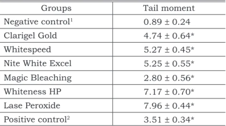
The single cell gel (comet) assay was used to measure DNA damage in CHO cells in vitro. DNA strand breaks were represented by the mean tail moment for 50 comets/sample. As seen in Table 1, the assay was able to detect significant increase in tail moment (MMS-treated cells) compared with the negative control. Furthermore, all compounds tested induced strand breaks in DNA, being the strongest effect observed at the highest dose of the hydrogen peroxide (Whiteness HP and Lase Peroxide, at a 35% concentration). On the other hand, Magic bleaching induced the lowest level of DNA breakage when compared to the other prod-ucts evaluated.
DISCUSSION
The aim of this study was to evaluate the geno-toxic damage dental bleaching agents-induced on CHO cells in vitro. The investigation was conducted utilizing the single cell gel (comet) assay. The single cell gel (comet) assay in its alkaline version is a rapid, simple, and reliable biochemical technique for evaluating DNA damage in mammalian cells28. The basic principle of the single cell gel (comet) assay is the migration of DNA in an agarose ma-trix under electrophoretic conditions. When viewed under a microscope, a cell has the appearance of a comet, with a head (the nuclear region) and a tail containing DNA fragments or strands migrating in the direction of the anode. Our own recent studies have demonstrated that the single cell gel (comet) assay is a suitable tool to investigate genotoxicity of compounds used in dental practice20,23,25.
TABLE 1 - Mean ± Standard deviation of DNA damage (tail moment) in Chinese hamster ovary (CHO) cells treated with dental bleaching agents (n = 3).
Groups Tail moment
Negative control1 0.89 ± 0.24
Clarigel Gold 4.74 ± 0.64*
Whitespeed 5.27 ± 0.45*
Nite White Excel 5.25 ± 0.55*
Magic Bleaching 2.80 ± 0.56*
Whiteness HP 7.17 ± 0.70*
Lase Peroxide 7.96 ± 0.44*
Positive control2 3.51 ± 0.34*
*p < 0.05 when compared to the negative control group.
In vitro studies are simple, inexpensive to per-form, provide a significant amount of information, can be conducted under controlled conditions, and may elucidate the mechanisms of cellular toxicity9. The results obtained from in vitro assays might be indicative of the effects observed in vivo. It is important to notice that the alkaline version of the single cell gel (comet) assay used here is sensitive for a wide variety of DNA lesions. Among them are DNA strand breaks; alkali-labile sites lesions in-cluding abasic sites and incomplete repair sites28. Tail moment is a virtual measure calculated by the computerized image analysis system considering both the length of DNA migration in the comet tail and the tail intensity. This parameter is one of the best indices of induced DNA damage among the various parameters calculated by this method. Considering that alkylating agents are expected to be the most potent and abundant chemical DNA-damaging found in our environment15 we were able to employ, in this study, the MMS as a model for alkylation damage (positive control).
Many trials have suggested deleterious effects of dental bleaching agents upon penetration into the pulp chamber or even cytotoxicity3,10. Never-theless, genotoxicity studies are rare up to now30. Taking into consideration that the single cell gel (comet) assay is potentially a part of a battery of in vitro/in vivo assays used for regulatory sub-missions, we decided to apply this assay in this setting. The results clearly demonstrated that all dental bleaching compounds contributed to the DNA damage. Hydrogen peroxide, a component of the dental bleaching agents, is a molecule that easily goes through the cell membrane and is transformed in hydroxyl radicals by a non-enzy-matic process in the presence of metal ions (Fe2+ or Cu2+) occurring in the cytoplasm, known as the Haber-Weiss or Fenton reaction. Hydroxyl radicals, a potent-derived free radical species, can induce single-strand breaks, double-strand breaks, al-kali-labile sites and various species of oxidized purines and pyrimidines11,14,26. Other free oxygen radicals derived from hydrogen peroxide can also interact with DNA and induce a broad spectrum of DNA lesions6. In fact, it has been widely reported that oxygen reactive species derived from hydrogen peroxide induce DNA breakage, mutations, as well as carcinogenesis1,7,30.
We also noticed that the strongest effect ob-served was with the highest dose of hydrogen peroxide (Whiteness HP and Lase Peroxide, at a 35% concentration). Our findings are in accor-dance with previous studies showing bad effects of higher concentrations when compared to
low-er concentrations of bleaching agents3,10. Over-all, these data reinforce the need for caution in the use of bleaching agents since there exists a strong evidence of the relationship between DNA damage and carcinogenesis21. It has been estab-lished that the environment contains significant amounts of carcinogenic processes; the carcino-genic effect will increase proportionately to the amounts of carcinogen observed5. As different concentrations of solutions eventually yield the same color change, although following different rates16, bleaching agents of lower concentrations are better since they do minimize the side-effects produced by hydrogen peroxide. It is important to emphasize that the single cell gel (comet) assay does not necessarily predict the mutagenic poten-tial of agents27. One possible explanation for the absence of a close relationship to mutagenesis is that the effects seen in the single cell gel (comet) assay for dental bleaching agents may occur as a consequence of an error free DNA repair process. Thus, for a more detailed judgment on the geno-toxic potential of dental bleaching agents, a battery of tests is feasible.
Considering that dead cells may present highly damaged DNA, the Single Cell Gel (comet) Assay Expert Group recommends a concurrent assessment of cellular viability28. The CHO cells in the present study were tested for trypan blue exclusion and our results indicated that constant-ly more than 70% of cells excluded trypan. Fur-thermore, we excluded from the analysis comets that presented a “cloud” of DNA considering that these cells could represent dead cells, resulting from putative cytotoxic effects of dental bleaching agents rather than primary DNA-damage following a direct interaction between DNA and genotoxic agent24.
CONCLUSION
In summary, our results indicate that expo-sure to dental bleaching agents may be a factor that increases the level of DNA damage in CHO cells. A higher concentration of hydrogen peroxide produced higher noxious activities in the genome as detected by single cell gel (comet) assay.
ACKNOWLEDGMENTS
REFERENCES
1. Asad NR, Leitao AC. Effects of metal ion chelators on DNA strand breaks and inactivation by hydrogen peroxide in Escherichia coli: detection of iron-dependent lesions. J Bac-teriol 1991;173:2562-8.
2. Auletta A, Ashby J. Workshop on the relationship between short-term information and carcinogenicity; Williamsburg, Virginia, January 20-23, 1987. Environ Mol Mutagen 1988;11:135-45.
3. Benetti AR, Valera MC, Manzini MNG, Miranda CB, Bal-ducci I. In vitro penetration of bleaching agents into the pulp chamber. Int Endod J 2004;37:120-4.
4. Bjelland S, Seeberg E. Mutagenicity, toxicity and repair of DNA base damage induced by oxidation. Mutat Res 2003;29:37-80.
5. Crump KS, Hoel DG, Langley CH, Peto R. Fundamental carcinogenic processes and their implications for dose ex-trapolation. Cancer Res 1976;36:2973-9.
6. Daroui P, Desai SD, Li TK, Liu AA, Liu LF. Hydrogen per-oxide induces topoisomerase I-mediated DNA damage and cell death. J Biol Chem 2004;279:14587-94.
7. Demple H, Linn B, Hallbrook J, Linn S. Escherichia coli with mutants are hypersensitive to hydrogen peroxide. J Bacteriol 1983;153:1079-82.
8. Floyd RA. The effect of peroxides and free radicals on body tissues. J Am Dent Assoc 1997;128:375.
9. Geurtsen W. Substances released from dental resin composites and glass ionomer cements. Eur J Oral Sci 1998;106:687-95.
10. Gokay O, Mujdeci A, Algin E. Peroxide penetration into the pulp from whitening strips. J Endod 2004;30:887-9. 11. Haliwell B. Superoxide-dependent formation of
hy-droxyl radicals in the presence of iron chelates. FEBS Lett 1978;92:321-6.
12. Hartmann A, Agurell E, Beevers C, Brendler-Schwaab S, Burlinson B, Clay P et al. Recommendations for con-ducting the in vivo alkaline comet assay. Mutagenesis 2003;18:45-51.
13. Haywood VB, Heymann HO. Nightguard vital bleach-ing. Quintessence Int 1989;20:173.
14. Joenje H. Genetic toxicology of oxygen. Mutat Res 1989;219:193-208.
15. Kuehl DW, Serrano S, Naumann S. Identification of potentially mutagenic contaminants in the aquatic envi-ronmental by liquid chromatographic-thermospray mass spectrometric characterization of in vitro DNA adducts. J Chromatogr A 1994;684:1113-9.
16. Leonard RH, Sharma A, Haywood VB. Use of different concentrations of carbamide peroxide for bleaching teeth. Quintessence Int 1998;28:503-7.
17. Li Y. Tooth bleaching using peroxide-containing agents: current status of safety issues. Compendium 1998;19:783.
18. Magrath I, Litvak J. Cancer in developing countries: oppor-tunity and challenge. J Natl Cancer Inst 1993;85:862-74. 19. McKelvey-Martin VJ, Green MH, Schmezer P,
Pool-Zobel BL, De Meo MP, Collins A. The single cell gel electro-phoresis assay (comet assay): a European review. Mutat Res 1993;288:47-63.
20. Ribeiro DA, Bazo AP, Franchi CAS, Marques MEA, Salvadori DMF. Chlorhexidine induces DNA damage in rat peripheral leukocytes and oral mucosal cells. J Periodontal Res 2004;39:358-61.
21. Ribeiro DA, Marques MEA, Assis GF, Anzai A, Poleti ML, Salvadori DMF. No relationship between subchronic fluoride intake and DNA damage in Wistar rats. Caries Res 2004;38:576-9.
22. Ribeiro DA, Marques MEA, Salvadori DMF. Assess-ment of genetic damage induced by dental bleaching agents on mouse lymphoma cells by single cell gel (comet) assay. J Oral Rehabil 2005;32:766-71.
23. Ribeiro DA, Marques MEA, Salvadori DMF. Lack of genotoxicity of formocresol, paramonochlorophenol and calcium hydroxide on mammalian cells by comet assay. J Endod 2004;30:593-6.
24. Ribeiro DA, Pereira PC, Machado JM, Silva SB, Pessoa AW, Salvadori DM. Does toxoplasmosis cause DNA dam-age? An evaluation in isogenic mice under normal diet or dietary restriction. Mutat Res 2004;559:169-76.
25. Ribeiro DA, Scolastici C, Alves de Lima PL, Marques MEA, Salvadori DMF. Genotoxicity of antimicrobial en-dodontic compounds by single cell gel (comet) assay in Chinese hamster ovary (CHO) cells. Oral Surg Oral Med Oral Pathol Oral Radiol Endod 2005;99:637-40.
26. Sankaranarayanan K. Nature of spontaneous and ra-diation-induced mutations in mammalian in vitro systems and mechanisms of induction of mutations by radiation. Mutat Res 1991;258:75-97.
27. Speit G, Hanelt R, Helbig A, Seidel A, Hartmann A. Detection of DNA effects in human cells with the comet assay and their relevance for mutagenesis. Toxicol Lett 1996;88:91-8.
28. Tice RR, Agurell E, Anderson D, Burlinson B, Hart-mann A, Kobayashi H et al. Single cell gel/comet assay: guidelines for in vitro and in vivo genetic toxicology testing. Environ Mol Mutagen 2000;35:206-21.
29. Weitzman SA, Weitberg AB, Stossel TP, Schwartz J, Sklar G. Effects of hydrogen peroxide on oral carcinogen-esis in hamsters. J Periodontol 1986;57:685-8.
30. Zouain-Ferreira SL, Zouain-Ferreira TR, da Silva CR, Cervantes Dias KR, Caldeira-de-Araujo A, Bernardo-Filho M. Radiation induced-like effects of four home bleaching agents used for tooth whitening: effects on bacterial cul-tures with different capabilities of repairing deoxyribo-nucleic acid (DNA) damage. Cell Mol Biol (Noisy-le-grand) 2002;48:521-4.