Phenotypic Analysis of
Caenorhabditis elegans
Mark D. Mathew., Neal D. Mathew., Paul R. Ebert*
School of Biological Sciences, University of Queensland, St. Lucia Campus, Brisbane, Queensland, Australia
Abstract
Background: There are four main phenotypes that are assessed in whole organism studies of Caenorhabditis elegans; mortality, movement, fecundity and size. Procedures have been developed that focus on the digital analysis of some, but not all of these phenotypes and may be limited by expense and limited throughput. We have developed WormScan, an automated image acquisition system that allows quantitative analysis of each of these four phenotypes on standard NGM plates seeded withE. coli. This system is very easy to implement and has the capacity to be used in high-throughput analysis.
Methodology/Principal Findings:Our system employs a readily available consumer grade flatbed scanner. The method uses light stimulus from the scanner rather than physical stimulus to induce movement. With two sequential scans it is possible to quantify the induced phototactic response. To demonstrate the utility of the method, we measured the phenotypic response ofC. elegansto phosphine gas exposure. We found that stimulation of movement by the light of the scanner was equivalent to physical stimulation for the determination of mortality. WormScan also provided a quantitative assessment of health for the survivors. Habituation from light stimulation of continuous scans was similar to habituation caused by physical stimulus.
Conclusions/Significance: There are existing systems for the automated phenotypic data collection of C. elegans. The specific advantages of our method over existing systems are high-throughput assessment of a greater range of phenotypic endpoints including determination of mortality and quantification of the mobility of survivors. Our system is also inexpensive and very easy to implement. Even though we have focused on demonstrating the usefulness of WormScan in toxicology, it can be used in a wide range of additionalC. elegansstudies including lifespan determination, development, pathology and behavior. Moreover, we have even adapted the method to study other species of similar dimensions.
Citation:Mathew MD, Mathew ND, Ebert PR (2012) WormScan: A Technique for High-Throughput Phenotypic Analysis ofCaenorhabditis elegans. PLoS ONE 7(3): e33483. doi:10.1371/journal.pone.0033483
Editor:Ben Lehner, Centre for Genomic Regulation, Spain
ReceivedJanuary 24, 2012;AcceptedFebruary 15, 2012;PublishedMarch 23, 2012
Copyright:ß2012 Mathew et al. This is an open-access article distributed under the terms of the Creative Commons Attribution License, which permits unrestricted use, distribution, and reproduction in any medium, provided the original author and source are credited.
Funding:NDM was supported by an Australian Postgraduate Award scholarship. No other external funding sources for this study. The funders had no role in study design, data collection and analysis, decision to publish, or preparation of the manuscript.
Competing Interests:The authors have declared that no competing interests exist.
* E-mail: p.ebert@uq.edu.au
.These authors contributed equally to this work.
Introduction
Caenorhabditis elegansis an ideal genetic model organism that has been applied to a wide range of studies into toxicology [1], lifespan [2], development, neurobiology [3] and pathology [4]. These studies rely on four main whole organism phenotypes; movement [5,6,7,8,9,10], mortality [1,11], fecundity [12,13,14] and size [15,16]. While the small size ofC. elegansenhances its utility as an
in vivomodel organism it also complicates scoring of phenotypes. The WormScan technique overcomes the experimental bottle-neck associated with scoring phenotypes of large numbers of individuals.
Automated or high-throughput procedures have been devel-oped to analyze movement [9,17], mortality [18], fecundity [19] and size [20]. Death inC. elegans is manually determined by an animal’s inability to respond to a mechanical stimulus [21], often by touching with a ‘worm pick’. This assay is labor intensive and repetitive, making it a prime candidate for automation.
Addition-ally, current high-throughput methods require expensive or specialized equipment [17,22,23,24,25,26].
To improve data acquisition in whole organism studies we have developed WormScan as a low-cost, high-throughput screening method based on a flatbed scanner. This procedure produces images of sufficient quality for robust identification of nematodes and allows a large numbers of culture plates to be processed in parallel. Scanners have previously enabled automated counting of mammalian and bacterial cell colonies, as well as virus plaques [27,28,29,30]. Scanning does not allow the high-frame rate image capture of camera-based methods. However, most C. elegans
stimulus of the initial scan and even quantify the degree to which they move.
Methods
Nematodes
C. eleganswere maintained under standard conditions at 20uC on NGM agar containing E. coli (OP50). Age-synchronized nematode cultures were derived from eggs harvested from adult
C. elegansby exposure to bleach. Eggs were then left to hatch over-night in M9 buffer with aeration. Growth was initiated by feeding [34]. All assays were conducted with either the wild-type, Bristol isolate ofC. elegans(N2) or the phosphine-resistant mutant,pre-33
[35,36] that was generated in the N2 background.
Exposure to chemicals and phenotypic analysis
For assay, 9 ml of NGM agar was added to 5.5 cm petri plates to a depth of approximately 0.33 cm. Synchronized L1 nematodes were added to plates that had previously been seeded with OP50 bacteria. Phosphine gas was generated andC. eleganswere exposed across a linear concentration range, at 20uC as previously described [35,36,37]. After a recovery period of 48 hours, movement in response to light stimulus, mortality, and length were quantified for all individuals on each plate of nematodes. Fecundity was determined without phosphine exposure as previously described [35]. Progeny nematodes were allowed to grow to the young adult stage at which time the nematodes were easily distinguished from scanning artifacts. Lifespan was also determined without phosphine exposure by transferring a single L4 stage C. elegans to each well of 12 well tissue culture plates containing NGM that had been seeded with OP50 and 40mM 5-fluoro-29-deoxyuridine.
Image capture
An Epson Perfection V700 Photo Scanner was used for transmission scanning of C. elegans on agar plates. Other than the lifespan and fecundity experiments, nematodes to be scanned were cultured at a density of 30–150 individuals per 5.5 cm plate. Images were captured in 16-bit grayscale at a resolution of 2400 dpi and a rate of 2 frames/180 s. Scanned images were attained using Epson Scan software version 3.810. The dimensions of an image produced by the scanner were measured, which confirmed that the dpi rating matched the physical size of the generated image.
Image analysis
Image analysis relies on the FIJI implementation of ImageJ (http://rsbweb.nih.gov/ij/ version 1.46a) with the following additional plugins; image stabilization (http://www.cs.cmu.edu/,kangli/code/
Image_Stabilizer.html version 18/06/2010) and hysteresis (http:// imagejdocu.tudor.lu/doku.php?id = plugin:filter:edge_detection:start version 22/3/2011). The plugins included in the FIJI package that were used are Advanced Weka Segmentation [38] (version 17/11/ 2011) and AnalyzeSkeleton [39]. All data analysis was performed on a computer with 2.8 GHz quad-core Intel Core i7 processor and 16 GB of RAM. A detailed tutorial and scripts are provided in Tutorial S1.
The first step in any of the WormScan procedures is to identify worms within the raw data image and to align sequentially scanned images. To begin, the image segmentation software must be trained to distinguish worms from the background. This is achieved by manually outlining worms on a test image and then running the Advanced Weka Segmentation plugin with the following filters selected; Gaussian blur, mean, Lipschitz,
differ-ence of Gaussians, variance and structure. The following parameters also have to be set: a sigma range of 2 to 16 pixels, membrane patch and thickness of 1 and 19 pixels. After training, the segmentation plugin is applied to the raw image file. Bona fide nematodes are distinguished from segmentation noise through particle analysis. The image stabilizer plugin is used to ensure that sequential scanned images are properly aligned. Once this pre-processing is completed, one of the followed procedures is carried out depending on the phenotype to be analyzed.
For behavior analysis a difference image is calculated that corresponds to the area of worm movement. The difference image is then converted to a binary (black & white) image through Hysteresis thresholding. The regions occupied by each worm in the initial image are overlaid with the corresponding area of the difference image. The fraction of overlapping pixels within each worm region that are white corresponds to the movement of that nematode. A worm is scored as dead if movement is less than 10%. To calculate worm length, individual worms have their curved morphological skeleton calculated through AnalyzeSkeleton. The length of this skeleton corresponds to the length of a straightened worm.
An abbreviated algorithm can be used to count worms as entities on the plate that move between sequential scans. This is achieved by aligning sequentially scanned images and generating a difference image. The contrast of the resulting difference image is enhanced by hysteresis, after which particle analysis is performed. This adaptation of WormScan is applicable to experiments that require routine counting of live worms such as fecundity and longevity experiments.
Results
We demonstrate the utility of WormScan by quantifying the toxicological effects of exposure of C. elegans to phosphine gas. Additionally, we use our method to determine fecundity and lifespan in the absence of phosphine exposure as well as habituation to the light stimulus provided by the scanner. WormScan is also adaptable to other organisms of similar dimensions (Figure S1, S2).
Sequential scanned images are of sufficient resolution to visualizeC. elegansthat are greater than half a millimeter in length (Figure 1a,b). An adaptive local threshold is used to convert images to binary to allow clear segmentation of animals from the background. In these segmented images, C. elegans position is resolved through particle analysis (Figure 1c). The delineated positions of C. elegans are then evaluated against the calculated difference of the two scans. Where movement is quantified as the percent displacement of each nematode. A minimum displace-ment of 10% between scans is used as a threshold for mortality to ensure reproducibility (Figure 1c).
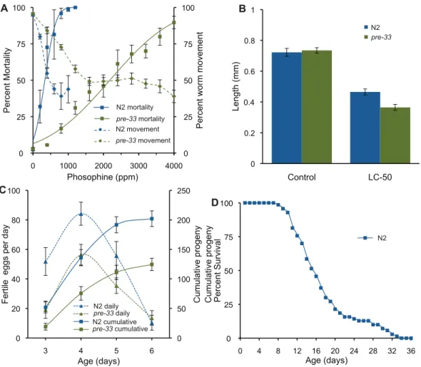
We used the flat bed scanner to compare mortality and behavior in response to phosphine exposure (Figure 2a). The mortality results are comparable to published data [36] and correspond closely with the observed behavioral inhibition.
Toxicity is sometimes reflected in altered growth parameters, which our algorithm is able to determine accurately. This is clearly seen in response to phosphine exposure, which results in growth inhibition of up to 50% in thepre-33mutant strain. It is interesting to note that resistance to phosphine induced mortality inpre-33is not reflected in a corresponding resistance to growth inhibition (Figure 2b).
of peak egg laying to be the same between the two strains within the resolution of the experiment. In contrast the cumulative number of eggs produced by N2 was nearly twice that of the mutant strain. The abbreviated algorithm was also used to monitor lifespan of wild-typeC. elegans. The longevity of the N2 strain was found to be equivalent to published data (Figure 2d) [40].
One potential worry regarding the technique is that the scanning procedure itself could influence the outcome of the assays. To test this possibility, we subjected worms to continuous scanning. This produced a classic habitation pattern to light stimulus similar to that of tap habitation (Figure 3) [41] with significant habituation occurring after the first scan. Additionally,
Continuous scanning over a period of 18 hours, did not induce mortality.
Discussion
WormScan is a readily available, high-throughput image system based on a flatbed scanner. Allowing for the quantification of the four main toxicological endpoints of C. elegans. The main advantages of this high-throughput automated method are ease of setup and low cost. This protocol will help reduce user bias associated with manual counting [42].
The flatbed scanner produces sufficient light intensity to induce negative phototaxis in C. elegans. This produced a classic
Figure 1. Processing and analysis of scanned images.(a) A 16-bit grey scale scan of a 5.5 cm petri dish containing N2 on NGM with OP50. Exposed to 200 ppm of phosphine for 24 hours with 48 hours of recovery. Only 4.5 by 4 mm crop is shown of the 5.5 cm. The black bar represents 1 mm, or 2400 dpi. (b) The sequential scan is taken 90 seconds after first. (c) Image segmentation with particle analysis, this distinguishes the nematodes from the background. Which are outlined with red lines, labeled W1–4. (d) Image difference of consecutive scans that has been thresholded with hysteresis. Where the worm positions are overlaid. This allows for calculation of both worm movement. Mortality is defined as less than less than 10% movement. Worm positions shown in red lines and labels commenting on level of movement.
habituation pattern similar to that of physical stimulus [41]. It has also been shown that exposure from intense blue violet light can induce mortality inC. elegans[32]. While the scanner uses broad spectrum white light rather than blue violet, it is of an intensity that warrants investigation of its effect on the worms. We found that continuous scanning over 18 hours did not cause mortality. Therefore, other than habituation effects, the light of the scanner is unlikely to interfere with the accuracy of extended assays that require repeated scanning.
Behavior in higher organisms is very complex and thus difficult to quantify. In contrast, C. elegans exhibit a simpler behavioral repertoire that consisting primarily of changes in rate or direction of movement [31]. WormScan is restricted to changes in rate of movement, which is suitable for automation of assays that rely on the ability to respond to a physical stimulus such as tapping [17] or a chemical attractant [6]. Behavior as an indicator of toxicity can be 25 to 100 times more sensitive than mortality [43]. In this regard, behavior was shown to be a sensitive assay of phosphine toxicity.
Previous methods for quantifying mortality have much lower throughput than WormScan. Our method provides a uniform
stimulus that induces a robust behavioral response great enough to allow accurate determination of mortality with high-throughput. Defining mortality as a threshold of less that 10% movement helps to eliminate false positives due to image noise. Using these parameters, mortality determination by WormScan is equivalent to that previously reported for phosphine exposure [35,36,37]. Using WormScan for lifespan determination eliminates the need for daily physical stimulation, which greatly reduces the potential for contamination.
WormScan has also been used to automate analysis of length and fecundity inC. elegans. Length ofC. eleganscan be determined by digitally straightening [44,45]. This algorithm has been adopted by WormScan to expand the range of phenotypes that can be assessed. We used this to accurately determine the inhibition of growth ofC. elegansdue to phosphine exposure.
The other favorable properties of a scanner over microscopes with CCD cameras is greater optical density [42] and superior depth of field [46]. This allows for translucent C. elegans to be resolved from the background media. However, the resolution is not sufficient to resolve L1 stage nematodes.
Figure 2. Toxicological end points ofC. elegans.(a) Movement reduction and induced mortality resulting from phosphine exposure. Square boxes indicate mean mortality for a given concentration. Error bars denote the standard error of means from three biological replicates. The corresponding solid line is a probit regression of mortality, calculated in Mathematica 8.0. Dashed lines with diamond boxes represent the mean movement of the replicated experiments, where the error bars represent 95% confidence interval. Overall the LC50of phosphine towards N2 is 337
andpre-33was found to be 2180 ppm. (b) Observed length differences between N2 andpre-33nematode strains after 48 hours recovery from phosphine exposure. Exposure was undertaken on L4 stage nematodes for 24 hours to phosphine at respective LC50phosphine concentrations or air
control. Error bars represent the standard error of mean from 3 biological replicates. (c) Fecundity of N2 andpre-33strains cultured in the absence of phosphine; the cumulative (___) or per day (—). Data was generated from three biological replicates of 6 worms each. The cumulative progeny on day six with 95% confidence is 202613.2 for N2 and 125610.5 forpre-33. (d) Lifespan of N2 cultured in the absence of phosphine. Mean lifespan was 17.360.6 from 3 trials of 48 nematodes per trial. Error is reported as the standard error of the means.
C. elegansmove at a rate of 0.5 mm/s [47], potentially resulting in significant movement between the required 90 seconds for a sequential scan. Current WormScan software can only measure movement up to one body length. This is not generally a problem as exposure to toxins and disease models often display phenotype of greatly decreased movement. Finer characteristics of nematode movement are not possible [48]. Which can be determined through low-throughput video microscope methods [9]. WormS-can and these low-throughput camera based methods are complementary.
Image analysis uses the open source software, ImageJ. This allows improvements to be implemented to overcome current limitations. For example, the current implementation of WormS-can requires non-overlapping nematodes for effective image analysis, which is achieved by limiting worm density to less than 150 individuals on a 5.5 cm diameter plate. However, worms can
be digitally untangled, which could allow a higher density of worms per plate to be analyzed in future studies [49]. Furthermore, ImageJ was adapted to allow WormScan to observe similar sized species.
C. elegansis a model organism that has been applied to a wide range of studies. However, manual phenotypic analysis is labor intensive and time consuming. WormScan automates high-throughput scoring of the most widely used whole-organism assays performed onC. elegans. This affordable, open platform will enable wide adoption with significant potential to reduce data variability between labs [9].
Supporting Information
Figure S1 Size quantification ofTribolium castaneum. (PDF)
Figure S2 Quantification ofTrichogramma. (PDF)
Tutorial S1 This tutorial package describes the minimal system requirements as well as how to access and set up the required open source software on your computer (in the Tutorial.docx file). The Tutorial.docx file also contains a step-by-step description of how to conduct the analyses described in the paper. To run the tutorial, you will use the included custom scripts (the 7 .ijm files) specific to the WormScan image analysis as well as the included set of demonstration images (the 5 .tif files). To begin the tutorial, open the Tutorial.docx file and follow the instructions. To Assist with trouble shooting, a folder labeled Sample_results is included as an example of the results you should expect to obtain from the analysis when you use the provided training file, RoiSet.zip. (ZIP)
Acknowledgments
We appreciate the assistance and advice of Andrew Tuck and David Schlipalius in the development of this method.
Author Contributions
Conceived and designed the experiments: MDM NDM PRE. Performed the experiments: MDM NDM. Analyzed the data: MDM NDM PRE. Contributed reagents/materials/analysis tools: PRE. Wrote the paper: NDM MDM PRE. Designed software: MDM.
References
1. Williams PL, Dusenbery DB (1988) Using the nematode Caenorhabditis elegans to predict mammalian acute lethality to metallic salts. Toxicology and Industrial Health 4: 469–478.
2. Lakowski B, Hekimi S (1998) The genetics of caloric restriction in Caenorhabditis elegans. Proceedings of the National Academy of Sciences of the United States of America 95: 13091–13096.
3. Brenner S (1974) The genetics of Caenorhabditis elegans. Genetics 77: 71–94. 4. Kaletta T, Hengartner MO (2006) Finding function in novel targets: C. elegans
as a model organism. Nature Reviews Drug Discovery 5: 387–398. 5. Chronis N, Zimmer M, Bargmann CI (2007) Microfluidics for in vivo imaging of
neuronal and behavioral activity in Caenorhabditis elegans. Nature Methods 4: 727–731.
6. Albrecht DR, Bargmann CI (2011) High-content behavioral analysis of Caenorhabditis elegans in precise spatiotemporal chemical environments. Nature Methods 8: 599–605.
7. Morgan PG, Cascorbi HF (1985) Effect of anesthetics and a convulsant on normal and mutant Caenorhabditis elegans. Anesthesiology 62: 738–744. 8. Miwa J, Tabuse Y, Furusawa M, Yamasaki H (1982) Tumor promoters
specifically and reversibly disturb development and behavior of Caenorhabditis elegans. Journal of cancer research and clinical oncology 104: 81–87. 9. Ramot D, Johnson BE, Berry TL, Carnell L, Goodman MB (2008) The Parallel
Worm Tracker: a platform for measuring average speed and drug-induced paralysis in nematodes. PLoS ONE 3: e2208.
10. Sznitman R, Gupta M, Hager GD, Arratia PE, Sznitman J (2010) Multi-environment model estimation for motility analysis of Caenorhabditis elegans. PLoS ONE 5: e11631.
11. Williams PL, Dusenbery DB (1990) Aquatic toxicity testing using the nematode, Caenorhabditis elegans. Environmental Toxicology and Chemistry 9: 1285–1290.
12. Harrington LA, Harley CB (1988) Effect of vitamin E on lifespan and reproduction in Caenorhabditis elegans. Mechanisms of ageing and develop-ment 43: 71–78.
13. Middendorf PJ, Dusenbery DB (1993) Fluoroacetic Acid Is a Potent and Specific Inhibitor of Reproduction in the Nematode Caenorhabditis elegans. Journal of nematology 25: 573–577.
14. Popham JD, Webster JM (1979) Cadmium toxicity in the free-living nematode, Caenorhabditis elegans. Environmental research 20: 183–191.
15. Traunspurger W, Haitzer M, Ho¨ss S, Beier S, Ahlf W, et al. (1997) Ecotoxicological assessment of aquatic sediments with Caenorhabditis elegans (nematoda) — a method for testing liquid medium and whole-sediment samples. Environmental Toxicology and Chemistry 16: 245–250.
16. Pulak R (2006) Techniques for analysis, sorting, and dispensing of C. elegans on the COPAS flow-sorting system. Methods in molecular biology (Clifton, NJ) 351: 275–286.
17. Swierczek NA, Giles AC, Rankin CH, Kerr RA (2011) High-throughput behavioral analysis in C. elegans. Nature Methods 8: 592–598.
Figure 3.C. elegansphototaxis habituation.A classic habitation pattern is observed with continuous scanning with a 90-second interval for 20 intervals. Temperature on the scanner surfaces was monitored and found to be 20uC. The light response was measured in triplicate with 30 worms/plate.
18. Gill MS, Olsen A, Sampayo JN, Lithgow GJ (2003) An automated high-throughput assay for survival of the nematode Caenorhabditis elegans. Free Radical Biology and Medicine 35: 558–565.
19. Boyd WA, McBride SJ, Rice JR, Snyder DW, Freedman JH (2010) A high-throughput method for assessing chemical toxicity using a Caenorhabditis elegans reproduction assay. Toxicology and Applied Pharmacology 245: 153–159.
20. Boyd WA, McBride SJ, Freedman JH (2007) Effects of Genetic Mutations and Chemical Exposures on Caenorhabditis elegans Feeding: Evaluation of a Novel, High-Throughput Screening Assay. PLoS ONE 2: e1259.
21. Van Voorhies WA (1992) Production of sperm reduces nematode lifespan. Nature 360: 456–458.
22. Doitsidou M, Flames N, Lee AC, Boyanov A, Hobert O (2008) Automated screening for mutants affecting dopaminergic-neuron specification in C. elegans. Nature Methods 5: 869–872.
23. Burns AR, Kwok TCY, Howard A, Houston E, Johanson K, et al. (2006) High-throughput screening of small molecules for bioactivity and target identification in Caenorhabditis elegans. Nature protocols 1: 1906–1914.
24. Boyd WA, Smith MV, Kissling GE, Freedman JH (2010) Medium- and high-throughput screening of neurotoxicants using C. elegans. Neurotoxicology and teratology 32: 68–73.
25. Rohde CB, Zeng F, Gonzalez-Rubio R, Angel M, Yanik MF (2007) Microfluidic system for on-chip high-throughput whole-animal sorting and screening at subcellular resolution. Proceedings of the National Academy of Sciences of the United States of America 104: 13891–13895.
26. Gosai SJ, Kwak JH, Luke CJ, Long OS, King DE, et al. (2010) Automated high-content live animal drug screening using C. elegans expressing the aggregation prone serpina1-antitrypsin Z. PloS one 5: e15460.
27. Dahle J, Kakar M, Steen HB, Kaalhus O (2004) Automated counting of mammalian cell colonies by means of a flat bed scanner and image processing. Cytometry Part A : the journal of the International Society for Analytical Cytology 60: 182–188.
28. Clarke ML, Burton RL, Hill AN, Litorja M, Nahm MH, et al. (2010) Low-cost, high-throughput, automated counting of bacterial colonies. Cytometry Part A : the journal of the International Society for Analytical Cytology 77: 790–797. 29. Sullivan K, Kloess J, Qian C, Bell D, Hay A, et al. (2012) High throughput virus
plaque quantitation using a flatbed scanner. Journal of virological methods 179: 81–89.
30. Bewes JM, Suchowerska N, McKenzie DR (2008) Automated cell colony counting and analysis using the circular Hough image transform algorithm (CHiTA). Physics in medicine and biology 53: 5991–6008.
31. Dhawan R, Dusenbery DB, Williams PL (1999) Comparison of lethality, reproduction, and behavior as toxicological endpoints in the nematode Caenorhabditis elegans. Journal of Toxicology and Environmental Health Part A 58: 451–462.
32. Edwards SL, Charlie NK, Milfort MC, Brown BS, Gravlin CN, et al. (2008) A Novel Molecular Solution for Ultraviolet Light Detection in Caenorhabditis elegans. PLoS Biology 6: e198.
33. Ward A, Liu J, Feng Z, Xu XZS (2008) Light-sensitive neurons and channels mediate phototaxis in C. elegans. Nature neuroscience 11: 916–922.
34. Stiernagle T (2006) Maintenance of C. elegans. WormBook : the online review of C elegans biology. pp 1–11.
35. Cheng Q, Valmas N, Reilly P, Collins P, Kopittke R, et al. (2003) Caenorhabditis elegans mutants resistant to phosphine toxicity show increased longevity and cross-resistance to the synergistic action of oxygen. Toxicological Sciences 73: 60.
36. Zuryn S, Kuang J, Ebert P (2007) Mitochondrial modulation of phosphine toxicity and resistance in Caenorhabditis elegans. Toxicological Sciences 102: 179–186.
37. Valmas N, Ebert P (2006) Comparative toxicity of fumigants and a phosphine synergist using a novel containment chamber for the safe generation of concentrated phosphine gas. PLoS ONE 1: 130.
38. Hall M, Frank E, Holmes G, Pfahringer B, Reutemann P, et al. (2009) The WEKA data mining software: an update. ACM SIGKDD Explorations Newsletter 11: 10–18.
39. Arganda-Carreras I, Ferna´ndez-Gonza´lez R, Mun˜oz-Barrutia A, Ortiz-De-Solorzano C (2010) 3D reconstruction of histological sections: Application to mammary gland tissue. Microscopy research and technique 73: 1019–1029. 40. Gems D, Riddle DL (2000) Defining wild-type life span in Caenorhabditis
elegans. The journals of gerontology Series A, Biological sciences and medical sciences 55: B215–219.
41. Rankin CH, Broster BS (1992) Factors affecting habituation and recovery from habituation in the nematode Caenorhabditis elegans. Behavioral neuroscience 106: 239–249.
42. Biston M-C, Corde S, Camus E, Marti-Battle R, Este`ve F, et al. (2003) An objective method to measure cell survival by computer-assisted image processing of numeric images of Petri dishes. Physics in medicine and biology 48: 1551–1563.
43. Anderson GL, Boyd WA, Williams PL (2001) Assessment of sublethal endpoints for toxicity testing with the nematode Caenorhabditis elegans. Environmental toxicology and chemistry / SETAC 20: 833–838.
44. Peng H, Long F, Liu X, Kim SK, Myers EW (2008) Straightening Caenorhabditis elegans images. Bioinformatics (Oxford, England) 24: 234–242. 45. Long F, Peng H, Liu X, Kim SK, Myers E (2009) A 3D digital atlas of C. elegans
and its application to single-cell analyses. Nature Methods 6: 667–672. 46. Herler J, Lipej L, Makovec T (2007) A simple technique for digital imaging of
live and preserved small fish specimens. Cybium 31: 39–44.
47. Park S, Hwang H, Nam S-W, Martinez F, Austin RH, et al. (2008) Enhanced Caenorhabditis elegans locomotion in a structured microfluidic environment. PLoS ONE 3: 10.1371/journal.pone.0002550.
48. Geng W, Cosman P, Berry CC, Feng Z, Schafer WR (2004) Automatic tracking, feature extraction and classification of C elegans phenotypes. IEEE transactions on bio-medical engineering 51: 1811–1820.