UNIVERSIDADE ESTADUAL PAULISTA
“JÚLIO DE MESQUISTA FILHO” UNESP
Faculdade de Medicina de Botucatu
Adriele Dandara Levorato
Avaliação da bioquímica sanguínea de
pacientes com paracoccidioidomicose,
tratados com cotrimoxazol ou itraconazol.
Orientador: Prof. Dr. Rinaldo Poncio Mendes
Botucatu/SP 2014
Dissertação apresentada ao
Adriele Dandara Levorato
Avaliação da bioquímica sanguínea de
pacientes com paracoccidioidomicose,
tratados com cotrimoxazol ou
itraconazol.
Orientador: Prof. Dr. Rinaldo Poncio Mendes
Botucatu/SP 2014
Dissertação apresentada ao
Programa de Pós-Graduação em Doenças Tropicais da Faculdade de Medicina de Botucatu, Universidade Estadual Paulista, para obtenção do título de mestre.
UNIVERSIDADE ESTADUAL PAULISTA “JÚLIO DE MESQUITA FILHO” UNESP
Levorato, Adriele Dandara.
Avaliação da bioquímica sanguínea de pacientes com paracoccidioidomicose, tratados com cotrimoxazol ou itraconazol / Adriele Dandara Levorato. - Botucatu, 2014
Dissertação (mestrado) - Universidade Estadual Paulista, Faculdade de Medicina de Botucatu
Orientador: Rinaldo Poncio Mendes Capes: 40101096
1. Paracoccidioides brasiliensis. 2. Paracoccidioidomicose - Tratamento. 3. Agentes antiinfecciosos. 4. Figado - Pesquisa. 5. Vias biliares - Pesquisa. 6. Distúrbios do metabolismo..
Palavras-chave: Alterações hepáticas e metabólicas; Bioquímica sanguínea; Cotrimoxazol; Itraconazol; Paracoccidioides brasiliensis.
“Que os vossos esforços desafiem as impossibilidades, lembrai-vos de que
Aos meus pais Dirço
Dirço
Dirço
Dirço e Luzia
e Luzia
e Luzia
e Luzia, que não mediram
esforços para a minha formação profissional, pelos abraços e
beijos todas às segundas-feiras, por sempre estarem me
esperando aos finais de semana. Obrigada por tudo. Amo
vocês.
À minha irmã Cleice
Cleice
Cleice
Cleice, pelo incentivo, apoio e palavras de conforto
durante os momentos difíceis durante esta caminhada. Amor por toda
a vida.
Ao meu namorado e grande amigo Diego
Diego
Diego
Diego, por
sempre me apoiar nas decisões a serem tomadas, por sempre
dizer “Calma, vai dar tudo certo” e que apesar da distância
manteve-se sempre presente através dos pensamentos. Te
amo.
Ao meu orientador, Dr. Rinaldo “Tiete” Poncio Mendes,
Dr. Rinaldo “Tiete” Poncio Mendes,
Dr. Rinaldo “Tiete” Poncio Mendes,
Dr. Rinaldo “Tiete” Poncio Mendes,
não apenas pela orientação deste trabalho,
mas pelos conselhos, paciência, ensinamentos e
por acreditar na minha capacidade profissional.
Muito obrigada.
Aos pacientes
pacientes
pacientes
pacientes atendidos no Ambulatório de Blastomicose –
A
A
A Deus
Deus
Deus
Deus, por guiar os meus passos, por mostrar os caminhos que devem ser seguidos
e pelo conforto durante os momentos que mais precisei.
Á Coordenação de Aperfeiçoamento de Pessoal de Nível Superior
Coordenação de Aperfeiçoamento de Pessoal de Nível Superior
Coordenação de Aperfeiçoamento de Pessoal de Nível Superior
Coordenação de Aperfeiçoamento de Pessoal de Nível Superior (CAPES) pela
concessão da bolsa de estudo.
A toda equipe de trabalho, Dr. Ricardo de Souza Cavalcante
Dr. Ricardo de Souza Cavalcante
Dr. Ricardo de Souza Cavalcante
Dr. Ricardo de Souza Cavalcante, Dra. Daniela
Dra. Daniela
Dra. Daniela
Dra. Daniela
Vanessa Moris
Vanessa Moris
Vanessa Moris
Vanessa Moris, Tatiane Fernanda Sylvestre
Tatiane Fernanda Sylvestre
Tatiane Fernanda Sylvestre
Tatiane Fernanda Sylvestre, Priscila Zacarias de Azevedo
Priscila Zacarias de Azevedo
Priscila Zacarias de Azevedo
Priscila Zacarias de Azevedo, Vanessa
Vanessa
Vanessa
Vanessa
Martinez Manfio
Martinez Manfio
Martinez Manfio
Martinez Manfio, Jéssi
Jéssi
Jéssi
Jéssica Cristina Bilizário Noguerol Andrade
ca Cristina Bilizário Noguerol Andrade
ca Cristina Bilizário Noguerol Andrade
ca Cristina Bilizário Noguerol Andrade, James Venturini
James Venturini
James Venturini
James Venturini e
Camila Marcheti
Camila Marcheti
Camila Marcheti
Camila Marcheti.
Aos colaboradores desse trabalho, Dra.
Dra.
Dra.
Dra. Daniela Vanessa Moris
Daniela Vanessa Moris
Daniela Vanessa Moris
Daniela Vanessa Moris, Tatiane Fernanda
Tatiane Fernanda
Tatiane Fernanda
Tatiane Fernanda
Sylvestre
Sylvestre
Sylvestre
Sylvestre, Priscila Zacarias de Azevedo
Priscila Zacarias de Azevedo
Priscila Zacarias de Azevedo
Priscila Zacarias de Azevedo, Dr. Ricardo de Souza Cavalcante
Dr. Ricardo de Souza Cavalcante
Dr. Ricardo de Souza Cavalcante
Dr. Ricardo de Souza Cavalcante, Vanessa
Vanessa
Vanessa
Vanessa
Martinez Manfio
Martinez Manfio
Martinez Manfio
Martinez Manfio e Jéssica Cristina B
Jéssica Cristina B
Jéssica Cristina B
Jéssica Cristina Bilizário Noguerol
ilizário Noguerol
ilizário Noguerol
ilizário Noguerol Andrade
Andrade
Andrade
Andrade, pela ajuda no
levantamento de todos os dados, paciência e apoio.
À amiga e profissional Daniela Vanessa Moris
Daniela Vanessa Moris
Daniela Vanessa Moris
Daniela Vanessa Moris, que iniciou o presente trabalho e
que me guiou para dar continuidade. Este mérito também é seu.
Aos amigos e pós-graduandos do Laboratório de Pesquisa de Doenças Tropicais -
LEPDT, Mariana Gatto
Mariana Gatto
Mariana Gatto
Mariana Gatto, Mariana Miziara
Mariana Miziara
Mariana Miziara
Mariana Miziara, Francilene Capel
Francilene Capel
Francilene Capel
Francilene Capel, Laura Mendes
Laura Mendes
Laura Mendes
Laura Mendes, Karen
Karen
Karen
Karen
Ingrid Tasca
Ingrid Tasca
Ingrid Tasca
Ingrid Tasca, Andréia Amaral
Andréia Amaral
Andréia Amaral
Andréia Amaral, Leonardo Medeiros
Leonardo Medeiros
Leonardo Medeiros
Leonardo Medeiros e Thaysa Carvalho
Thaysa Carvalho
Thaysa Carvalho
Thaysa Carvalho,,,, pela convivência,
reclamações e risos. Admiro cada um a seu modo.
Ao funcionário do LEPDT e amigo, Carlos Roberto Gonçalves de Lima
Carlos Roberto Gonçalves de Lima
Carlos Roberto Gonçalves de Lima
Carlos Roberto Gonçalves de Lima –
Carlinhos
Carlinhos
Carlinhos
Carlinhos, pela convivência, conselhos e apoio em todos os momentos.
Às amigas e companheiras de república Mariana Gatto
Mariana Gatto
Mariana Gatto
Mariana Gatto e Tatiane Fernanda
Tatiane Fernanda
Tatiane Fernanda
Tatiane Fernanda
Sylvestre
Sylvestre
Sylvestre
Sylvestre, por todo apoio concedido nos momentos de alegrias e tristezas, risos e pelo
companheirismo durante todos esses anos.
À minha amiga e companheira Tatiane Fernanda Sylvestre
Tatiane Fernanda Sylvestre
Tatiane Fernanda Sylvestre
Tatiane Fernanda Sylvestre, pelo convívio diário
desde o tempo da graduação em Araraquara. Você é muito especial.
Aos funcionários da Pós-Graduação e do Departamento de Doenças Tropicais e
Diagnóstico por Imagem, em especial Solange S. Cagliari
Solange S. Cagliari
Solange S. Cagliari
Solange S. Cagliari e Bruno Quirino Jorgetto
Bruno Quirino Jorgetto
Bruno Quirino Jorgetto
Bruno Quirino Jorgetto, pelo
auxílio e atenção em todos os momentos.
Á coordenadoria
coordenadoria
coordenadoria
coordenadoria e ao programa de Pós
Pós
Pós
Pós----Graduação em Doenças Tropicais
Graduação em Doenças Tropicais
Graduação em Doenças Tropicais
Graduação em Doenças Tropicais, pela
oportunidade de aprimorar meus conhecimentos e confiança em meu trabalho.
3. Results………...21
4. Discussion………....37
5. References………....44
Artigo 1 – Evaluation of hepatobiliary system in patients with paracoccidioidomycosis treated with trimethoprim-sulfamethoxazole or itraconazole………..94
Artigo 2 – Evolução de reações de fase aguda e de variáveis metabólicas em pacientes com paracoccidoidomicose tratados com cotrimoxazol ou itraconazol...95
V) Anexo ...96
Resumo
Estudos sobre as alterações da bioquímica sanguínea na paracoccidioidomicose
(PCM), antes e durante o tratamento, são escassos e em geral apresentam casuísticas
pequenas e curto tempo de seguimento. Assim, foi realizado um estudo prospectivo com
200 pacientes com PCM submetidos à avaliação das alterações hepatobiliares, metabólicas
e reações de fase aguda à admissão e durante o seguimento do tratamento com
cotrimoxazol (CMX) ou itraconazol (ITC). Dos 200 pacientes, 149 apresentaram a forma
crônica (FC) e 51 a forma aguda/subaguda (FA); 31 foram tratados com ITC e 169 com
CMX. Avaliações clínicas e da bioquímica sanguínea foram realizadas antes de se iniciar o
tratamento, definido como momento 0 (M0) e, a seguir, periodicamente, até que os
pacientes apresentassem cura clínica, definida pelo desaparecimento da sintomatologia
apresentada à admissão e redução da velocidade de hemossedimentação (VHS) a valores
normais (M3), que ocorreu após 18 a 23 semanas de tratamento. Entre estes dois
momentos, os pacientes foram avaliados a intervalos definidos em semanas, em relação ao
início do tratamento: M1: 4 a 6 e M2: 7 a 10. Os níveis séricos de bilirrubina conjugada
(BC), bilirrubina total (BT), aspartato aminotransferase (AST), alanina aminotransferase
(ALT), fosfatase alcalina (ϜᾹ), gama-glutamiltransferase (ɣ-GT), triglicerídes, glicose,
colesterol total, lípides totais, cálcio, ácido úrico, fósforo, proteínas totais e frações,
mucoproteínas, α1-glicoproteína ácida e eletroforese de proteínas séricas foram dosados no
Laboratório Central do Hospital das Clínicas da Faculdade de Medicina de Botucatu -
UNESP. Os dados foram apresentados como razões entre o valor absoluto e o limite
superior de normalidade para as variáveis que avaliaram a função hepatobiliar, e com os
limites superiores e, ou, inferiores para as variáveis metabólicas e reações de fase aguda. A
avaliação da função hepática revelou que na admissão do paciente, como decorrência da
enquanto BT, BC e ɣ-GT apresentaram as maiores elevações. Entre as três variáveis com
alterações mais prevalentes, ϜᾹ e ɣ-GT predominaram na FA enquanto ALT não diferiu
segundo forma clínica; BC e ɣ-GT apresentaram maior intensidade na FA enquanto a BT
não diferiu segundo forma clínica. Após introdução do tratamento antifúngico,
observou-se alteração de todas as variáveis; ALT e ϜᾹ foram as variáveis com maior incidência de
elevação; enquanto CB revelou a maior intensidade de elevação. Entre as duas variáveis
com maior incidência, ϜᾹ apresentou maior incidência de elevação na FA que na FC,
enquanto, a incidência de elevação de ALT não diferiu segundo forma clínica. A
incidência de elevação de ϜᾹ e ɣ-GT não variou segundo antifúngico; a elevação dos
níveis séricos de BC e BT foram mais frequentes em pacientes tratados com ITC do que
com CMX. As alterações do tipo hepatocelular predominaram em pacientes tratados com
CMX enquanto, os do tipo colestático leve em doentes que receberam ITC. Os pacientes
que receberam CMX tiveram normalizados os níveis séricos de ALT, BC, FA e ɣ-GT e
reduzidos os de AST e BT durante o tratamento, enquanto, os que receberam ITC
mantiveram elevados os níveis séricos de ALT, BT, BC, ϜᾹ e ɣ-GT, e tiveram reduzidos
apenas os de AST. A avaliação das alterações metabólicas e reações de fase aguda revelou
que na admissão ao Serviço a prevalência de alterações laboratoriais variou de 3,0% para
os níveis séricos elevados de ácido úrico a 98,0% para os níveis de ɣ-globulina em
pacientes com a FA enquanto, aqueles com a FC apresentaram uma variação de 3,5% para
os níveis séricos diminuídos de glicose a 99,3% para os níveis elevados de gama-globulina.
Após introdução do antifúngico, a incidência de alterações variou de 3,2% para os níveis
séricos diminuídos de fósforo a 15,9% para os níveis séricos elevados de triglicérides. Os
pacientes com a FC apresentaram maior incidência de alterações dos níveis séricos de
presente estudo revelou que são muito complexas as alterações da função hepatobiliar e
metabólicas, que devem ser avaliadas e monitoradas durante o tratamento da PCM.
Summary
Studies on the alterations in blood biochemistry in paracoccidioidomycosis (PCM),
before and during treatment are scarce and generally have small patients number and short
follow-up time. Thus, a prospective study was conducted with 200 patients with PCM
submitted to the evaluation of hepatobiliary function, metabolic and acute phase reactions
alterations at admission and during follow-up of treatment with cotrimoxazole (CMX) or
itraconazole (ITC). Of the 200 patients, 149 presented a chronic form (CF), and 51 the
acute subacute form (AF), 31 were treated with ITC, and 169 were treated with CMX.
Clinical and blood biochemistry were performed before starting the treatment, defined as
time 0 (M0), and then periodically until the patients had clinical cure, defined as the
disappearance of the symptomatology presented at admission and regression of the
erythrocyte sedimentation rate to normal (M3), which occurred after 18 to 23 weeks of
treatment. Between these two moments, the patients were evaluated at intervals defined in
weeks, compared to the beginning of treatment: M1: 4-6 and M2:7-10. The serum levels of
conjugated bilirubin (CB), total bilirubin (TB), aspartate aminotransferase (AST), alanine
aminotransferase (ALT), alkaline phosphatase (ϜᾹ), gamma- glutamyltransferase (ɣ-GT),
triglycerides, glucose, total cholesterol, total lipids, calcium, uric acid, phosphorus, total
proteins and fractions, mucoproteins, α1-acid glycoprotein and serum protein
electrophoresis were measured at the Central Laboratory of the Hospital of the Faculty of
Medicine of Botucatu - UNESP. Data were presented as ratios between the absolute value
and the upper limit of normality for the variables that assessed hepatobiliary function, and
with upper and/or lower limit of the metabolic variables and acute phase reactions. The
evaluation of hepatobiliary function revealed that the patient's admission as a result of the
PCM that serum levels of ϜᾹ and ɣ- GT were the variables with the highest prevalence,
more frequent alterations, the ϜᾹ and ɣ-GT predominated in AF, while ALT did not differ
by clinical form; ɣ-GT and CB showed higher intensity in AF, while BT did not differ by
clinical form. After introduction of antifungal treatment, we observed changes in all
variables, ALT and ϜᾹ were the variables with the highest incidence of elevation, while
CB had the highest intensity of elevation. Between the two variables with the highest
incidence, ϜᾹ showed a higher incidence of elevation in AF than in CF, while the
incidence elevation of ALT did not differ by clinical form. The incidence of elevated ϜᾹ
and ɣ-GT serum levels did not vary according antifungal; elevation of serum levels of CB
and TB were more frequent in patients treated with ITC than those CMX. Alterations of
hepatocellular type predominated in patients treated with CMX, while the mild cholestatic
type in patients receiving ITC. Patients who had received CMX normalized serum levels of
ALT, CB, ALP and ɣ-GT and low the AST and TB, whereas those who received ITC
remained with elevated serum levels of ALT, TB, CB, ALP and ɣ-GT, and had only
reduced the AST. The evaluation of metabolic alterations and acute phase reactions
revealed that at admission to Service the prevalence of laboratory abnormalities ranged
from 3.0% to elevated uric acid serum levels to 98.0% for ɣ-globulin serum levels in
patients with AF, while those with CF varied from 3.5% glucose serum levels decreased to
99.3 % for the high levels of gamma- globulin. After introduction of antifungal, the
incidence of alterations ranged from 3.2% for phosphorus serum levels decreased to 15.9
% for elevated serum levels of triglycerides. Patients with CF had higher incidence of
alterations in serum levels of total cholesterol, triglycerides, glucose, calcium and uric acid
than those patients with AF. The present study revealed that are very complex the
alterations of hepatobiliary function and metabolics, which must be evaluated and
AD Levorato 2 sido descritos na América Central, e no México, motivo pelo qual deixava de ser
unicamente sul-americana. Além disso, já se iniciava uma nova abordagem na sistemática
de fungos, ligando-se o nome da doença ao de seu agente etiológico.
Uma das principais características desse fungo imperfeito é o dimorfismo térmico.
Em tecidos humanos e em culturas mantidas a 37°C apresenta-se na fase leveduriforme,
sua forma patogênica, com parede rica em a-glucana. Em temperatura ambiente
apresenta-se na faapresenta-se filamentosa, sua forma infectante, que ocorre em natureza, com parede rica em
b-glucana.2
Apesar de existirem áreas endêmicas bem definidas para este patógeno, o nicho
ecológico de sua fase sapróbia continua mal caracterizado, devido ao pequeno número de
vezes em que o fungo foi isolado do ambiente 7,8 ao longo período de latência da doença 9 e
ao pequeno número de relatos da doença em animais domésticos 9 ou selvagens11,12. Em
1986, Naiff et al.13 relataram o isolamento de P. brasiliensis em tatus (Dasypus
novemcinctus) estudados em Tucuruí - Pará, área considerada não-endêmica para esta
doença.
Admite-se que a infecção seja adquirida quando propágulos da fase micelial do
fungo são inalados, instalando-se nos alvéolos pulmonares. A seguir, o fungo passa à fase
leveduriforme, transformação considerada fundamental para que se estabeleça a infecção
14,15. O fungo pode, então, se disseminar por via hematogênica e/ou linfática para qualquer
parte do organismo.14 A penetração cutânea direta do fungo é muito rara, apesar de ter sido
AD Levorato 3 Ao entrar no organismo, P. brasiliensis pode ser destruído imediatamente ou
multiplicar-se, produzindo uma lesão de inoculação e se disseminando para linfonodos
regionais. Desta forma, constitui-se o complexo primário, formado pelo polo
parenquimatoso, pela linfangite ascendente e pelo pólo ganglionar. A infecção
paracoccidióidica pode regredir ou progredir, dependendo de fatores ligados ao fungo e ao
hospedeiro. A regressão pode ser acompanhada de destruição de todos os fungos,
formando-se cicatriz local estéril ou se acompanhar da persistência de focos quiescentes,
com fungos viáveis. A progressão da infecção determina o aparecimento de sinais e
sintomas, o que caracteriza a doença ativa 16. Os focos latentes podem apresentar
reativação posterior, denominada reinfecção endógena, e levar à doença ativa.
As manifestações clínicas da PCM se relacionam, em geral, ao comprometimento
de pulmões, pele, mucosa das vias aerodigestivas superiores, adrenais e de órgãos ricos no
sistema fagocítico mononuclear, tais como fígado, baço, linfonodos e medula óssea 14. No
entanto, deve-se registrar que a PCM pode comprometer qualquer órgão, aparelho ou
sistema.
A PCM se apresenta sob três formas clínicas principais: forma aguda ou subaguda,
forma crônica e forma residual. A forma aguda ou subaguda, também chamada forma
juvenil, é responsável por 20 a 25% dos casos, acomete em geral crianças, adolescentes e
adultos jovens, caracteriza-se por apresentar instalação mais rápida, de algumas semanas a
poucos meses e apresentar envolvimento predominante do sistema reticuloendotelial, isto
é, baço, fígado, nódulos linfáticos e medula óssea. Nessa forma clínica as manifestações
pulmonares são raras e a presença de lesões de mucosa das vias aerodigestivas superiores é
pouco frequente. Nos tecidos são encontrados muitos fungos em multiplicação e
granulomas em geral frouxos. A forma crônica ou do adulto ocorre em 75% a 80% dos
AD Levorato 4 clínica de longa duração, com frequência acima de seis meses. As manifestações
pulmonares são muito frequentes e em geral associadas ao comprometimento de outros
órgãos, tais como mucosa das vias aerodigestivas superiores, pele e, por vezes, adrenais.
As formas residuais, também denominadas sequelas, são observadas após tratamento e se
caracterizam pelas manifestações clínicas ligadas às sequelas observadas após tratamento,
entre as quais se destacam fibrose e enfisema pulmonar e síndrome de Addison.17,18
Como não é doença de notificação compulsória no Brasil, a real prevalência da
PCM não pode ser calculada. Coutinho et al.19 estudaram 3181 óbitos por PCM no Brasil,
entre 1980 e 1995, e demonstraram a grande magnitude e a baixa visibilidade dessa
micose, destacando que constituía a oitava causa de mortalidade por doença
predominantemente crônica ou repetitiva, entre as infecciosas e parasitárias, e a mais
elevada taxa de mortalidade entre as micoses sistêmicas. Os autores também relataram que
a taxa média de mortalidade anual era de 1,45 para um milhão de habitantes. No Estado de
São Paulo, a paracoccidioidomicose passou a ser de notificação obrigatória a partir de
junho de 2009.
A infecção é muito mais frequente que a doença, pois muitos indivíduos se
infectam com o fungo, mantêm focos latentes por toda a vida e nunca adoecem. Inquérito
realizado em Pratânia (SP), por meio de reações intradérmicas com paracoccidioidina e
histoplasmina, no mesmo indivíduo, revelou que 51,2% da população eram, ao mesmo
tempo, paracoccidioidino-positivos e histoplasmino-negativos, o que demonstra elevada
prevalência de infecção paracoccidióidica, pois os outros fungos que poderiam propiciar
reação cruzada com antígenos de P. brasiliensis não têm sido identificados na região. Esse
mesmo estudo revelou que, entre crianças de 5 a 13 anos de idade, essa prevalência era
AD Levorato 5 A PCM é observada em pacientes que tiveram ou se encontram em contato direto e
prolongado com o solo, como os trabalhadores rurais. 3,14,21,22 Predomina em indivíduos do
sexo masculino, com razão de masculinidade de 7,2:1, e é mais prevalente na faixa etária
entre 30 e 59. 3,21,22,23 O estrogênio pode explicar a menor prevalência de PCM em
pacientes do sexo feminino, pois retarda ou impede as transições micélio-levedura e
conídio-levedura, necessárias para que o fungo atinja a fase patogênica, o que pode
explicar o reduzido número de mulheres afetadas pela doença na idade adulta e a
observação da mesma prevalência em ambos os sexos em pacientes com idade inferior a 13
anos.22,24
Os doentes com PCM revelam comprometimento imune celular específico, isto é,
resposta deficiente a antígenos de P. brasiliensis, mas não aos de outros agentes
infecciosos.25 Falhas na apresentação do antígeno, excesso de antígenos e de
imunocomplexos circulantes e deficiência de receptores para interleucina-2 (IL-2) parecem
estar ligados a uma resposta insatisfatória das células T. O comprometimento da resposta
das células T se acompanha de diminuição da atividade fungicida dos macrófagos.
Portanto, os pacientes apresentam comprometimento do perfil Th1 da resposta imune
celular, com baixos níveis de TNF-a, IFN-g e IL-2, associado à manutenção ou à elevação
da produção de IL-5, IL-10 e TGF-b, que caracteriza a exacerbação do perfil Th2. 26
A produção de anticorpos específicos se encontra aumentada em pacientes com as
formas aguda e crônica da PCM. 27 No entanto, além de facilitar a opsonização de células fúngicas, não se conhece outra ação dos anticorpos na defesa do hospedeiro. Além disso,
deve-se registrar que a gravidade da doença guarda relação inversa com a resposta imune
AD Levorato 6 1.1. Aspectos laboratoriais da paracoccidioidomicose
Sendo a PCM uma doença sistêmica, além de cuidadosas ananmese e exame físico,
uma série de exames complementares deve ser solicitada, antes do tratamento e durante o
seguimento de cada paciente visando a avaliação da gravidade do paciente, assim a
resposta ao tratamento. Esses exames compreendem hemograma completo, velocidade de
hemossedimentação, provas bioquímicas hepáticas, renais e metabólicas e eletroforese de
proteínas. 30
O hemograma no momento do diagnóstico da PCM revela em geral anemia
normocítica e normocrômica. Leucocitose pode ser observada com maior frequência nos
pacientes com a forma clínica crônica grave. A eosinofilia é a alteração mais frequente da
PCM, principalmente em pacientes com a forma aguda/subaguda. A velocidade de
hemossedimentação está aumentada na maior parte dos casos, normalizando-se durante o
tratamento. 31-33
Os exames bioquímicos realizados antes da introdução do tratamento antifúngico
refletem alterações induzidas pela PCM que devem se normalizar após terapêutica eficaz.
Por outro lado, há exames que se revelam normais antes da introdução do tratamento e que
se alteram após a utilização do antifúngico, refletindo sua toxicidade. Entre eles
destacam-se os que avaliam fígado e rins.
Assim, esse estudo bioquímico contribui de forma relevante para uma avaliação
mais completa do paciente e de sua resposta ao tratamento. Apesar de sua importância,
poucos são as publicações que avaliaram a bioquímica do sangue de pacientes com PCM e
são raros os estudos que abordam o seguimento após introdução do tratamento
AD Levorato 7 Os níveis séricos de mucoproteínas, α1-glicoproteína ácida e proteína C reativa
também se encontram elevados e tendem à normalização durante o tratamento. A
eletroforese de proteínas séricas apresenta diminuição dos níveis de albumina e elevação
dos níveis da γ-globulina, estas alterações revertem-se com o tratamento.32,34 Um estudo com 30 pacientes com a forma crônica e 12 com a forma aguda, revelou o aumento dos
níveis de α2-globulina e ɣ-globulina na forma crônica e níveis de proteínas totais, α1
-globulina, α2-globulina e ɣ- globulina na forma aguda/subaguda. 35
Nogueira et al, monitoraram dados laboratoriais à admissão e durante o seguimento
da terapia de 38 crianças com paracoccidioidomicose com idade até 14 nos, tratados por
24-30 meses com derivado sulfamídico ou cetoconazol. Os exames revelaram anemia,
eosinofilia, bilirrubinas e aminotransferases elevadas até três meses de tratamento,
hipoalbuminemia, até seis meses e leucócitos, velocidade de hemossedimentação
normalizaram-se após nove a 12 meses de tratamento. 36
1.2. Tratamento da paracoccidioidomicose
O tratamento da PCM consiste em duas fases: inicial ou de ataque e de manutenção.
O tratamento de ataque persiste até que se observem cura clínica e normalização da
velocidade de hemossedimentação. O tratamento de manutenção deve ser mantido até um
ano após as curas micológica, radiológica e sorológica. As drogas eficazes contra a PCM
compreendem três grupos: anfotericina B, do grupo dos antibióticos poliênicos;
sulfadiazina e associação sulfametoxazol-trimetoprim; e azólicos, entre os quais se
destacam o cetoconazol, derivado imidazólico e o itraconazol, derivado triazólico de
AD Levorato 8 A associação sulfametoxazol-trimetoprim também denominada cotrimoxazol
(CMX), revelou-se muito eficaz no tratamento da PCM. Queixas de intolerância gástrica
são relatadas em alguns casos, sendo necessária a substituição da medicação. A
hepatotoxicidade é observada com frequência, com discreto aumento dos níveis séricos de
aminotransferases, bilirrubinas, fosfatase alcalina e γ-glutamiltransferase. Além disso,
também se observa alterações da função renal, com discreto aumento dos níveis séricos de
ureia e creatinina. O CMX apresenta vantagem de ser distribuído gratuitamente pelos
serviços ligados ao Ministério da Saúde e Secretaria de Saúde do Estado de São Paulo,
para tratamento de doenças bacterianas e da PCM. Assim, os resultados satisfatórios
observados no tratamento da PCM, associada à sua distribuição gratuita, fizeram com que
o CMX se tornasse a droga de escolha no tratamento da maior parte dos pacientes com essa
micose sistêmica. 37
O itraconazol (ITC), na maioria dos casos relatados na literatura, é uma excelente
opção no tratamento da PCM, devido sua eficácia e tolerabilidade. Derivado triazólico de
primeira geração com uma afinidade seletiva pelo citocromo P-450 da célula fúngica, esta
característica permite que pequenas concentrações do itraconazol dificultem a biossíntese
do ergosterol, sem interferir nas substâncias dependentes do citocromo P-450 humano,
AD Levorato 9 2. Objetivos
2.1. Objetivo geral
1. Avaliar a bioquímica sanguínea de pacientes com paracoccidioidomicose (PCM)
antes e após a introdução do tratamento antifúngico.
AD Levorato 10 5. Lacaz CS, Porto E, Martins JEC. Micologia Médica. In: Paracoccidoidomicose. 8ª
ed. São Paulo, Brasil: Editora Sarvier, 1991; p. 248-61.
6. Ajello L. Paracoccidioidomycosis: a historical review. In: Paracoccidioidomycosis.
PAHO Sci. Publ.; 1972. p.254.
7. Albornoz MB. Isolation of Paracoccidioides brasiliensis from rural soil in
Venezuela. Sabouraudia 1971; 9: 248-53.
8. Franco M, Bagagli E, Scapolio S, Lacaz CS. A critical analysis of isolation of
Paracoccidioides brasiliensis from soil. Med Mycol 2000; 38: 185 – 91.
9. Restrepo A. Morphological aspects of Paracoccidioides brasiliensis in lumph
nodes: implications for the prolonged latency of paracoccidioidomycosis? Med
Mycol 2000; 38: 317-22.
10.Ricci G, Mota FT, Wakamatsu A, Serafim RC, Borra RC, Franco M. Canine
paracoccidioidomycosis. Med Mycol 2004; 42: 379-83.
11.Grose E, Tamsitt JR. Paracoccidioides brasiliensis recovered from the intestinal
tract of three bats (Artibeus lituratus) in Colombia, S.A. Sabouraudia 1965; 4(2):
124-5.
12.Costa EO, Diniz LSM, Fava Netto C, Arruda C, Dagli MLZ. Delayed
hypersensitivity test with paracoccidioidin in captive Latin America wild mammals.
J Med Vet Mycol 1995; 33: 39-42.
13.Naiff RD, Ferreira LCL, Barrett TV, Naiff MF, Arias JR. Paracoccidioidomicose
enzoótica em tatus (Dasypus novemcinctus) no estado do Pará. Rev Inst Med Trop
São Paulo 1986; 28(1): 19-27.
14.Franco M. Host-parasite relationships in paracoccidioidomycosis. J Med Vet Mycol
AD Levorato 11 15.Camargo ZP, Franco MF. Current knowledge on pathogenesis and
immunodiagnosis of paracoccidioidomycosis. Rev Iberoam Micol 2000; 17:41-48.
16.Franco M, Montenegro MR, Mendes RP, Marques SA, Dillon NL, Mota NGS.
Paracoccidioidomycosis: a recently proposed classification of its clinical forms.
Rev Soc Bras Med Trop 1987; 20(2): 129-32.
17.Mendes RP. The gamut of clinical manifestations. In: Franco M, Lacaz CS,
Restrepo-Moreno A, Del Negro G. Paracoccidioidomycosis. Boca Raton: CRC
Press; 1994. p. 233-58.
18.Manual de Vigilância e Controle da Paracoccidioidomicose. Secretaria de Estado
da Sáude - Coordenadoria de Controle de Doenças - Centro de Vigilância
Epidemiológica “Prof. Alexandre Vranjac”. São Paulo, 2008.
19.Coutinho Z F, Silva D, Lazéra M, Petri V, Oliveira R M, Sabroza P C, et al.
Paracoccidioidomycosis mortality in Brazil (1980-1995). Cad Saúde Pública 2002;
18(5): 1441-54.
20.Rodrigues CC. Avaliação da infecção por Histoplasma capsulatum por meio de
reações intradérmicas em moradores da zona urbana e rural do Município de
Pratânia (SP) [tese]. Pós Graduação em Doenças Tropicais: Faculdade de Medicina
de Botucatu / UNESP; 2004.
21.Giraldo R, Restrepo A, Gutiérrez F, Robledo M, Londoño F, Hernández H, et al.
Pathogenesis of paracoccidioidomycosis: a model based on the study of 46
pacients. Mycopathologia 1976; 58(2): 63-70.
22.Restrepo A, Salazar ME, Cano LE, Stover EP, Feldman D, Stevens DA. Estrogens
inhibit mycelium-to-yeast transformation in the fungus Paracoccidioides
brasiliensis: implications for resistance of females to paracoccidioidomycosis.
AD Levorato 12 23.Moreto TC, Marques MEA, Oliveira MLSC, Moris DV, Carvalho LR, Mendes RP.
Trans R Soc Trop Med Hyg 2011; 105: 473– 478
24.Borges-Walmsley MI, Chen D, Shu X, Walmsley AR. The pathobiology of
Paracoccidioides brasiliensis. Microbiologia 2002; 10(2): 80-7.
25.Benard G, Hong MA, Del Negro GMB, Batista L, Shikanai-Yasuda MA, Duarte
AJS. Antigen-specific immunosupression in paracoccidioidomycosis. Am J Trop
Med Hyg 1996; 54(1): 7-12.
26.Benard G, Romano CC, Cacere CR, Juvenale M, Mendes-Giannini MJS, Duarte
AJS. Imbalance of IL-2, IFN-g and IL-10 secretion in the immunosupression
associated with human paracoccidioidomycosis. Cytokine 2001; 13(4): 248-52.
27.Bueno JP, Mendes-Giannini MJS, Del Negro GMB, Assis CM, Takiguti CK,
Shikanai-Yasuda MA. IgG, IgM and IgA antibody response for the diagnosis and
follow-up of paracoccidioidomycosis: comparison of counter
immunoelectrophoresis and complement fixation. J Med Vet Mycol 1997; 35:
213-17.
28.Mota NGS, Rezkallah-Iwasso MT, Peraçoli MTS, Audi RC, Mendes RP,
Marcondes J, et al. Correlation between cell-mediated immunity and clinical forms
of paracoccidioidomycosis. Trans R Soc Trop Med Hyg 1985; 79: 765-72.
29.Biagioni L, Souza MJ, Chamma LG, Mendes RP, Marques SA, Mota NGS, Franco
M. Serology of paracoccidioidomycosis. II. Correlation between class-specific
antibodies and clinical forms of the disease. Trans R Soc Trop Med Hyg 1984; 78:
617 – 21.
30.Shikanai-Yasuda, Telles Filho FQ, Mendes RP, Colombo AL, Moretti ML.
AD Levorato 13 31.Cunha AC, Almeida FP, Lacaz CS. O hemograma e o mielograma na granulomatose
paracoccidióidica (Blastomicose Sul-Americana). An Fac Med 1948; 24-26.
32.Shikanai-Yasuda MA, Higaki Y, Uip DE, Mori NS, Del Negro G, et al.
Comprometimento da medula óssea e eosinofilia na paracoccidioidomicose. Rev
Inst Med Trop 1982; 34:85.
33.Martinez R, Fiorillo AM. Efeito do tratamento da blastomicose sul-americana
(paracoccidioidomicose) sobre parametros do metabolismo do ferro. Rev Inst Med
Trop 1978; 20:190.
34.Fiorillo AM, Takaoka L, Fernandes LAR. Estudo das proteínas séricas na
blastomicose sul-americana. Rev Soc Bras Med Trop 1972; 6:117.
35.Marquez AD, Moreira APV, Leonello PC, Nakanishi FA, Itano EN. Serum proteins
and fractions, HDL-cholesterol and total IgG and IgE levels in cases of acute and
chronic paracoccidioidomycosis. Rev Soc Bras Med Trop 2009; 42(3): 245-49.
36.Nogueira MGS, Andrade GMQ, Tonelli E, Diniz SN, Goes AM, Cisalpino PS.
Aspectos laboratoriais evolutivos de crianças em tratamento da
paracoccidioidomicose. Rev Soc Bras Med Trop 2006; 39(5): 478-83.
37.Barbosa W, Vasconcellos WMP. Ação da sulfametoxazol associada ao trimetoprim
na terapêutica da blastomicose sul-americana. Rev Patol Trop 1973; 3(2): 329-39.
38.Naranjo et al. Treatment of paracoccidioidomycosis with itraconazole. J Med Vet
Mycol 1990; 28:67-76.
39.Mendes RP, Barraviera B, Souza LR, Pereira PCM, Marcondes-Machado J, Franco
MF, et al. Evaluation of itraconazole in the treatment of paracoccidioidomycosis
II.
II.
AD Levorato 14 II) Artigo 1 - Evaluation of hepatobiliary system in patients with
paracoccidioidomycosis treated with trimethoprim-sulfamethoxazole or
itraconazole.
Adriele D. Levoratoa, Daniela V. Morisb, Ricardo de Souza Cavalcantea, Tatiane F.
Sylvestrea, Priscila Z. de Azevedoa, Lídia R Carvalhoc, Rinaldo P. Mendesa.
a Tropical Disease Area, Faculdade de Medicina de Botucatu- São Paulo State
University-UNESP, Brazil.
b Universidade do Oeste Paulista – UNOESTE, Brazil.
c Bioestatisc Department, Biosciences Institute, São Paulo State University-UNESP,
Brazil.
Abstract
A prospective study was performed in 200 paracoccidioidomycosis (PCM) patients,
51 of whom with the acute/subacute form (AF) and 149 with the chronic form (CF),
submitted to the evaluation of the hepatobiliary system at admission and during the
follow-up treatment with the trimethoprim-sulfamethoxazole combination - SXT or itraconazole -
ITC. This study aimed to better evaluate the involvement of the hepatobiliary system in
PCM and the effect of these antifungal compounds on the hepatobiliary system. Clinical
and blood chemistry evaluations were performed before treatment (moment 0 – M0) and
periodically until the patients had reached clinical cure and normal erythrocyte
sedimentation rate (moment 3 - M3). Serum levels of conjugated bilirubin (CB), total
bilirubin (TB), aspartate aminotransferase (AST), alanine aminotransferase (ALT), alkaline
phosphatase (ALP) and gamma-glutamyltransferase (ɣ-GT) were evaluated. At admission,
AD Levorato 15 the prevalence of lab tests with elevated values ranged from 6.2% for TB to 32.6% for
ϒ-GT. After treatment, the incidence of elevated serum levels ranged from 8.5% for TB to
33.0% for ALT. This incidence was higher in the AF than in the CF for the variables CB,
AST, ALP and ϒ-GT; however, the intensity of the elevations was higher in the CF. The
predominant pattern of the alterations differed as to antifungal compound: hepatocellular in
patients treated with SXT (64.7%) and mixed/mild with ITC (60.0%). The course of the
alterations during the treatment showed regression to normal values in patients receiving
SXT and persistence with ITC. Our findings contribute to a safe follow-up of PCM
patients under treatment.
1. Introduction
Paracoccidioidomycosis (PCM), an endemic disease in Latin America, is a systemic
mycosis caused by thermally dimorphic fungi of the Paracoccidioides brasiliensis and
Paracoccidioides lutzii complexes (1,2,3). When not appropriately treated, PCM
progressively worsens and may end in the death of the patient.
A Brazilian study on death certificates 1980 – 1995 revealed a mortality rate from
PCM of 1.45/1,000,000 inhabitants and identifies this mycosis as the eighth leading cause
of death by predominantly chronic or recurrent infectious and parasitic diseases (4). A
study performed in São Paulo State (Brazil) between 1985 and 2005 reported a PCM
mortality rate of 2.66/1.000.000 inhabitants, 1.58 of them from the disease and 1.08 from
associated causes, and in the Botucatu region (São Paulo State), these numbers reach 8.73,
4.89 and 3.84, respectively, demonstrating the hyper-endemic status of this area and the
AD Levorato 16 Itraconazole (ITC) and the trimethoprim-sulfamethoxazole combination (SXT) are
the main antifungal compounds used in PCM treatment, which should be maintained for a
long time (6-17).
To better evaluate every PCM patient and to monitor the treatment, several
laboratory tests are necessary: acute phase reactions, erythrocyte sedimentation rate, blood
chemistry evaluating the hepatobiliary and kidney system, and metabolic alterations.
However, few studies have evaluated the hepatobiliary function of PCM patients and are
limited by small patient numbers and short follow-up periods. (7,8,9,10,11,13,15,17).
Thus, this study aimed to better evaluate the hepatobiliary system of PCM patients
at admission before introduction of the treatment as well as to evaluate the effects of the
antifungal compounds ITC and SXT on these functional parameters.
2. Patients and Methods
This study was performed at the Infectious Diseases Service of the University
Hospital of the Faculdade de Medicina de Botucatu, São Paulo State University (UNESP).
2.1. Study design
A prospective study was conducted in 200 PCM patients submitted to the
evaluation of hepatobiliary system at admission at our institution and during the treatment
follow-up with the SXT or ITC. One hundred forty-nine patients presented the chronic
form (CF), and 51 the acute/subacute form (AF). Thirty-one patients were treated with ITC
AD Levorato 17 formulation once a day at a dose of 200 mg, and SXT was administered with 400 mg of
sulfamethoxazole and 80 mg of trimethoprim using 3 tablets twice each day throughout the
study period.
Clinical and blood chemistry evaluations were performed before starting the
treatment, defined as moment 0 (M0), and periodically until the patients exhibited clinical
cure and regression of the erythrocyte sedimentation rate (ESR) to normal values, called
the final moment- M3 (18to 23 weeks). Clinical cure was defined as the disappearance of
the symptomatology previously exhibited by the patient. Between these two moments, the
patients were evaluated at intervals defined in relationship to the number of weeks of
treatment: M1: 4 to 6; M2: 7 to 10.
The study protocol was reviewed and approved by the institutional ethics
committee.
2.2. Study population
The criteria for enrollment in the study included an age of 12 years or more and
PCM confirmed by both a suggestive clinical picture and the identification of typical P.
brasiliensis yeast forms in clinical materials (confirmed cases) or a suggestive clinical
picture associated with serum antibodies determined by the double agar gel
immunodiffusion test (probable cases). The patients included in the study were admitted
between 1988 and 2011. Patients were excluded if they had other systemic diseases of
infectious, inflammatory or neoplastic etiology; if they were pregnant or lactating; if they
had a previous history of hypersensitivity or severe side effects to azoles or SXT; or if they
were taking medications that affect the pharmacokinetics of these antifungal compounds or
AD Levorato 18 2.3. Classification of clinical forms
The classification of the clinical forms was carried out according to Mendes (17)
and was done by the infectious diseases medical doctor responsible for attending the
patients (6).
2.4. Outcomes
The prevalence, incidence, intensity and course of the hepatobiliary alterations were
evaluated for patients treated with each antifungal compound. Comparisons between the
groups were also performed.
2.5. Methods
Clinical information from each patient was obtained from the corresponding card,
which was filled based on the specific record and from the attending physicians during the
follow-up.
All of the laboratory data were collected at the Central Laboratory of the University
Hospital. Serum levels of conjugated bilirubin (CB), total bilirubin (TB), aspartate
aminotransferase (AST), alanine aminotransferase (ALT), alkaline phosphatase (ALP) and
ɣ-glutamyltransferase (ɣ-GT) were the parameters measured to evaluate the hepatobiliary
AD Levorato 19 2.6. Prevalence and Incidence
Prevalence indicates the percentage of patients with altered variables at admission
and incidence the percentage of patients who developed alterations during the period of
study, i.e., variables initially normal but altered after introduction of the antifungal
compound or variables initially altered which increased the alterations after treatment.
2.7. Criteria of drug-induced liver injury
The criteria of drug-induced disorders used in the present study were proposed in
an international consensus with a small modification and has been briefly presented (18).
All of the liver tests were expressed as a ratio between the value measured and the
upper limit of the normal range (N). In the evaluation of its intensity the highest value
observed after introduction of the antifungal compound was used. When the increase in the
liver test results was between N and 2N, the finding was considered an “abnormality”, as it
was suggested by the consensus (17), or a mild injury.
Liver injury was classified as hepatocellular, cholestatic or mixed according to the
biochemical profile. Hepatocellular injury was defined as an increase of ALT alone or R
≥5, where R is the ratio between the serum activity of ALT and the serum activity of ALP,
measured at the same time and expressed as multiple of N. Cholestatic injury was defined
as the increase of ALP above 2N alone or R≤2. Mixed injury was diagnosed when both
ALT (above 2N) and ALP were increased and when R was between 2 and 5 (2<R<5). Mild
mixed injury was diagnosed when both ALT and FA were between 1.1 and 1.9 and when
AD Levorato 20 The erythrocyte sedimentation rate (ESR) was determined by the Westergreen
method employing blood anticoagulated with EDTA. The results were measured as
millimeters in the first hour, and the upper limits of the reference values for men and
women, respectively, are as follows: a) below age 50 years: 15 and 20; b) between ages 50
and 85 years: 20 and 30; and c) above 85 years: 30 and 42 (19,20).
2.8. Statistical analysis
All of the variables were submitted to a statistical analysis. The treatment groups
were compared by the Kruskal-Wallis test for ordinal measurements and the chi-square test
or Fisher´s exact test for categorical measurements. The Wilcoxon test was performed for
the analysis of paired samples. The t test was performed for the analysis of unpaired
samples. Comparison of moments between clinical forms and treatment were used the tests
of Friedman and Mann-Whitney. Significance was set at p ≤ 0.05. These analysis were
AD Levorato 21 3. Results
3.1. Evaluation of homogeneity all the groups.
The patients treated with ITC did not differ from those who receveid SXT as to age,
gender, clinical form and severity (Table 1).
Table 1. Distribution of 200 paracoccidioidomycosis patients evaluated for hepatobiliary system as to age, gender, clinical form and treatment.
Treatment p value
ITC (n=31) CMX (n=169)
Age (years) 42,6 ±15,1 43,0±14,5 0,91
Male gender (%) 84,3 88,7 0,49
Clinical form (%)
Acute 34,3 24,0 0,10
Chronic 65,7 76,0
Acute form (%) 0,45
Severe 28,2 21,6
0,41
Moderate 6,2 2,4
Chronic form (%)
Mild 6,2 3,6
Moderate 53,2 56,9
Severe 6,2 15,6
ITC – itraconazole; CMX – cotrimoxazole; Comparison of frequencies: chi-square test; Comparison of means: Unpaired t test.
The results were be presented using tables with the number of individuals evaluated
AD Levorato 22 3.2. The evaluation of hepatobiliary system upon admittance to service (M0).
The prevalence of lab tests with elevated values ranged from 6.2% for TB serum
levels to 32.6% for ϒ-GT serum levels (Table 2).
Table 2- Prevalence of increased serum levels of tests evaluating the hepatobiliary
system of patients with active paracoccidioidomycosis before treatment
(moment M0).
Variable Patients (n) Prevalence (%)
AST 181 12.7BC
ALT 181 18.2BC
TB 178 6.2C
CB 175 12.0BC
ALP 181 26.0AB
ɣ-GT 181 32.6A
Teste de Cochran (p=0.00001). TB-total bilirrubins; CB-conjugated bilirubin;
AST-aspartate aminotransferase; ALT-alanine aminotransferase; ALP-alkaline phosphatase; ɣ-GT- ɣ-glutamyltransferase. Capital letters compare frequencies in the same column; frequencies with the same letter do not differ (p>0.05);
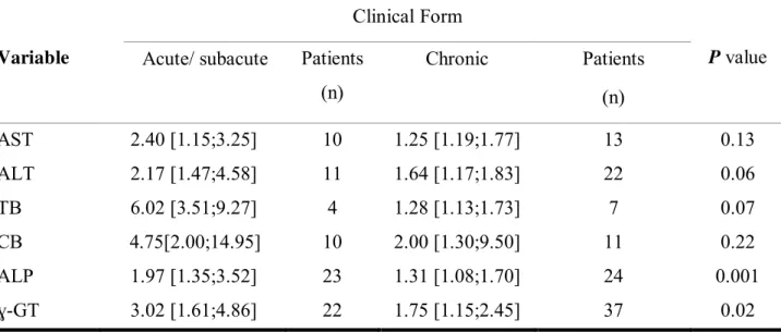
AD Levorato 23 A comparison of the elevated variables among the two clinical forms of PCM
revealed that the median AST and CB values did not differ between the acute/subacute
form (AF) and the chronic form (CF). However, the median ALP and ϒ-GT values were
higher in AF patients than in CF patients, while ALT and TB values tended to be higher in
AF patients (Table 3).
Table 3- Median, 1st and 3rd quartiles of the increased serum levels of tests which evaluate the hepatobiliary system of patients with active paracoccidioidomycosis before
treatment (moment M0), as to clinical forms.
Variable
Clinical Form
P value Acute/ subacute Patients
(n)
Chronic Patients
(n)
AST 2.40 [1.15;3.25] 10 1.25 [1.19;1.77] 13 0.13
ALT 2.17 [1.47;4.58] 11 1.64 [1.17;1.83] 22 0.06
TB 6.02 [3.51;9.27] 4 1.28 [1.13;1.73] 7 0.07
CB 4.75[2.00;14.95] 10 2.00 [1.30;9.50] 11 0.22
ALP 1.97 [1.35;3.52] 23 1.31 [1.08;1.70] 24 0.001
ɣ-GT 3.02 [1.61;4.86] 22 1.75 [1.15;2.45] 37 0.02
[ ] – 1st and 3rd quartiles. TB – total bilirrubins; CB – conjugated bilirubin; AST – aspartate aminotransferase; ALT
AD Levorato 24 The prevalence of evaluated serum levels of CB, ALP and ɣ-GT was higher in AF
patients than in CF patients (Table 4).
Table 4- Prevalence of elevated serum levels of variables evaluating the hepatobiliary
system before treatment as to clinical form.
Variable Acute/subacute form Chronic form Level of
significance Patients (n) Alterations (%) Patients (n) Alterations (%)
CB 46 21.7 129 8.5 p<0.05
TB 46 8.7 132 5.3 p=0.20
AST 46 21.7 135 9.6 0.05< p <0.10
ALT 46 23.9 135 16.3 p>0.10
ALP 46 50.0 135 17.8 p<0.001
ɣ-GT 46 47.8 135 24.7 p<0.02
CB – conjugated bilirrubina; TB- total bilirrubina; AST- aspartate aminotransferase; ALT- alanina aminotransferase; ALP- alkaline phophatase; ɣ-GT- ɣ- glutamyltransferase; n-number of patients.
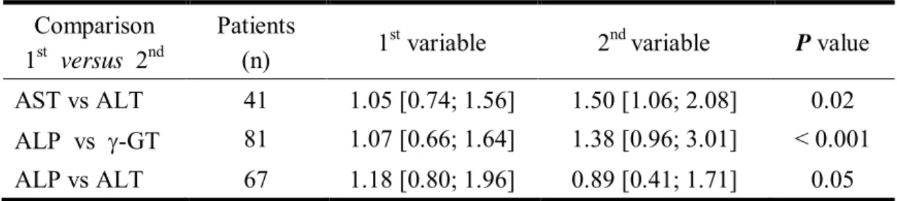
Furthermore, comparison variables, two by two, showed that the ALT elevations
were higher than those of AST, ɣ-GT than the ALP, and finally, the ALP than the ALT
AD Levorato 25 Table 5- Comparative study of the intensity of variables evaluating the hepatobiliary
system of patients with active paracoccidioidomycosis before treatment
(moment M0). Data presented as median, 1st and 3rd quartiles. Paired-sample
testing of continuous data (Wilcoxon test).
Comparison
1st versus 2nd Patients (n) 1st variable 2nd variable P value
AST vs ALT 41 1.05 [0.74; 1.56] 1.50 [1.06; 2.08] 0.02 ALP vs g-GT 81 1.07 [0.66; 1.64] 1.38 [0.96; 3.01] < 0.001 ALP vs ALT 67 1.18 [0.80; 1.96] 0.89 [0.41; 1.71] 0.05
AST- aspartate aminotransferase; ALT – alanine aminotransferase; ALP – alkaline phosphatase; GT - ɣ-glutamyltransferase; N – number of patients.
Comparing the prevalence of the biochemical markers of the hepatobiliary function
reveals that ALT tended to be higher than AST, that ϒ-GT tended to be higher than ALP
and that ALP tended to be higher than ALT (Figure 1).
Figure 1 – Comparative study of the prevalence of variables evaluating the hepatobiliary system of patients
with active paracoccidioidomycosis before treatment (moment M0). Paired-sample testing of dichotomous
data (McNemar test). AST – aspartate aminotransferase; ALT – alanine aminotransferase; AL –alkaline phosphatase; ϒ-GT-glutamyltransferase. 0 20 40 60 80 100 120 140 160 180
AST vs ALT ALP vs g-GT ALP vs TGP
Both normal
Both increased
1st increased and 2nd normal
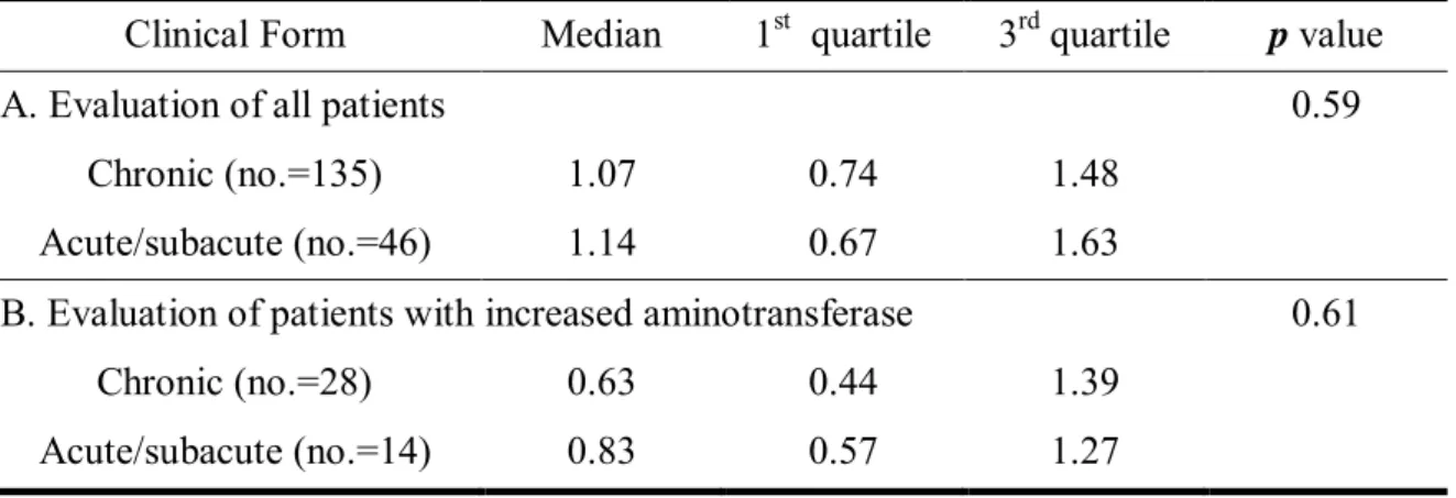
AD Levorato 26 The DeRitis quotient, evaluated in 181 patients, was slightly higher than 1.0 in both
AF patients and CF patients. No difference was observed according to clinical form (Table
4). However, when only the 42 patients with elevated aminotransferases were evaluated,
values lower than 1.0 were observed, indicating a disproportionate increase in ALT
compared with AST; this observation did not vary as to clinical form (Table 6).
Table 6 – DeRitis quotient, presented as median, 1st and 3rd quartiles, of patients with active paracoccidioidomycosis, evaluated before treatment (moment M0), as to
clinical form. A. Evaluation in 181 patients. B. Evaluation of the 42 patients
showing increased aminotransferases serum levels.
Clinical Form Median 1st quartile 3rd quartile p value
A. Evaluation of all patients 0.59
Chronic (no.=135) 1.07 0.74 1.48
Acute/subacute (no.=46) 1.14 0.67 1.63
B. Evaluation of patients with increased aminotransferase 0.61
Chronic (no.=28) 0.63 0.44 1.39
Acute/subacute (no.=14) 0.83 0.57 1.27
3.3. Hepatobiliary system course in patients with acute/sub-acute and chronic forms
of PCM.
Antifungal therapy resulted in AST normalization both in AF patients and in CF
AD Levorato 27 Table 7 – Follow-up of aspartate aminotransferase serum levels of
paracoccidioidomycosis-patients treated with itraconazole (ITC) or the
trimethoprim-sulfametoxazole combination (SXT) as to moment and clinical
form. Evaluation of patients with increased serum levels at admission. Data
presented as median, 1st and 3rd quartiles. Friedman test.
Clinical Form Treatment
Acute/subacute (n=11) Chronic (n=14) SXT (n=19) ITC (n=6) M0 1.35 [1.19;2.65]A 1.25 [1.2;1.76]A 1.35 [1.20;0.75]A 1.30 [1.15;2.75]A
M1 0.80 [0.55;1.36]B 0.55 [0.50;0.61]B 0.60 [ 0.54;1.19]AB 1.20 [0.90;1.40]AB
M2 0.90 [0.67;1.56]AB 0.83 [0.55;1.20]AB 0.90 [0.67;1.62]AB 1.05 [0.61;2.71]AB M3 0.97 [0.43;1.16]B 0.78 [0.36;1.05]B 0.90 [0.46;1.19]B 1.05 [0.35;1.05]B
p value ˂0.001 ˂0.001 0.01 0.04
[ ] – 1st and 3rd. Capital letters compare frequencies in the same column; frequencies with the same letter
do not differ (p>0.05); frequencies with different letters indicate statistically significant difference (p≤0.05).
M0 – before treatment, M1 – 4 to 6; M2 – 7 to 10; and M3: clinical cure and regression of the erythrocyte
sedimentation rate to normal values (18-23) weeks after beginning treatment; SXT – trimethoprim-sulfamethoxazole combination; ITC – itraconazole; N – number of patients.
Patients treated with SXT exhibited a reduction in the AST serum levels to normal
values at M3, whereas those treated with ITC exhibited a significant reduction at M3 but
continued to exhibited AST values above normal (Table 7).
Antifungal therapy led to ALT normalization at M1 and M2 but only in PCM
patients with CF. At M3, ALT serum levels tended to increase, exhibiting slightly elevated
levels at M0/M2. AF patients continued to exhibit elevated ALT levels at M3, which did not
AD Levorato 28 Table 8 – Follow-up of alanine aminotransferase serum levels of
paracoccidioidomycosis-patients treated with itraconazole (ITC) or the
trimethoprim-sulfametoxazole combination (SXT) as to moment and clinical
form. Evaluation of patients with increased serum levels at admission. Data
presented as median, 1st and 3rd quartiles. Friedman test.
Clinical Form Treatment
Acute/subacute (n=12) Chronic (n=22) SXT (n=28) ITC (n=6) M0 2.06 [1.50;4.28] 1.64 [1.17;1.83]A 1.69 [1.29;2.25]A 1.78 [1.56;2.17]
M1 2.14 [ 1.17;4.04] 0.72 [0.61;0.83]B 0.78 [0.61;1.33]AB 1.50 [1.11;2.13]
M2 1.11 [0.64;2.39] 0,78 [0.41;1.33]B 0.89 [0.41;1.39]B 1,11 [0.92;3.16] M3 1.24 [0.67;2.68] 0.97[0.61;1.28]AB 1.06 [0.67;1.44]AB 1.28 [0.61;1.50]
P
value 0.18 0.007 0.005 0.32
[ ] - 1st and 3rd quartiles Capital letters compare frequencies in the same column; frequencies with the
same letter do not differ (p>0.05); frequencies with different letters indicate statistically significant difference
(p£0.05). M0- before treatment, M1:4 to 6, M2: 7 to 10 and M3: clinical cure and regression of the erythrocyte
sedimentation rate to normal values (18-23) after beginning treatment. SXT - trimethoprim-sulfamethoxazole combination. ITC - itraconazole; N-number of patients
Patients treated with SXT exhibited a reduction in the ALT serum levels to slightly
above-normal values at M3, whereas those treated with ITC exhibited no significant
reduction at any moment (Table 8).
Antifungal therapy resulted in the progressive regression of TB serum levels and
normalization at M3 but only in patients with AF. Patients with CF continued to exhibit
elevated TB levels at all timepoint, and the values did not differ among the timepoints
AD Levorato 29 Table 9 – Follow-up of total bilirrubins serum levels of
paracoccidioidomycosis-patients treated with itraconazole (ITC) or the trimethoprim-sulfametoxazole combination
(SXT) as to moment and clinical form. Evaluation of patients with increased serum levels
at admission. Data presented as median, 1st and 3rd quartiles. Friedman test.
Clinical Form Treatment
Acute/subacute (n=5) Chronic (n=7) SXT (n=8) ITC (n=4) M0 5.80 [1.16;7.76]A 1.28 [1.12;1.73] 1.39 [1.05;3.05]A 3.51 [1.21;9.05]
M1 1.06 [0.61;3.00]AB 0.65 [0.50;1.30] 0.65 [0.40;2.80]AB 0.82 [0.58;1.18]
M2 0.70 [0.50;1.00]AB 1.00 [0.75;1.93] 1.00 [0.90;1.08]AB 0.50 [0.50;0.80]
M3 0.50 [0.28;1.52]B 1.00 [0.52;1.23] 0.55 [0.40;1.15]B 1.19 [0.60;1.67] P
value 0.019 0.19 0.04 0.15
[ ] - 1st and 3rd quartiles Capital letters compare frequencies in the same column; frequencies with the
same letter do not differ (p>0.05); frequencies with different letters indicate statistically significant difference
(p£0.05). M0- before treatment, M1:4 to 6, M2: 7 to 10 and M3: clinical cure and regression of the erythrocyte
sedimentation rate to normal values (18-23) weeks after beginning treatment. SXT: trimethoprim-sulfamethoxazole combination. ITC: itraconazole; N-number of patients.
Patients treated with SXT exhibited a progressive reduction and normalization of
TB serum levels at M3, whereas those treated with ITC exhibited no significant reduction
at any moment (Table 9).
Antifungal therapy led to the progressive regression of CB serum levels and
normalization at M2 but only in patients with CF. Patients with AF continued to exhibit
AD Levorato 30 Table 10 – Follow-up of conjugated bilirrubins serum levels of
paracoccidioidomycosis-patients treated with itraconazole (ITC) or the trimethoprim-sulfametoxazole
combination (SXT) as to moment and clinical form. Evaluation of patients with
increased serum levels at admission. Data presented as median, 1st and 3rd
quartiles. Friedman test.
Clinical Form Treatment
Acute/subacute (n=11) Chronic (n=13) SXT (n=17) ITC (n=7) M0 3.00 [2.00;14.04] 2.00 [1.28;5.50]A 2.33 [1.3;11.96]A 3.00 [1.48;9.23]
M1 1.67 [0.61;5.81] 1.50 [0.50;6.25]AB 0.80 [0.50;7.25]B 3,25 [2.00;4.00]
M2 1.67 [0.46;2.46] 0.80 [0.50;6.67]B 0.80 [0.35;2.45]B 1.00 [0.65;2.55] M3 2.00 [0.46;7.45] 1.00 [0.00;1.88]B 0.60 [0.04;2.13]B 2.35 [1.48;4.75]
P
value 0.14 0.004 ˂0.001 0.51
[ ] - 1st and 3rd quartiles Capital letters compare frequencies in the same column; frequencies with the same letter do
not differ (p>0.05); frequencies with different letters indicate statistically significant difference (p£0.05). ). M0- before
treatment, M1:4 to 6, M2: 7 to 10 and M3: clinical cure and regression of the erythrocyte sedimentation rate to normal
values (18-23) weeks after beginning treatment. SXT: trimethoprim-sulfamethoxazole combination. ITC: itraconazole; N-number of patients.
Patients treated with SXT exhibited a normalization of CB serum levels at M2,
whereas those treated with ITC did not exhibit a significant reduction at any moment
(Table 10).
Antifungal therapy led to progressive regression of ALP serum levels, with slightly
elevated values at M3 but only in patients with AF. Patients with CF exhibited progressive
AD Levorato 31 Table 11 – Follow-up of alkaline phosphatase serum levels of
paracoccidioidomycosis-patients treated with itraconazole (ITC) or the trimethoprim-sulfametoxazole
combination (SXT) as to moment and clinical form. Evaluation of patients with
increased serum levels at admission. Data presented as median, 1st and 3rd
quartiles. Friedman test.
Clinical Form Treatment
Acute/subacute (n=29) Chronic (n=26) SXT (n=44) ITC (n=8) M0 2.18 [1.33;3.68]A 1.22 [1.08;1.68]A 1.53 [1.16;2.26]A 1.30 [1.10;2.24]
M1 1.75 [1.24;3.13]AB 1.09 [0.95;1.33]B 1.25 [0.98;1.84]AB 1.30 [1.10;2.24]
M2 1.27 [0.81;1.47]B 1.10 [0.91;1.26]B 1.13 [0.81;1.37]BC 1.30 [1.10;2.24] M3 1.09 [0.74;2.05]B 0.87 [0.72;1.21]B 1.05 [0.71;1.61]C 1.30 [1.10;2.24]
P
value ˂0.001 0.001 ˂0.001 0.65
[ ] - 1st and 3rd quartiles Capital letters compare frequencies in the same column; frequencies with the same letter do
not differ (p>0.05); frequencies with different letters indicate statistically significant difference (p£0.05). ). M0- before
treatment, M1:4 to 6, M2: 7 to 10 and M3: clinical cure and regression of the erythrocyte sedimentation rate to normal
values (18-23) weeks after beginning treatment. SXT: trimethoprim-sulfamethoxazole combination. ITC: itraconazole; N-number of patients.
Patients treated with SXT exhibited a progressive reduction of ALP serum levels
with slightly elevated values at M3, whereas those treated with ITC exhibited no significant
reduction at any moment (Table 11).
Antifungal therapy led to progressive regression and normalization of ϒ-GT serum
levels at M3 but only in patients with AF. Patients with CF exhibited serum levels