DOI: 10.4025/actascibiolsci.v33i4.8091
Anatomy of the male reproductive system of
Phrynops geoffroanus
(Testudines: Chelidae)
Silvia Rosana Pagliarini Cabral, Lia Raquel de Souza Santos, Lilian Franco-Belussi, Rodrigo Zieri, Carlos Eduardo Saranz Zago and Classius de Oliveira*
Departamento de Biologia, Universidade Estadual Paulista "Júlio de Mesquita Filho”,Rua Cristóvão Colombo, 2265, 15054-000, São José do Rio Preto, São Paulo, Brazil. *Author for correspondence. E-mail: classius@ibilce.unesp.br
ABSTRACT. The reproductive system of male Phrynops geoffroanus adults is macroscopically described and the variation in testicular biometry is evaluated. A pair of oval testes is connected by the efferent ductules to the epididymis, which continue as deferent ducts, which emerge in the penis. The volume of the gonads showed the highest averages during spring and summer months. GSI varied significantly throughout the year, with the highest averages observed in the months that correspond to the end of spring and the beginning of summer, when the reproduction of the species takes place, and the lowest averages were seen in winter, suggesting a cyclical testicular activity.
Keywords: Testudines, Phrynops geoffroanus, reproductive system,testes, epididymis, efferent ductules, deferent ducts, penis.
RESUMO. Anatomia do aparelho reprodutor masculino de Phrynops geoffroanus (Testudines: Chelidae). O sistema reprodutor de machos adultos de Phrynops geoffroanus é descrito macroscopicamente e a variação da biometria testicular é avaliada. Um par de testículos ovais está conectado pelos dúctulos eferentes aos epidídimos, que se continuam como ductos deferentes, e que por sua vez desembocam no pênis. O volume das gônadas apresentou as maiores médias durante os meses de primavera e verão. O IGS variou significativamente ao longo do ano, com maiores médias observadas nos meses que correspondem ao final da primavera e início do verão, quando ocorre a reprodução da espécie, e as menores médias durante o inverno, sugerindo uma atividade testicular cíclica. Palavras-chave: Testudines, Phrynops geoffroanus, sistema genital, testículos, epidídimos, dúctulos
eferentes, ducto deferente, pênis.
Introduction
The urogenital system of adult chelonians consists, in both sexes, of a pair of kidneys and ureters, a urinary bladder, a pair of accessory vesicles, gonads and cloaca. In males, the sexual structures consist of a pair of testes, efferent ductules, epididymis, deferent ducts, penis; and in young individuals, remaining oviducts (ROMER; PARSONS, 1985; HILDEBRAND, 1995). The testes are spherical or oval, white-yellowish in color, and are connected ventrally to the kidneys by the mesorchium. The efferent ductules start from each testicle, forming the epididymal ducts after converging, which are elongated and convoluted (CABRAL et al., 2011). At the root of the penis are bulbs of the corpus cavernosum, a mass of spongy tissue responsible for penile eversion (ASCHLEY, 1969). The corpora cavernosa are located in the ventral wall of the cloaca, and have the shape of longitudinal crests separated by a groove (ROMER; PARSONS, 1985).
The species Phrynops geoffroanus is widely distributed in neotropical regions (Colombian Amazon, Venezuela, the Guyanas, Uruguay, northern Argentina, and Brazil from north to south) (GUIX et al., 1989; McCORD et al., 2001) and, as other chelonians, are oviparous with internal fertilization and have a reproductive system adapted to that condition. In males, the reproductive system features epididymis, responsible for temporary spermatozoa storage, and intromittent organs (penis) to transfer gametes to the body of females (ROMER; PARSONS, 1985).
their reproductive behavior (MOLINA, 1996; SOUSA; ABE, 2001) and the effect of temperature on determining sex (VOGT; BULL, 1982; VENEZUELA et al., 1997), especially in captive animals. In the present study, the male reproductive system of Phrynops geoffroanus (Testudines: Pleurodira, Chelidae) is described based on biometric data of the testes and the animal, in order to check for the existence of individual and seasonal anatomical variations.
Material and methods
The animals were collected through manual fishing using hooks, during monthly excursions to Felicidade stream, (S 20º 46’ 20.6”, W 49º 21’ 18.0”), an affluent of the Preto river, within the city limits of São José do Rio Preto, Estado de São Paulo, Brazil. A total of 16 adults males of Phrynops
geoffroanus were captured (Collection license:
061/2005-RAN/IBAMA) between April 2005 and May 2006, with four animals sampled per season of the year.
The animals were euthanized using high doses of ketamine hydrochloride (AURICCHIO; SALOMÃO, 2002). Additionally, 5 mL of ethanol was injected in the brain through the foramen magnum, assuring euthanasia. Biometric data such as maximum carapace and plastron length were obtained using a tape measure with 1 mm graduation, and total weight was obtained using a precision scale with 1 g accuracy.
The animals were opened using a vibrating bone saw, appropriate for osteotomy. After removal of the plastron, the organs of the urogenital system were documented in photographs (Nikon, Coolpix 8700) and described anatomically.
The testes were weighed in an analytical balance accurate to 0.001 g and measured (cm) using a precision metal pachymeter (0.05 mm). Gonad volume (V) was estimated according to the formula V = ¾ x π x a x b2, in which a = gonad length (longer axis), and b = width (shorter axis). Testicle and body weight were used to calculate the gonadosomatic index (GSI = total testicle mass/total body mass x 100).
To analyze the variation in GSI over the seasons of the year, the Kruskal-Wallis test was applied, and the One-Way ANOVA was used to analyze the variance among gonad volumes. Biometric data (weight, length and width) from the gonads were compared, between the two sides, throughout the year using the Wilcoxon test.
Results
The male reproductive system of Phrynops
geoffroanus consists of a pair of testes, from which
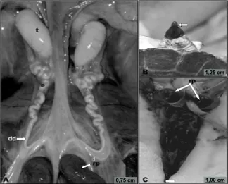
narrow efferent ductules emerge to form the epididymis, which uncoil in the caudal portion and continue on to the deferent duct, all of which are involved by the urogenital mesentery. In the cloacal region, the deferent duct lead into a groove at the base of the penis (Figure 1).
Figure 1. Male reproductive system of Phrynops geoffroanus. A. Overall view of the reproductive system: tetes (t); epididymis (e); deferent duct (dd), after the line, root of penis (rp). B. Everted penis, with free extremity and penial groove (arrowhead) C. Penis: notice the intense pigmentation; root of penis (rp) consisting of the bulbs of the corpora cavernosa and distal extremity (arrow).
The copulatory organ is a mass of richly vascularized tissue, with intense pigmentation from the base to the extremity of the penis, which is eversible through the cloaca by the action of erector muscles and blood inflow. It is a quite developed organ, positioned longitudinally to the ventral wall of the cloaca, which tapers towards its final free portion (Figure 1B and C). At the base of the penis are two dark-colored bulbous structures, known as the bulbs of the corpora cavernosa, one on each side, and also involved in the eversion and retraction processes of the penis (Figure 1A and C). Medially between the bulbs is the urinary bladder, positioned bilaterally from the accessory urinary vesicles, which are organs exclusive to the urinary tract, and with the rectum, posterior in the sagittal plane.
The testes are located ventrally to the kidneys, in the anterior portion of the pelvic region (Figure 1A). Each testicle is an oval organ, white-yellowish in color, with conspicuous testicular vascularization through the testicular tunica. They are coated by the
1,00 cm 0,75 cm
Seasonal anatomical variation in P. geoffroanus 489
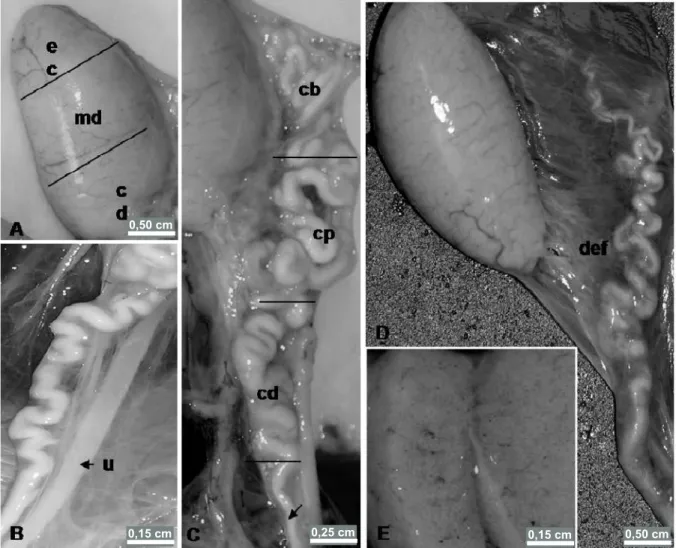
testicular tunica albuginea, a fibrous layer, and kept in place by a serous membrane lining named the mesorchium. Testes are connected to the epididymis by the efferent ductules, a bundle of small ductules, slightly sinuous, which meet and lead into the epididymal duct (Figure 2D). With regard to the internal organization of testicular parenchyma, the presence of mediastinum or testicular septa was not identified, giving an aspect of a mass of seminiferous tubules (Figure 2E).
The epididymis is an elongated structure consisting of a coiled duct, whitish in color, increasing in caliber towards the caudal region (Figures 1A and 2C and D). It meets the testes through the mesorchium, which binds both organs to the dorsal wall of the body. For the sake of description, the epididymis can be divided into three
regions: cranial region, medial region and caudal region. (Figure 2C). The epididymis are not connected to the testes in a similar fashion throughout their length; the main attachments are between the medial and caudal regions of the testicle with the cranial and medial regions of the epididymis. The anterior extremity of the epididymis possess a blind-ending epididymal duct (Figure 2D). The efferent ductules and epididymal ducts are bound by peritoneal conjunctive tissue. In the distal end of the epididymis, the epididymal duct becomes less coiled and its caliber is noticeably reduced, through which sperm is conducted (Figure 2D). Parallel to the epididymal ducts and deferent ducts are the ureters, which lead into the cloaca (Figure 2B).
Figure 2. Male reproductive organs of Phrynops geoffroanus. A. testes defining the regions: cranial region (cr), medial region (m) and caudal region (cd). B. Portion of the epididymis, deferent duct and urinary duct – ureter (u), which leads into the cloaca. C. Epididymis, where are indicated: cranial region (cr), medial region (m) and caudal region (cd). and anterior portion of the deferent duct (arrowhead). D. Testicle and epididymis lined by the gonadal mesentery and connecting them to a bundle of efferent ductules (def); epididymal duct, increasing in caliber from the head towards the tail. E. Sectioned testicle showing the absence of septa and testicular mediastinum, confining testicular parenchyma uniformly.
0,50 cm
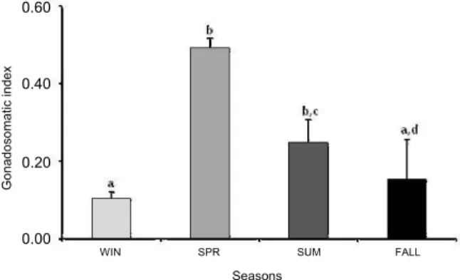
As for GSI, it was similar in spring (late September, October, November and December), averaging X = 0.49 ± 0.38, and summer (late December, January, February and March), averaging X =0.25 ± 0.06 (H = 10.65; p = 0.23); however, it differed statistically when compared to fall (X = 0.15 ± 0.1) and winter (X = 0.10 ± 0.01), which were similar to one another (H = 10.65; p = 0.00; p = 0.01 and p = 0.44, respectively) (Figure 3). High averages were also observed during the summer, but there was no statistically significant difference with fall and winter (H = 10.65; p = 0.21 and p = 0.06) (Figure 3). Gonad volume showed the highest averages during spring and summer months, but no difference was detected among the evaluated seasons (F = 2.41; p = 0.12) (Figure 4).
Figure 3. Variation in the gonadosomatic index (GSI) of Phrynops geoffroanus during its reproductive cycle. Means ± Standard deviation. Same letters mean there is no statistical difference (p = 0.05). WIN: Winter, SPR: Spring, SUM: Summer, FALL: Fall.
Figure 4. Variation in testicular volume of Phrynops geoffroanus during its reproductive cycle. Means ± Standard deviation. There was no statistical difference among the seasons of the year. WIN: Winter, SPR: Spring, SUM: Summer, FALL: Fall.
The right and left testes did not show any significant difference in their dimensions (weight, length and width) (Wilcoxon test: weight T = 61, p = 0.71; length T = 41, p = 0.75; width T = 31, p = 0.5).
Discussion
The male reproductive system of Phrynops
geoffroanus shows an anatomical pattern similar to
those described for other reptiles in its group (ORR, 1986; RIEPPEL, 2000; HICKMAN JR. et al., 2004). In these animals, as in all other amniotic vertebrates, fertilization is internal and the male reproductive system shows modifications related to that condition, with epididymis for sperm storage and copulatory organs to transfer these sperm, thus guaranteeing the success of fertilization (ROMER; PARSONS, 1985).
In P. geoffroanus, the testes are ovoid-shaped and
whitish in color, differently from the shape and color described for other Testudines, which are spherical and yellowish (ASCHLEY, 1969; GOULART, 2004). In general, the testicular morphology observed in P. geoffroanus follows the anatomical pattern described for all other reptiles (ORR, 1986; HILDEBRAND, 1995; POUGH et al., 2003). As in Iguana iguana, whose testes are rounded and are connected laterally to the epididymis, which in turn are connected to the deferent duct (FERREIRA et al., 2002).
The gonadosomatic index varied significantly among the months in analysis, with the highest averages observed during the spring and early summer, coinciding with the with reproductive period described for the species (MOLINA, 1998; MOLINA; GOMES, 1998; SOUSA; ABE, 2001).
The seminiferous or spermatic ducts, efferent ductules, epididymis and deferent ducts are derived from archinephric duct, which is an organ for the transport of both urine and sperm in anamniotic vertebrates (ROMER; PARSONS, 1985; ORR, 1986). In amniotic species the duct came to be used only for gamete transportation in males, with the passage of urine done via the ureters. This anatomical-functional pattern is confirmed by the complete anatomical separation of both pathways, as described for P. geoffroanus.
The efferent ductules are a network of small ducts that conduct sperm from the testes to the epididymis (ASCHLEY, 1969; ROMER; PARSONS, 1985, CABRAL et al., 2011). In
P. geoffroanus, the efferent ductules are connected to
the epididymis at the medial region and caudal extremity of the organ, whereas in Crysemys picta this connection is not continuous throughout the epididymis, being restricted only to the medial region (HOLMES; GIST, 2004). In C. picta (HOLMES; GIST, 2004), as in P. geoffroanus as well, the ducts are connected in a network pattern.
WIN SPR SUM FALL
WIN SPR SUM FALL
Seasons
Seasons
Gona
dos
o
m
a
tic ind
e
x
0.60
0.40
0.20
0.00
Testiculçar volume
(cm
3)
200
150
100
50
Seasonal anatomical variation in P. geoffroanus 491
In Trachemys scripta and C. picta, the epididymis
are coiled and dark in color (ASCHLEY, 1969; HOLMES; GIST, 2004). The occurrence of pigmentation in reproductive organs is also described in anurans, for which pigmentation has been reported in the testicular tunica (OLIVEIRA et al.,2002; OLIVEIRA; ZIERI, 2005; ZIERI et al., 2007; FRANCO-BELUSSI et al., 2009). In
P. geoffroanus, the presence of pigmentation was
observed in the penis, but was not described in the other reproductive organs. The epididymis, as in
T. scripta and C. picta (ASCHLEY, 1969; HOLMES;
GIST, 2004), are also coiled structures, but whitish and attached to the testes and kidneys.
The development of a copulatory organ is common among amniotic animals. Those who undergo internal fertilization do not have a typical penis and the spermatozoa are transferred to the females by gonopods and claspers, as in fish (ROMER; PARSONS, 1985). In amphibians, the cloaca is everted and serves as a sperm transfer structure (RIEPPEL, 2000).
In reptiles, an intromittent or copulatory organ is observed in all living species, with the exception of
Sphenodon sp. In snakes and lacertilians, exclusive
paired structures developed along the lateral wall of the cloaca – the hemipenis, regarded as an autapomorphy of that group (RIEPPEL, 2000; POUGH et al., 2003).
Chelonians, crocodilians, and some groups of basal birds have a single penis on the ventral wall of the cloaca. Similarities occur in the initial development stages of the copulatory organs in reptiles and mammals, which suggests that the standard of differentiation of a single penis is a basal trait in Amniota (REINAND; PIEAU, 1998 apud RIEPPEL, 2000). The same author considers the structure of the penis to be a trait that unites Testudines (Anapsida) and crocodilians (Diapsida), regarded as the morphological precursors of mammalian penis (HICKMAN JR. et al., 2004; KELLY, 2004).
Larson (1998) and Pough et al. (2003) consider that the presence of a penis is a primitive condition, and that the absence of copulatory organs in
Sphenodon sp. and in most birds should be regarded
as secondary.
The penis of P. geoffroanus is an organ with dark pigmentation, positioned longitudinally from the ventral wall of the cloaca and tapering off in its final portion. In the base of the penis are two dark-colored structures, one on each side, which are involved in the processes of eversion and retraction of the penis. The morphology of the penis is similar to that described by Ashley (1969) and Goulart
(2004) for chelonians. According to these authors, the corpora cavernosa are composed of fibrous-spongy tissue and between the longitudinal crests of these corpora is a groove; in the external extremity is the glans penis. During erection, the corpora cavernosa fill with blood, which distends the penis, and the groove forms a tube in which sperm is transported.
The morphological modifications in the reproductive system related to the success in internal fertilization remained in the majority of amniotic vertebrate males. Testudines constitute one of the oldest groups of living reptiles, and the first to feature these structures.
Conclusion
The male reproductive system of P. geoffroanus is, generally, similar to that of other Testudines, consisting of a pair of testes, efferent ductules, epididymis, deferent ducts and penis. No biometric variations occured between the left and right gonads, as the GSI varied throughout the year, with higher averages observed in spring and summer, and the lowest averages observed during winter.
Acknowledgements
The authors are thankful to Marcelo Dalpasquale for help with the translation of the manuscript and Brazilian Coordination for the Improvement of Higher Education Personel – CAPES (fellowship to S.R.P.C).
References
ASCHLEY, L. M. Laboratory anatomy of the turtle, 1st ed. Dubuque: WM. C. Brown Company Publishers, 1969. p. 32-36.
AURICCHIO, P.; SALOMÃO, M. Técnicas de coleta e preparação de vertebrados para fins científicos e didáticos. São Paulo: Instituto Pau Brasil de História Natural, 2002.
CABRAL, S. R. P.; ZIERI, R.; FRANCO-BELUSSI, L.; SANTOS, L. R. S.; ZAGO, C. E. S.; TABOGA, S. R.; OLIVEIRA, C. Morphological changes of the epididymis and description of the excurrent ducts of Phrynops
geoffroanus (Testudines: Chelidae) during the reproductive
cycle. The Anatomical Record, v. 294, n. 1, p. 145-155, 2011.
CHRISTIANSEN, J. L.; DUNHAN, A. E. Reproduction of the yellow mud turtle (Kinosternon flavescens flavescens) in New Mexico. Herpetologica, v. 28, n. 2, p. 130-137, 1972. FERREIRA, A.; LAURA, I. A.; DOLDER, H. Reproductive cycle of male green iguanas, Iguana iguana
FRANCO-BELUSSI, L.; ZIERI, R.; SANTOS, L. R. S.; MORESCO, R. M.; OLIVEIRA, C. Pigmentation in anuran testes: anatomical pattern and variation. The Anatomical Record, v. 292, n. 2, p. 178-182, 2009. GOULART, C. E. S. Herpetologia, herpetocultura e medicina de répteis. 1. ed. Rio de Janeiro: L. F. Livros de Veterinária, 2004. p. 37-56.
GUIX, J. C., SALVATTI, M.; PERONI, M. A.; LIMA-VERDE, J. S. Aspectos da reprodução de Phrynops geoffroanus (Schweigger, 1812) em cativeiro (Testudines, Chelidae). Grupo de Estudos Ecológicos, 1989. (Série Documentos 1, p. 1-19).
HICKMAN JR., C. P.; ROBERTS, L. S.; LARSONS, A. Princípios integrados de zoologia. 11. ed. Rio de Janeiro: Guanabara Koogan, 2004. p. 531-538.
HILDEBRAND, M. Análise da estrutura dos vertebrados. São Paulo: Atheneu, 1995. p. 700.
HOLMES, H. J.; GIST, D. H. Excurrent duct system of the male turtle Crysemys picta. Journal of Morphology, v. 261, n. 3, p. 312-322, 2004.
KELLY, D. A. Turtles and mammals penis designs are anatomically convergent. The Royal Society London – Biological letters, v. 271, p. 93-95, 2004.
LARSON, P. L. The Theropod reproductive system. Gaia, v. 15, n. 15, p. 389-397, 1998.
MAHMOUD, I. Y.; CYRUS, R. V. The testicular cycle of the common snapping turtle, Chelydra serpentina, in Wisconsin. Herpetologica. v. 48, n. 2, p. 193-201, 1992. MAHMOUD, I. Y.; KLICKA, J. Seasonal gonadal changes in kinosternid turtles. Journal of Herpetology, v. 6, n. 3-4, p. 183-189, 1972.
McCORD, W. P.; JOSEPH-OUNI, M.; LAMAR, W. W. A taxonomic reevaluation of Phrynops (Testudines Chelidae) with the description of two new genera and a new species of Batrachemys. Revista de Biología Tropical, v.49, n. 2, 715-764, 2001.
MOLINA, F. B. Mating behavior of captive Geoffroy’s side-necked turtles, Phrynops geoffroanus (Testudines: Chelidae). Herpetological Natural History, v. 4, n. 2, p. 155-160, 1996.
MOLINA, F. B. Comportamento e biologia reprodutiva dos cágados Phrynops geoffroanus, Acanthochelis radiolata e
Acanthochelis spixii (Testudines, Chelidae) em cativeiro.
Revista Brasileira de Etologia, special number, p. 25-40, 1998.
MOLINA, F. B.; GOMES, N. Incubação artificial e processos de eclosão em Trachemis dorbinyi. Revista Brasileira de Zoologia, v. 15, n. 1, p. 135-143, 1998. OLIVEIRA, C.; ZANETONI, C.; ZIERI, R. Morphological observations on the testes of Physalaemus
curvieri (Amphibia, Anura). Revista Chilena de
Anatomia, v. 20, n. 3, p. 263-268, 2002.
OLIVEIRA, C.; ZIERI, R. Pigmentação testicular em
Physalaemus nattereri (Steindachner) (Amphibia, Anura)
com observações anatômicas sobre o sistema pigmentar extracutâneo. Revista Brasileira de Zoologia, v. 22, n. 2, p. 454-460, 2005.
ORR, R. T. Biologia dos vertebrados. 5. ed. São Paulo: Livraria Rocca Ltda., 1986. p. 508.
POUHG, F. H.; JANIS, M. C.; HEISER, B. J. A vida dos vertebrados. 3. ed. São Paulo: Atheneu. 2003. p. 270-290.
RIEPPEL, O. Turtles as diapsid reptiles. Zoologica Scripta, v. 29, n. 3, p. 199-212, 2000.
ROMER, S.; PARSONS, T. S. Anatomia comparada dos vertebrados. 5. ed. São Paulo: Atheneu, 1985. SOUSA, F. L.; ABE, A. S. Population structure and reproductive aspects of the freswater turtle, Phrynops
geoffroanus, inhabiting an urban river in souththeastern
Brazil. Studies on Neotropical Fauna and Environment, v. 36, n. 1, p. 57-62, 2001.
VENEZUELA, N.; BOTERO, R.; MARTINES, E. Field study of sex determination in Podocnemis expansa from Colombian Amazonia. Herpetologica, v. 53, n. 3, p. 339-398, 1997.
VOGT, R. C.; BULL, J. J. Temperature controlled sex-determination in turtles: ecological and behavorial aspects. Herpetologica, v. 38, n. 1, p. 156-164, 1982.
ZIERI, R.; TABOGA, S. R.; OLIVEIRA, C. Melanocytes in the testes of Eupemphix nattereri (Anura, Leiuperidae): histological, etereological and ultrastructural aspects. The Anatomical Record, v. 290, n. 7, p. 795-800, 2007.
Received on August 29, 2009. Accepted on April 17, 2010.