Locus in B Cell Development
Patty B. Garcia1, Amie Cai1, Jamie G. Bates2, Hector Nolla1, Mark S. Schlissel1*
1Department of Molecular and Cell Biology, University of California, Berkeley, California, United States of America,2Department of Biochemistry, Stanford University School of Medicine, Stanford, California, United States of America
Abstract
Regulated expression of miRNAs influences development in a wide variety of contexts. We report here that miR290-5p (100049710) and miR292-5p (100049711) are induced at the pre-B stage of murine B cell development and that they influence assembly of the Igklight chain gene (243469) by contributing to the activation of germline Igktranscription (kGT). We found that upon forced over-expression of miR290-5p/292-5p in Abelson Murine Leukemia Virus (AMuLV) transformed pro-B cells, two known activators ofkGT, E2A (21423) and NF-kB (19697), show increased chromosomal binding to the kappaintronic enhancer. Conversely, knockdown of miR290-5p/292-5p in AMuLV pro-B cells blunts drug-induced activation of kGT. Furthermore, miR290-5p/292-5p knockdown also diminishes kGT activation, but not Rag1/2 (19373, 19374) expression, in an IL-7 dependent primary pro-B cell culture system. In addition, we identified a deficiency inkGT induction in miR290 cluster knockout mice. We hypothesize that increased expression of miR290-5p and miR292-5p contributes to the induction ofkGT at the pre-B stage of B cell development through increased binding of NF-kB and E2A tokappalocus regulatory sequences.
Citation:Garcia PB, Cai A, Bates JG, Nolla H, Schlissel MS (2012) miR290-5p/292-5p Activate the ImmunoglobulinkappaLocus in B Cell Development. PLoS ONE 7(8): e43805. doi:10.1371/journal.pone.0043805
Editor:Sebastian D. Fugmann, National Institute on Aging, United States of America
ReceivedJune 20, 2012;AcceptedJuly 26, 2012;PublishedAugust 23, 2012
Copyright:ß2012 Garcia et al. This is an open-access article distributed under the terms of the Creative Commons Attribution License, which permits unrestricted use, distribution, and reproduction in any medium, provided the original author and source are credited.
Funding:This work was funded in part by NIH RO1 HL48702 (MSS, http://grants.nih.gov/grants/oer.htm) and an NSF Predoctoral Fellowship (PG, http://www. nsfgrfp.org/). The funders had no role in study design, data collection and analysis, decision to publish, or preparation of the manuscript.
Competing Interests:The authors have declared that no competing interests exist. * E-mail: [email protected]
Introduction
Recent work implicates microRNAs (miRNAs) in the regulation of B cell development [1] [2,3]. miRNAs are small non-coding RNAs, approximately 20–25 nucleotides in length processed from longer precursors, that exert sequence-targeted post-transcription-al repression of target transcripts [4,5]. Primary miRNA transcripts are processed in the nucleus by an RNaseIII enzyme Drosha (14000), then exported to the cytoplasm for further processing by a second such enzyme, Dicer [4] (192119). Dicer selects a mature,22 nt miRNA strand that serves as effector in
the RNA-induced silencing complex (RISC) to regulate target transcripts. Mice with B cell lineage-specific deletion of Dicer exhibit a developmental block at the pro-B stage of development [1]. This finding implicates the miRNA pathway and its effector members as playing an essential role at this stage and highlight the important function of miRNAs at the pro-B to pre-B transition, a critical checkpoint in B cell development. Although some miRNAs and their functions have been described [2,3] further studies are needed to thoroughly identify miRNAs regulating B cell develop-ment.
miR290-5p and miR292-5p are members of the miR290 polycistronic cluster [6]. The miR290 cluster is expressed as a single transcript encoding seven miRNAs. miR290-5p/292-5p share the seed sequence CUCAAA similar to miR291-5p (100049715, 100124471), AUCAAA. This indicates that they are similar in function. The remaining miR290 cluster members that share the seed sequence AAGUCC are expressed under different contexts. These miRNAs are robustly expressed together in
eutherian embryonic stem cells and have therefore been called the Early Embryonic microRNA Cluster (EEmiRC) [6]. Generally the CUCAAA miR290 cluster members and the AAGUCC members are not thought to overlap functionally.
The miR290 cluster germline knockout displays partially penetrant embryonic lethality in which homozygotes survive gestation at only 7% of the predicted Mendelian ratio [7]. Medeiros et al. hypothesize that the phenotype is partially penetrant in part due to the mixed background in their studies (129/C57BL6). Additionally, they point out that other miRNA deletions result in partially penetrant phenotypes, possibly due to random fluctuations of gene expression levels in the absence of the miRNAs. They further speculate that this is the case in the miR290 cluster deletion. A role for miR290 cluster members in lymphoid cells has not been described.
unrearrangedkappalocus prior to rearrangement generating what are known as germline kappa transcripts (kGT) [13]. The
appearance of these transcripts indicates a kappalocus that is in an open chromatin state, available for access by the V(D)J recombinase proteins, Rag1 and Rag2 [14].Kappalocus activation is tightly regulated during B cell development.
The activation and rearrangement of thekappalocus requires twokappalocus enhancers, the kappa intronic enhancer (Eki) and
the 39 kappa enhancer (39Ek) [15]. Transcription factors that
enhancekappalocus activation through binding to these enhancers include E2A, which binds to both enhancers [16], NF-kB, which
binds Eki [17,18], and Pax5 (18507), which binds 39Ekand has
been shown to be necessary for kappa rearrangements [19]. Binding of these transcription factors to thekappalocus enhancers is regulated as cells develop in the bone marrow. Signaling through the IL-7 receptor and pre-B Cell Receptor (pre-BCR) concomitantly direct transcription factors to bind either enhancer, thereby activatingkGT andkapparearrangements [20,21]. While
much is known about signaling through these pathways, nothing is known about the role miRNAs play inkappalocus activation at the pro-B to pre-B transition.
We sought to identify miRNAs that play a role in the pro-B to pre-B transition using AMuLV-transformed pro-B cells as a model system. AMuLV transformation immortalizes developing B cells at the pro-B to large pre-B stages of B cell development [22,23]. The immortalized cells are blocked in differentiation and receive survival signals from constitutive v-Abl [24] kinase activity, independent of IL7 (16196) cytokine signals [25]. Studies by our lab and others have shown that inactivation of the constitutive Abl kinase activity, by the small molecule Abl-kinase inhibitor STI571 (also known as Gleevec), in AMuLV cell lines results in G1 cell cycle arrest, progression to a pre-B-like stage, and eventual apoptosis [25]. The transcripts induced upon STI571 treatment include pre-B stage-specific genes such as SpiB (272382), IRF4 (16364), Rag1/2, andkGT, confirming that Abl kinase activity
blocks B cell differentiation. While pre-B stage-specific genes have been identified in this system, miRNAs specific to this stage have yet to be described.
In this study, we screened for miRNAs that are upregulated in STI571-treated AMuLV-transformed B cells and that pro-mote B cell differentiation. Our screen identified two miRNAs, miR290-5p and miR292-5p, that originate from the miR290 Cluster transcript and share an identical seed-sequence. miR290-5p and miR292-miR290-5p are independently able to induce expression of
kGT expression as well as increase DNA binding activity of NF-kB and E2A. These transcription factors bind directly to their
cognate sites in the intronickappaenhancer. We propose a novel role for miR290-5p and miR292-5p in the activation of thekappa locus during B cell development.
Results
microRNA Screen in AMuLV proB Cells
To identify miRNAs that may be of importance at the pro-to-pre-B cell transition of B cell development we analyzed RNA purified from AMuLV-transformed pro-B cell lines. When AMuLV-transformed pro-B cells are treated with STI571, they progress to a pre-B-like state of development [25]. To broadly identify miRNAs that change in expression levels upon the addition of STI571, we used the Exiqon microarray platform to screen for such miRNA populations in three independent AMuLV transformants (E2A+/+, 22028, 63212). We cultured each line in the presence or absence of STI571 for 12 hrs (2.5mM), purified
total RNA from these samples, and subjected them to microarray analysis (GSE38331).
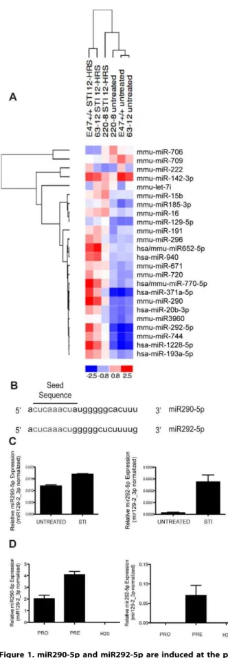
The general trend observed was an increase in expression of miRNAs upon STI571 treatment. However, there were a few miRNAs that decreased in expression upon STI571 treatment (Fig. 1a). Two of the miRNAs identified in the screen as having expression consistently and significantly increased across all three independently transformed AMuLV-transformed pro-B cell lines (Fig. 1a) were miR290-5p and miR292-5p. These miRNAs are members of the murine miR290 polycistronic cluster, previously reported to be expressed only in gonads of both sexes, but not in other tissues examined [7]. They share the same seed sequence (Fig. 1b.) indicating a possible functional redundancy within pre-B cells.
To validate the results of the microarray, we cultured E2A+/+ AMuLV cells in the presence or absence of STI571 for 12 hrs (2.5mM). Using specific Taqman qPCR assays, we confirmed that both miR290-5p and miR292-5p display a modest increase in expression upon STI571 treatment (Fig. 1c).
We next asked whether these miRNAs displayed a similar pattern of regulated expression in wild-type primary developing pro- and pre-B cells. We sorted pro-B (B220+, CD43+, IgM2) and pre-B (B220+, CD432, IgM2) cells from wild-type mouse bone marrow by flow cytometry, purified total RNA, and performed qPCR for quantification of miR290-5p and miR292-5p. We found that the increase in expression we observed in STI571 treated AMuLV-transformed pro-B cells, was mirrored in primary pro-B and pre-B cells (Fig. 1d). A third member of the miR290 polycistronic cluster with a similar seed sequence as miR290-5p/ 292-5p, miR291-5p, is also induced; however other members of the miR290 cluster with an alternate seed sequence did not increase upon STI571 treatment (Figure S1).
Over-expression of miR290-5p or miR292-5p Induces Germline IgkTranscription in AMuLV Cells
Previous studies have shown that the addition of STI571 to AMuLV-transformed pro-B cells induces expression of genes characteristic of the pre-B cell stage in development [25]. Since these miRNAs increase in expression upon STI571 treatment, we hypothesized that they may play a role in regulating developmen-tal progression at the pre-B cell stage. In order to probe the role that these miRNAs might play at this drug-induced developmental transition, we cloned each miRNA in its genomic context into an MSCV-based IRES-CD2 marked retroviral expression vector. We transduced the E2A+/+ AMuLV cell line with each miRNA vector or an empty vector control and asked whether the miRNA-overexpressing cells activate transcription of the germline Igklocus
transcript,kGT. The activation of the Igk locus is a hallmark of
the pre-B stage of development that is tightly correlated with the activation of V-to-Jk rearrangement [13]. Upon stable
over-expression of either miR290-5p or mir292-5p, we observed an induction inkGT (Fig. 2a), but not to the levels seen in wild-type
AMuLV-transformed pro-B cells treated with STI571. However, when we treated the miRNA-transduced cell lines with STI571 (12 hrs, 2.5mM), we saw a super-induction of kGT (Fig. 2a).
These data indicate that either miRNA is able to independently act upon a pathway that results in activation of the germline Igk
locus. In contrast, neither miRNA altered the induced or basal levels ofRag1transcription in these same cells (Fig. 2b).
miRNA Knockdown Partially Blunts the Induction ofkGT upon v-Abl Inhibition
an artifact of over-expression, we generated miRNA sponge constructs to perform knockdown experiments. These sponge constructs were generated by independently cloning three imperfect miRNA binding sites for each miRNA into the
Figure 1. miR290-5p and miR292-5p are induced at the pro-B to pre-B transition.1A. Heat-map representing levels of miRNAs from a microarray analysis of RNA purified from the indicated AMuLV cell lines cultured in the absence or presence of STI571 (2.5mM, 12 hr). Performed once in three independently transformed cell lines. 1B.
Schematic depiction of the shared seed sequence of the mature miR290-5p and miR292-5p microRNAs. 1C. qPCR analysis of miR290-5p or miR292-5p expression levels in RNA purified from E2A+/+AMuLV cells cultured in the absence or presence of STI571 (2.5mM, 12 hr). Data was normalized to the expression of miR129-2_3p. Error bars represent range for replicate qPCR reactions. The data shown is from one representative experiment of three biological replicates. 1D. qPCR analysis of miR290-5p or miR292-5p in primary wild-type pro-B (B220+, CD43+, IgM2) cells or pre-B (B220+, CD432, IgM2) cells. Data was normalized to the expression of miR129-2_3p. Error bars represent range for replicate qPCR reactions. Data shown is from one representative experiment, of three independent sort experiments. doi:10.1371/journal.pone.0043805.g001
Figure 2. Over-expression of miR290-5p or miR292-5p induces
kGT expression in AMuLV cells.2A, B. qPCR analysis of (A)kGT or
(B) Rag1 expression in RNA purified from E2A+/+AMuLV cells over-expressing either miR290-5p or miR292-5p, cultured in the absence or presence of STI571 (2.5mM, 12 hr). The (A)kGT qPCR y-axis reflects two
different scales. Data was normalized to the expression ofHprt. Error bars represent range for replicate qPCR reactions. Asterisk represents P value,0.05. The P value was derived by the Student’s T test. Data shown is from one representative experiment of at least four individual experiments.
39UTR of a GFP (7011691) cDNA [24]. We also cloned a sponge construct for a different miRNA, miR129-2_3p (723953), to use as a negative control. miR129-2_3p is expressed at similar levels as miR290-5p and miR292-5p at the pre-B stage, and is not induced at the pro-to pre-B stage in primary B cells or upon STI571 treatment of AMuLV-transformed pro-B cells (data not shown). We generated stable AMuLV-transformed cell lines transduced with retroviral vectors expressing these sponge constructs to examine their effects onkGT expression.
To first confirm that the sponge constructs were engaging with the target miRNA, we stably transduced AMuLV cells with GFP miRNA-sponge constructs marked with a dual tomato red cDNA. The expression of the tandem tomato red cDNA was independent of miRNA sponge regulation and serves for normalization against the variable GFP expression. Upon STI treatment (42 hrs, 1mM) we observed a decrease in GFP expression relative to tomato red expression, in the miR290-5p or miR292-5p sponges, suggesting that the sponges were engaging with the target miRNAs (Figure S2).
We then examined the sponge effects onkGT expression. We
cultured the stable GFP sponge cell lines with STI571 (12 hrs, 2.5mM) and observed a normal induction of kGT in the
negative control sponge cell line (Fig. 3a). However, we observed a blunting of normal kGT induction in the
miR290-5p or miR292-miR290-5p sponge lines (Fig. 3a). This blunting of kGT
induction upon knockdown of endogenous drug-induced miR290-5p or miR292-5p indicates that these miRNAs partially contribute to the activation of the kappa locus upon STI571 treatment of AMuLV-transformed cells.
miRNA Knockdown Partially BluntskGT Induction in Primary Cell Culture
To further test the role of miR290-5p and miR292-5p in the activation of the kappa locus observed in our AMuLV model system, we performed similar sponge knockdown experiments in a primary developing B cell culture system. Cultured primary pro-B cells proliferate in the presence of high concentrations of rIL7 but do not differentiate, while at low concentrations of rIL7, the cells exit the cell cycle and progress to the pre-B cell stage of development [20]. We harvested bone marrow from wild-type mice and cultured cells in 2 ng/ml rIL7 for 5 days to expand the pro-B cell population. We then retrovirally transduced the expanded pro-B cell culture with either the negative control, miR290-5p, or miR292-5p sponges. After an additional 24 hours in rIL7 culture, we harvested a portion of each population as a ‘‘0 hour’’ time-point. We then washed the remaining cells and re-cultured them in the absence of rIL7 for 24 hr.
In cells transduced with the negative control sponge, there was normal induction of kGT upon IL-7 withdrawal as expected
(Fig. 3b). However, in the presence of the miR290-5p or miR292-5p sponges, IL7 withdrawal resulted in a significantly diminished induction ofkGT expression (Fig. 3b). We observed
indistinguish-able induction ofRag1 gene expression upon IL7 withdrawals in miR292-5p as compared to miR129-2_3p negative control sponge cultures (Fig. 3c). Together these data indicate that miR290-5p and miR292-5p independently contribute to the induction ofkGT
expression in primary pre-B cells.
miR290-5p and miR292-5p Enhance DNA Binding Activity of E2A and NF-kB
In an effort to elucidate the pathway through which miR290-5p or miR292-5p induce kGT, we examined transcription factors
known to be involved in the induction ofkGT by direct binding to
either the 39kappaenhancer (39Ek) or thekappaintronic enhancer
(Eki). Among them, E2A and NF-kB bind Eki while E2A can also
bind 39Ek[16,18,18]. To determine if miR290-5p/292-5p
over-expression increases over-expression of E2A, we performed qPCR. We did not observe an increase in E2A mRNA levels upon miR290-5p/miR292-5p over-expression. (Figure S3).
Since both E2A and NF-kB binding activity can be regulated
post-transcriptionally, we went on to ask whether miR290-5p/ 292-5p regulate the induction ofkGT by inducing DNA binding
activity of E2A and NF-kB to their target sequences in thekappa
locus. To approach this we performed Chromatin Immunopre-cipitation (ChIP).
It was reported previously that upon IL7 withdrawal, the amount of bound E2A increases at Eki [20] and knockdown of
miR290-5p or mir292-5p upon IL7 withdrawal blunts kGT
induction as shown above. So we asked if miR290-5p or miR292-5p over-expression enhances E2A binding to Eki in vivo. We
made use of the HF4 AMuLV-transformed pro-B cell line generated from a mouse that expresses a FLAG-tagged E2A knocked into the E2A locus [26,27]. We stably expressed miR129-2_3p, miR290-5p, or miR292-5p in this cell line and performed ChIP with anti-FLAG or anti-IgG antibodies. Using qPCR to analyze the precipitated DNA, we observed increased E2A-FLAG binding to Eki in both miR290-5p and miR292-5p
over-expressing cells (Fig. 4a). These data indicate that miR290-5p or miR292-5p expression induces binding of E2A to Eki within the kappalocus.
The gene encoding a known inhibitor of E2A activity, ID2 [28] (15902), has predicted binding sites for miR290-5p/miR292-5p in its 39UTR. We sought to determine if miR290-5p/292-5p directly repress the ID2 39UTR using a dual-luciferase assay. A luciferase mRNA fused to the ID2 39UTR was not directly repressed by these miRNAs (Figure S4).
NF-kB, like E2A, binds Eki [18]. We went on to examine
whether NF-kB binding activity increases when these two
miRNAs are over-expressed in E2A+/+ AMuLV lines cultured with STI (1mM, 16 hr). We stably expressed miR129-2-_3p,
miR290-5p, or miR292-5p in this cell line and performed ChIP with anti-p50 (18033) (an NF-kB subunit) or anti-IgG antibodies.
We observed increased p50 binding to Eki in both miR290-5p and
miR292-5p over-expressing cells and this binding was further increased in the presence of STI571 (Fig. 4b). These data indicate that miR290-5p or miR292-5p expression induces binding of
NF-kB to Eki within thekappalocus.
To determine if factors upstream of NF-kB are affected by
miR290-5p/miR292-5p expression, we examined IkBa(18035), a
known inhibitor of NF-kB [29]. Protein levels for IkBa are
repressed upon miR290-5p or miR292-5p over-expression to similar levels as observed in the STI571-treated control sample. This indicates that members of this pathway may be regulated by miR290-5p/292-5p (Fig. 4c).
We used a dual-luciferase assay to determine whether miR290-5p/292-5p can directly repress the IkBa 39UTR. Luciferase
activity was not repressed in cells overexpressing miR290-5p/292-5p by fusion of its cDNA to the IkBa39UTR (Fig. 4d).
The miR290 Cluster in B Cell Development
IgM2) populations. We found a moderate increase in the percentage of pre-B cells in the homozygous miR290 knockout mice, as compared to the wild-type mice (p = 0.05) (Fig. 5a, b). To confirm our miRNA knockdown results that implicated both miR290-5p and miR292-5p as playing a role in activating the kappalocus, we used the miR290 cluster knockout mice to analyze
kGT expression in pro-B, large pre-B (B220+, CD432, IgM2
FSC2Hi), and small pre-B cell (B220+, CD432, IgM2FSC-Lo) populations. We observed a bluntedkGT induction in the small
pre-B population of miR290 cluster knockout mice (Fig. 5c). Interestingly, we also observed a blunted Rag1 induction in the small pre-B population of miR290 cluster knockout mice (Fig. 5d). This was unexpected since we did not observe an effect on Rag1 in
the pro-B culture experiments. This difference might be attribut-able to the other miR290 cluster members also deleted in the miR290 knockout.
Together, these data indicate that members of the miR290 polycistronic cluster, miR290-5p and miR292-5p are involved in the activation of the kappa locus as indicated by expression of
kGT.
Discussion
This study demonstrates that developing B cells induce expression of miR290-5p and miR292-5p at the pre-B stage to fully activate the germline Igklocus, a critical event in the pro-to-Figure 3.kGT induction is blunted upon knockdown of miR290-5p or miR292-5p.3A. qPCR analysis ofkGT expression in RNA purified from
E2A+/+AMuLV cells expressing a miR290-5p or miR292-5p sponge knockdown construct, and cultured in the presence of STI571 (2.5mM) for the indicated lengths of time. Data was normalized to the expression ofHprt. Error bars represent range for replicate qPCR reactions. Asterisk represents P value,0.05. The P value was derived by the Student’s T test. These data are from one representative experiment of at least four independently performed experiments. 3B, C. qPCR analysis of (B)kGT or (C) Rag1 expression in RNA purified from wild-type primary pro-B cells transduced with
either a miR129-2_3p control knockdown sponge, or a miR290-5p or miR292-5p knockdown sponge. Wild-type primary pro-B cells were cultured in rIL7 (2 ng/ml) for 5 days and then transduced with indicated sponge construct. Marker positive cells were then sorted and IL7 withdrawn from culture for 24 hrs before RNA was harvested. qPCR measures (B)kGT or (C) Rag1 expression in cells before or after the withdrawal of IL7. Data was
normalized toHprtexpression levels. Error bars represent range for replicate qPCR reactions. Asterisk represents P value,0.05. The P value was derived by the Student’s T test. These data show one representative experiment of at least four independently performed experiments.
pre-B cell transition. A modest induction of miR290-5p/292-5p occurs in response to STI571-inhibition of v-abl in transformed pro-B cell cultures, as well as in response to IL7 attenuation in primary B cell cultures. It is possible that miR290-5p/miR292-5p induction upon STI571-treatment of Abelson cells and IL7 attenuation in primary B cell cultures, is a consequence of either cell cycle arrest or activation of the apoptosis pathway known to occur under these circumstances. We reported previously, however, that activation of kGT and Rag1 transcription in
STI571-treated Abelson cells is independent of both processes [25]. In addition, preliminary experiments show that miR290-5p/ 292-5p are induced at an earlier time point than the onset of cell cycle arrest in STI-571 treated Abelson cells, therefore, making it unlikely that this miRNA cluster is induced as consequence of cell cycle arrest.
The modest miR290-5p/292-5p induction may be a reflection of the partial role these miRNAs play in fullkGT activation. NF-kB and E2A showed increased binding to Ekiin vivo, in response
to miR290-5p/292-5p expression. Using over-expression, we found that miR290-5p/292-5p contribute to full activation of
kGT. Knockdown strategies showed that miR290-5p/292-5p are
necessary for full activation ofkGT. In miR290 cluster knockout
mice we observe diminishedkGT levels in both pro-B and pre-B
cells. Additional pre-B stage signals, such as other miRNAs, may be required to induce full expression ofkGT [4]. v-abl inhibition
or IL7 attenuation likely induce additional signaling pathways, aside from miRNAs, that are synergistic with miR290-5p/292-5p, and necessary for fullkGT induction.
Previous work has shown that kGT are correlated with the
activation of IgkLC rearrangement [13], and may be required for
progression from pre-B to immature B cells. We observed a deficiency in kGT induction in knockdown experiments from
AMuLV-transformed pro-B cells and primary B cell cultures. In the miR290 cluster knockout mice we observed a modest accumulation of pre-B cells and a general blunting ofkGT levels.
Together, these observations support our conclusion that members of the miR290 polycistronic cluster play a role in the develop-mental activation of kGT expression.
A miRNA-regulated Pathway for the Activation of the IgkappaLocus
Previous work showed that IL7 attenuation allows for both expression of Rag1/2 as well as induction ofkGT through E2A
binding to Eki [20]. Interestingly, we did not observe an increase
in E2A mRNA levels upon miR290-5p/292-5p over-expression. We propose that E2A activation, upon IL7 attenuation, is regulated through miR290-5p/292-5p expression. We note that miR290-5p/292-5p does not regulate Rag1/2 expression in cell culture systems. Rag1 induction remained intact in both the AMuLV pro-B cells as well as the IL7-dependent primary B cell Figure 4. Over-expression of miR290-5p or miR292-5p induces
activation of E2A and NF-kB.4A. ChIP analysis of HF4 AMuLV cells
expressing His-FLAG-E2A, at endogenous levels, and over-expressing either miR129-2_3p, miR290-5p, or mir292-5p. Chromatin samples were immunoprecipitated with anti-FLAG or IgG control antibody. Relative enrichment of bound DNA over input was determined by subjecting precipitates to qPCR with primers specific to the Eki binding region for
E2A. This data shows one representative experiment of two indepen-dent experiments. 4B. ChIP analysis of E2A+/+ AMuLV cells over-expressing either miR129-2_3p, miR290-5p, or mir292-5p, in the absence or presence of STI571 (1mM, 16 hr). Chromatin samples were
immunoprecipitated with anti-p50 or IgG control antibody. Relative enrichment of bound DNA over input was determined by subjecting precipitates to qPCR with primers specific to the Eki binding region for
NF-kB. This data shows one representative experiment of two
independent experiments. 4C. Immunoblot analysis of E2A+/+AMuLV cells expressing either an empty vector control, miR290-5p, or miR292-5p and cultured in either the absence or presence of STI571 (2.5mM, 12 hr). Immunoblot was probed with anti-IkBa and anti-Tubulin
antibodies. 4D. Luciferase assay of total cell lysates from HEK293 cells transiently transfected with either a wild-type IkBa39UTR reporter or a
mutant IkBa39UTR reporter along with a scrambled miRNA, mir290-5p
or miR292-5p. Error bars represent range for biological replicate luciferase reactions. Data shows one representative experiment of at least three independent experiments.
cultures, upon knockdown of miR290-5p or mir292-5p, while
kGT induction was blunted. In miR290-5p/292-5p
over-express-ing cell lines we did not observe kappa locus rearrangements, despitekGT induction (data not shown). This may be explained
by the lack of Rag1/2 activation when miR290-5p/292-5p are over-expressed in AMuLV pro-B cells in the absence of STI571. However, in the miR290 cluster knockout mice we observe a blunting of Rag1 induction at the pro-B, large pre-B, and small pre-B stages. This different effect on Rag1 may be attributed to the other miRNA members of the miR290 cluster also deleted in the miR290 cluster knockout mouse. However, aside from miR291-5p, we could not detect their expression in sorted pre-B cells of wild-type mice. Therefore, we propose that loss of IL7 signaling activates miR290-5p/292-5p, that leads to induction ofkGT.
NF-kB, has been shown, by our group and others, to be highly
expressed at the pre-B stage [30,31]. We reported a correlation between the expression of both NF-kB and its inhibitor, IkBa,
withkGT and light chain gene rearrangements. Furthermore, we
reported that RAG1/2 expression is independent from NF-kB
expression in pre-B cells. Activation of NF-kB is due in part to
miR290-5p/292-5p induction at the pre-B stage.
The NF-kB inhibitor, IkBa[29], is an interesting candidate for
direct targeting by miR290-5p/292-5p. In our studies we observed a decrease in IkBa protein expression upon miR290-5p/292-5p
over-expression. Despite having predicted miR290-5p/miR292-5p binding sites in its 39UTR, a luciferase mRNA fused to the IkBa 39UTR was not directly repressed by these miRNAs. It is
important to note that IkBa has a predicted miR290-5p/292-5p
binding site in exon 4 that has not been examined. Likewise, a known inhibitor of E2A, ID2 [28], has predicted binding sites for miR290-5p/miR292-5p in its 39UTR, but the ID2 39UTR was not repressed by the miRNAs. It is possible that the target of miR290-5p/miR292-5p is an unknown inhibitor of one or both of these transcription factors, or a factor that is further upstream in the pathway. Additional studies are necessary to identify the relevant direct targets of these miRNAs in pre-B cells.
miRNAs in B Cell Development
In this study, we show a role for miRNAs at the pre-B stage in B cell development. Several groups have shown that miRNAs are essential for the pro-to-pre-B transition, supporting our postulate that miRNAs regulate different stages of B cell development. Notably, Koralov [1] and colleagues deleted Dicer, an essential miRNA processing enzyme, in AMuLV-transformed pro-B cells and identified a set of upregulated transcripts. One such example, Bim (12125), a pro-apoptotic gene, was identified as a target of miR17-92 (723905) at the pro-B stage [1,3,32]. These studies provide a foundation in support of the AMuLV-transformed pro-B Figure 5. Germline knockout of the miR290 cluster affects the
pre-B cell population.5A. FACS analysis of 6 week old miR290 cluster knockout or wild-type mice. FACS plot reflects CD19 enriched cells gated on IgM negative cells. B220+, CD43+correspond to pro-B cells and B220+, CD432correspond to pre-B cells.; WT: pro-B, 5.72%, pre-B 84.1%; KO: pro-B, 2.47%, pre-B 91.1%. This experiment represents one of four independent experiments. Each experiment was with at least one
mouse per genotype. 5B. Plot of the percentages of CD19+, IgM2pro-B (B220+, CD43+) and pre-B (B220+, CD432) for each independent knockout and wild-type mouse analyzed as in Figure 5A. Average percentages are WT: pro-B, 6.05%, pre-B 85.3%; KO: pro-B, 3.4%, pre-B 90.6%. Line represents the average percentage for each population and the P value was derived by the Student’s T test. The average was derived from at least four mice per genotype. 5C, D qPCR analysis of (C)
kGT or (D) Rag1 levels in RNA purified from flow-sorted pro-B (B220+,
CD43+, IgM2), large pre-B (B220+, CD432, IgM2, FSC-Hi), and small pre-B (B220+, CD432, IgM2, FSC-Lo) cells. Mice were 6 week old miR290 cluster knockout or wild-type mice. The data was normalized to Hprtexpression. Error bars represent range for replicate qPCR reactions. Here we show qPCR data from a representative mouse from each genotype. This experiment has been repeated with four mice per genotype and data shown is representative of three out of four miR290 knockout mice.
cell model system for identifying miRNAs. They also support that miRNAs may regulate other transitions in B cell development such as the pre-B to immature-B transition that we observe.
Many miRNAs, including those expressed at the pro-to-pre-B transition, are described as being master regulators of key pathways. For example, miR150 (387168) was identified as a robust regulator at the pro-to-pre-B transition [2,33]. B lineage-specific miR150 transgenic mice display a block at the pro-to-pre-B transition. Additionally, the miR17-92 polycistronic cluster knockout mouse has a block at the pro-to-pre-B transition in B cell development [3], also serving as an example of master regulatory miRNA potential in B cell development.
miR290-5p/292-5p knockdown, in either STI571-treated AMuLV-transformed pro-B cells or in an IL7 regulated primary B cell culture system, and germline-deletion of the miR290 cluster in mice, result in a blunting of kGT induction. Blunting rather
than ablation of kGT induction indicates that these miRNAs
contribute to, but are not essential for, this process. This is different than the robust effect of miR150 described above. This finding, together with the accumulation of the pre-B population in the miR290 cluster knockout mouse, underscores the potential of low-expressing miRNAs, such as miR290-5p/292-5p, that behave as modulators of pathways in developmental systems.
miR290 Cluster and its Members
As described above, the miR290 cluster germline knockout has a partially penetrant embryonic lethality in which homozygotes survive gestation at only 7% of the predicted Mendelian ratio. Homozygous knockout mice that die in-utero have two major abnormal phenotypes. At E.10.5, 16% of homozygous knockouts develop outside of the yolk sac, while 40% of homozygotes have severe developmental defects including reduced somite number and delayed chorioallantoic attachment, among other things [7]. These defects are obvious contributors to the reduced survival potential of homozygotes, and emphasize the importance of the miR290 cluster in development. Our studies are the first to describe a phenotype in lymphoid cells in the miR290 cluster knockout mice.
In our study, miR290-5p/292-5p are thought to be similar in function since they share the seed sequence CUCAAA. This means that they are functionally redundant and target the same targets. The miR290 cluster members that share the seed sequence AAGUCC are expressed under different contexts. The differential expression of the CUCAAA and AAGUCC members in B cells may be attributed to differential maturation of the miRNAs in the miR290 cluster. Differential maturation may be due to pri-miRNA accessibility to Drosha, determined by the miR290 cluster tertiary structure, as is the case with the miR17,92 cluster [34],
among other possibilities.
Most recently, a role for the seed sequence-AAGUCC miRNAs but not seed sequence-CUCAAA miRNAs, was described in regulating DNA methyltransferases [35], the genes involved in apoptosis. In Dicer deletion studies of ESC [36], and in the regulation of the G1/S transition of the cell cycle in ESC. The AAGUCC miRNAs are characterized as proliferation-regulating miRNAs [37]. It is worth noting that in the ESC, the miR290 cluster AAGUCC members play the role of master regulators and strongly regulate proliferation. In our study we observe a more modulatory role for the CUCAAA members of this cluster, miR290-5p and miR292-5p. Interestingly, the AAGUCC miR-NAs, including miR* counterparts miR290-3p and miR292-3p, are not detected in our system (Figure S1). There are few validated targets for miR290-5p or miR292-5p, but this group may include
p16 [38] (12578) and Lats1 (16798), two genes implicated in cell cycling.
In summary, we identified two miRNAs, miR290-5p and miR292-5p, that are induced in pre-B cells. They increase binding activity of E2A and NF-kB to chromosomal Eki sequences and
modulate induction ofkGT expression.kGT expression is critical
to activation of thekappalocus for rearrangement of a functional immunoglobulin light chain. Our studies have uncovered a novel role for miR290-5p and miR292-5p in this key developmental process.
Materials and Methods
Ethics Statement
All mouse experimentation was approved by the Animal Care and Use Committee of the University of California at Berkeley (Protocol#R253-0313BR). The handling of the animals was in accordance with this protocol.
Mice
xmiR290 cluster 2/2 mice, on a C57BL/6 and 129 mixed background, were a gift from the Sharp and Jaenisch Labs at the Koch Institute (MIT). C57BL/6 mice were purchased from Jackson Laboratories.
Cell Culture and Retroviral Infections
Phoenix cells [24] were grown in Dulbecco’s modified Eagle’s medium (DMEM) supplemented with heat-inactivated 10% fetal calf serum, and sodium pyruvate (800mM). AMuLV cell lines E2A+/+, 220-8 [25], and 63-12 [39] were cultured in RPMI supplemented with heat-inactivated 5% fetal calf serum, penicillin (100mg/ml), 2 mM L-Glutamine, and 2-mercaptoethanol (50mM).
MSCV-iRES-hCD2-miR30 was previously described [40]. The miR30 cassette was replaced with either miR129-2_3p, miR290-5p, or miR292-5p. The corresponding miRNAs* were mutated for each pre-miRNA. Primers used for miRNA cloning were as follows:
miR129-2_3p 59
ACTGCTCGACCCCCTTCGAGC-TACCCTTAT,
miR129-2_3P 39 ACTGGCGGCCGCTCCCCCTGTGGTA-CAAAGTC;
miR290-5p 59ATCGGGATCCTCTGGGCGCGAGTGA, miR290-5p 39ATCGGTCGACGCTCAAGAGAGGG; miR292-5p 59ATCGGGATCCGTGGAGGCCCTCTCT, miR292-5p 39ATCGGTCGACCCAAGCCGCGGAG. Sponges were generated from long oligos with imperfect binding sites for miRNAs and were annealed and cloned into V26-MSCV-PIG. Long oligos used to generate each miRNA sponge contain three consecutive copies of binding sites for the corresponding miRNA with each binding site separated by a CCGG stuffer sequence. A bulge for imperfect binding was created correspond-ing to positions 9–12 of the miRNA. Bindcorrespond-ing sites were as follows: miR129-2_3p: ATGCTTTTTGTTTGAAGGGCTT, miR290-5p: AAAGTGCCCCACCCGTTTGAGT, miR292-miR290-5p: CAAAA-GAGCCAAACGTTTGAGT.
primary bone marrow cells (six bones from one mouse) were transduced with 2 ml viral supernatant by spinfection (2500RPM, 2 Hr., 32C). After spinfection, volume was brought up to 4 ml with RPMI (complete) and cultured.
ChIP
Chromatin Immunoprecipitation (ChIP) was conducted as previously described [26]. Recovered DNA was resuspended in 100ml water. Primers used for PCR analysis of recovered DNA
were as follows: Eki F’TTAAGGCCTGTCCATACAGT; Eki
R’ATGTTTGGGAGTCTGAACAC; NF-kB
F’AC-CTCTGTCACCCAAGAGTTGGC; NF-kB
R’ACAGGGCCT-TAAGCCAGGGTC.
qRT-PCR
Total RNA was isolated with Trizol Reagent (Invitrogen) and reverse transcription was performed with random hexamers and Moloney MLV reverse Transcriptase (MMLV) (Invitrogen). Quantitative reverse transcription PCR (qRT-PCR) was per-formed with Jumpstart Taq(Sigma), EvaGreen (Biotium), on an ABI 7300 Thermocycler (Applied Biosystems). PCR amplification was as follows: 95C 3 min, 95C 30 sec, 60C 30 sec, 72C 30 sec (data collection), for 40 cycles. Primers used in this study were previously described [40]. Primers used forkGT were as follows:
kGT 59: GGACGTTCGGTGGAGGC. kGT 39: GGAAGATGGATACAGTTGGT.
miRNA reverse transcription was conducted as previously described [40], using 50 ng of total RNA and miRNA-specific reverse-transcription primer (Assay IDs 001184, 002590, 001055, ABI). miRNA qRT-PCR was conducted as previously described [40], using miRNA-specific Taqman primer sets included in miRNA-specific assays.
Flow Cytometry and FACS Sorting Analysis
Total bone marrow was harvested from mice and red blood cells were depleted with EryLyse Buffer (0.14 M NH4Cl, 20 mM Hepes). Cells were stained with anti-CD19 MACS microbeads (Miltenyi Biotec) and were CD19+ enriched using MACS MS columns (Miltenyi Biotec). CD19+ cells were then stained using the following antibodies: B220-PeCy5 (clone RA3-6B2, BD), CD43-Pe (clone S7, BD), IgM-FITC (clone II/41, eBiosciences), IgD-Biotin (clone 11–26, eBiosciences), Streptavidin-PeCy7 (BD). Stained cells were resuspended in FACS buffer (5% BSA, 10 mM Hepes) for cell sorting.
Supporting Information
Figure S1 Expression of alternative miR290 cluster members. qPCR analysis of miR291-5p, miR290-3p, and miR292-3p expression levels in RNA purified from primary wild-type pro-B (B220+, CD43+, IgM2) or pre-B (B220+, CD432, IgM2) cells. Data was normalized to the expression of miR129-2_3p. Error bars represent range for replicate qPCR reactions. Data shows one representative experiment of at least three independent experiments.
(TIF)
Figure S2 miRNAs engage with knockdown-sponge construct. FACS analysis of sponge marker, GFP, expression upon STI571 (1mM, 42 hr) induction of miRNAs. E2A+/+ AMuLV cells were stably transduced with tandem tomato marked sponge constructs for miR129-2_3p, mir290-5p, or miR292-5p. Cells were gated on the tomato positive population and were analyzed for GFP expression upon STI571 treatment. (Grey fill, untreated cell line; Black line, STI-treated cell line).
(TIF)
Figure S3 Over-expression of miR290-5p or miR292-5p do not decrease E2A mRNA expression.QPCR analysis of E2A+/+AMuLV cells expressing either an empty vector control, miR290-5p, or miR292-5p for (A) E12 or (B) E47.
(TIF)
Figure S4 miR290-5p or miR292-5p do not directly repress the ID2 39UTR.Luciferase assay of total cell lysates from HEK293 cells transiently transfected with either a wild-type ID2 39UTR reporter or a mutant ID2 39UTR reporter along with a scramble miRNA, mir290-5p or miR292-5p. Error bars represent range for biological replicate luciferase reactions. Data shown is of one experiment representative of at least three independent experiments.
(TIF)
Acknowledgments
We would like to acknowledge technical assistance from Dan Huang and Min Cho and valuable discussions with members of the Schlissel lab. We would also like to acknowledge Lea Medeiros from the Jaenisch lab (MIT) for sharing with us the miR290 cluster knockout mice.
Author Contributions
Conceived and designed the experiments: PBG MS. Performed the experiments: PBG AC HN. Analyzed the data: PBG AC MS. Contributed reagents/materials/analysis tools: PBG JGB HN MS. Wrote the paper: PBG MS.
References
1. Koralov SB, Muljo SA, Galler GR, Krek A, Chakraborty T, et al. (2008) Dicer ablation affects antibody diversity and cell survival in the B lymphocyte lineage. Cell 132(5): 860–874.
2. Xiao C, Calado DP, Galler G, Thai TH, Patterson HC, et al. (2007) MiR-150 controls B cell differentiation by targeting the transcription factor c-myb. Cell 131(1): 146–159.
3. Ventura A, Young AG, Winslow MM, Lintault L, Meissner A, et al. (2008) Targeted deletion reveals essential and overlapping functions of the miR-17 through 92 family of miRNA clusters. Cell 132(5): 875–886.
4. Filipowicz W, Bhattacharyya SN, Sonenberg N (2008) Mechanisms of post-transcriptional regulation by microRNAs: Are the answers in sight? Nat Rev Genet 9(2): 102–114.
5. Bartel DP (2009) MicroRNAs: Target recognition and regulatory functions. Cell 136(2): 215–233.
6. Houbaviy HB, Dennis L, Jaenisch R, Sharp PA (2005) Characterization of a highly variable eutherian microRNA gene. RNA 11(8): 1245–1257.
7. Medeiros LA, Dennis LM, Gill ME, Houbaviy H, Markoulaki S, et al. (2011) Mir-290–295 deficiency in mice results in partially penetrant embryonic lethality and germ cell defects. Proc Natl Acad Sci U S A 108(34): 14163–14168. 8. Cooper MD, Alder MN (2006) The evolution of adaptive immune systems. Cell
124(4): 815–822.
9. Schatz DG, Swanson PC (2011) V(D)J recombination: Mechanisms of initiation. Annu Rev Genet 45: 167–202.
10. Lam KP, Kuhn R, Rajewsky K (1997) In vivo ablation of surface immunoglobulin on mature B cells by inducible gene targeting results in rapid cell death. Cell 90(6): 1073–1083.
11. Constantinescu A, Schlissel MS (1997) Changes in locus-specific V(D)J recombinase activity induced by immunoglobulin gene products during B cell development. J Exp Med 185(4): 609–620.
13. Schlissel MS, Baltimore D (1989) Activation of immunoglobulin kappa gene rearrangement correlates with induction of germline kappa gene transcription. Cell 58(5): 1001–1007.
14. McDevit DC, Perkins L, Atchison ML, Nikolajczyk BS (2005) The ig kappa 3’ enhancer is activated by gradients of chromatin accessibility and protein association. J Immunol 174(5): 2834–2842.
15. Inlay M, Alt FW, Baltimore D, Xu Y (2002) Essential roles of the kappa light chain intronic enhancer and 3’ enhancer in kappa rearrangement and demethylation. Nat Immunol 3(5): 463–468.
16. Inlay MA, Tian H, Lin T, Xu Y (2004) Important roles for E protein binding sites within the immunoglobulin kappa chain intronic enhancer in activating vkappa jkappa rearrangement. J Exp Med 200(9): 1205–1211.
17. Sen R, Baltimore D (1986) Multiple nuclear factors interact with the immunoglobulin enhancer sequences. Cell 46(5): 705–716.
18. Shaffer AL, Peng A, Schlissel MS (1997) In vivo occupancy of the kappa light chain enhancers in primary pro- and pre-B cells: A model for kappa locus activation. Immunity 6(2): 131–143.
19. Geier JK, Schlissel MS (2006) Pre-BCR signals and the control of ig gene rearrangements. Semin Immunol 18(1): 31–39.
20. Johnson K, Hashimshony T, Sawai CM, Pongubala JM, Skok JA, et al. (2008) Regulation of immunoglobulin light-chain recombination by the transcription factor IRF-4 and the attenuation of interleukin-7 signaling. Immunity 28(3): 335–345.
21. Mandal M, Powers SE, Maienschein-Cline M, Bartom ET, Hamel KM, et al. (2011) Epigenetic repression of the igk locus by STAT5-mediated recruitment of the histone methyltransferase Ezh2. Nat Immunol 12(12): 1212–1220. 22. Chen YY, Wang LC, Huang MS, Rosenberg N (1994) An active v-abl protein
tyrosine kinase blocks immunoglobulin light-chain gene rearrangement. Genes Dev 8(6): 688–697.
23. Klug CA, Gerety SJ, Shah PC, Chen YY, Rice NR, et al. (1994) The v-abl tyrosine kinase negatively regulates NF-kappa B/Rel factors and blocks kappa gene transcription in pre-B lymphocytes. Genes Dev 8(6): 678–687. 24. Swift S, Lorens J, Achacoso P, Nolan GP (2001) Rapid production of
retroviruses for efficient gene delivery to mammalian cells using 293T cell-based systems. Curr Protoc Immunol Chapter 10: Unit 10.17C.
25. Muljo SA, Schlissel MS (2003) A small molecule abl kinase inhibitor induces differentiation of abelson virus-transformed pre-B cell lines. Nat Immunol 4(1): 31–37.
26. Kuo TC, Chavarria-Smith JE, Huang D, Schlissel MS (2011) Forced expression of cyclin-dependent kinase 6 confers resistance of pro-B acute lymphocytic leukemia to gleevec treatment. Mol Cell Biol 31(13): 2566–2576.
27. Greenbaum S, Zhuang Y (2002) Identification of E2A target genes in B lymphocyte development by using a gene tagging-based chromatin immuno-precipitation system. Proc Natl Acad Sci U S A 99(23): 15030–15035. 28. Kee BL (2009) E and ID proteins branch out. Nat Rev Immunol 9(3): 175–184. 29. Chiao PJ, Miyamoto S, Verma IM (1994) Autoregulation of I kappa B alpha
activity. Proc Natl Acad Sci U S A 91(1): 28–32.
30. Cadera EJ, Wan F, Amin RH, Nolla H, Lenardo MJ, et al. (2009) NF-kappaB activity marks cells engaged in receptor editing. J Exp Med 206(8): 1803–1816. 31. Derudder E, Cadera EJ, Vahl JC, Wang J, Fox CJ, et al. (2009) Development of immunoglobulin lambda-chain-positive B cells, but not editing of immunoglob-ulin kappa-chain, depends on NF-kappaB signals. Nat Immunol 10(6): 647–654. 32. Xiao C, Srinivasan L, Calado DP, Patterson HC, Zhang B, et al. (2008) Lymphoproliferative disease and autoimmunity in mice with increased miR-17– 92 expression in lymphocytes. Nat Immunol 9(4): 405–414.
33. Zhou B, Wang S, Mayr C, Bartel DP, Lodish HF (2007) miR-150, a microRNA expressed in mature B and T cells, blocks early B cell development when expressed prematurely. Proc Natl Acad Sci U S A 104(17): 7080–7085. 34. Chaulk SG, Thede GL, Kent OA, Xu Z, Gesner EM, et al. (2011) Role of
pri-miRNA tertiary structure in miR-17,92 pri-miRNA biogenesis. RNA Biol 8(6): 1105–1114.
35. Sinkkonen L, Hugenschmidt T, Berninger P, Gaidatzis D, Mohn F, et al. (2008) MicroRNAs control de novo DNA methylation through regulation of transcriptional repressors in mouse embryonic stem cells. Nat Struct Mol Biol 15(3): 259–267.
36. Zheng GX, Ravi A, Calabrese JM, Medeiros LA, Kirak O, et al. (2011) A latent pro-survival function for the mir-290–295 cluster in mouse embryonic stem cells. PLoS Genet 7(5): e1002054.
37. Wang Y, Baskerville S, Shenoy A, Babiarz JE, Baehner L, et al. (2008) Embryonic stem cell-specific microRNAs regulate the G1-S transition and promote rapid proliferation. Nat Genet 40(12): 1478–1483.
38. Rizzo M, Evangelista M, Mariani L, Simili M, Rainaldi G, et al. (2011) Immortalized mouse embryo fibroblasts are resistant to miR-290-induced senescence regardless of p53 status. Physiol Genomics 43(20): 1153–1159. 39. Alt F, Rosenberg N, Lewis S, Thomas E, Baltimore D (1981) Organization and
reorganization of immunoglobulin genes in A-MULV-transformed cells: Rearrangement of heavy but not light chain genes. Cell 27(2 Pt 1): 381–390. 40. Amin RH, Schlissel MS (2008) Foxo1 directly regulates the transcription of