TESE DE DOUTORADO
DIAGNÓSTICO GENÉTICO DE DUAS FAMÍLIAS COM CASOS
DE ACIDOSE TUBULAR RENAL DISTAL POR MEIO DE
WHOLE-‐EXOME SEQUENCING
PAULA CRISTINA DE BARROS PEREIRA
Paula Cristina de Barros Pereira
DIAGNÓSTICO GENÉTICO DE DUAS FAMÍLIAS COM CASOS
DE ACIDOSE TUBULAR RENAL DISTAL POR MEIO DE
WHOLE-‐EXOME SEQUENCING
Tese de Doutorado apresentada ao Programa de Pós-‐ Graduação em Ciências da Saúde da Faculdade de Medicina da Universidade Federal de Minas Gerais, como requisito parcial para obtenção do grau de Doutor.
Área de Concentração: Medicina Molecular
Orientadora: Profª. Dra. Ana Cristina Simões e Silva
Professora Titular do Departamento de Pediatria -‐ Faculdade de Medicina da UFMG
Co-‐Orientadora: Prof. Dra. Débora Marques de
Miranda
Professora Adjunta do Departamento de Pediatria -‐ Faculdade de Medicina da UFMG
! , , 3 4 5 678# 97 5
: 5 * ; <
5 &( /
= >? 5
@ A B5 < "
8 * @ C 2 ( 6 @ $
D # * E > % & >; * F G
< " * ; GG " &( / GGG
< " G @)
UNIVERSIDADE FEDERAL DE MINAS GERAIS
Reitor: Prof. Clélio Campolina Diniz
Vice-‐Reitora: Profª Rocksane de Carvalho Norton
Pró-‐reitor de Pós-‐graduação: Prof. Ricardo Santiago Gomez Pró-‐reitor de Pesquisa: Prof. Renato de Lima Santos
FACULDADE DE MEDICINA
Diretor: Prof. Francisco José Penna Vice-‐diretor: Prof. Tarcizo Afonso Nunes
PROGRAMA DE PÓS-‐GRADUAÇÃO EM CIÊNCIAS DA SAÚDE ÁREA DE CONCENTRAÇÃO: MEDICINA MOLECULAR
Coordenador: Prof. Luiz Armando Cunha De Marco Sub-‐coordenador: Prof. Débora Marques de Miranda Colegiado:
AGRADECIMENTOS
À Professora Ana Cristina Simões e Silva, minha orientadora, pela confiança ao longo dessa jornada; grande exemplo de competência e profissionalismo.
À Professora Débora Marques de Miranda, minha co-‐orientadora, pela credibilidade e apoio a todo o tempo.
À Flávia Melo pela amizade, disponibilidade e imensa paciência.
À turma da bancada (em especial Nayra, Tininha, Paty e Lu) por toda ajuda, torcida e companheirismo.
“A tarefa não é tanto ver aquilo que ninguém viu, mas pensar o que ninguém ainda pensou sobre aquilo que todo mundo vê.”
RESUMO
Acidose tubular renal (ATR) distal ou ATR tipo 1 compreende um grupo heterogêneo de afecções resultantes da disfunção dos túbulos distais, que pode levar a um déficit de crescimento, nefrocalcinose, raquitismo e, raramente, doença renal crônica. Essa tubulopatia pode ser transmitida tanto na forma autossômica dominante quanto na autossômica recessiva. A forma dominante da doença ocorre tipicamente na adolescência ou na idade adulta, enquanto a variante recessiva desenvolve-‐se predominantemente na infância.
O objetivo deste estudo foi detectar e caracterizar as alterações genéticas presentes em duas famílias, não relacionadas, de pacientes portadores de ATR distal através da metodologia de whole-‐exome sequencing (WES). A familia 1 é composta por uma menina e seu irmão que apresentavam ATR distal e audição normal, enquanto na familia 2 havia duas irmãs gêmeas com ATR distal associada a surdez neurosensorial. A técnica de WES foi realizada em duas amostras agrupadas e, para confirmar os resultados, foi utilizado o método de sequenciamento de Sanger. Duas mutações foram identificadas nos genes ATP6V0A4 e ATP6V1B1: uma mutação nova no exon 13 do gene ATP6V0A4 (c.1232G>T) e uma mutação no exon 12 do gene ATP6V1B1 que já foi previamente descrita na literatura (c.1149_1152insC).
Nosso estudo indica que os resultados obtidos com o whole-‐exome sequencing podem ser úteis para o diagnóstico e a abordagem clinica de pacientes com ATR distal, especialmente porque, além de ser uma doença Mendeliana, é rara e com herança complexa.
Nossos resultados confirmaram o valor do whole-‐exome sequencing para o estudo de doenças raras e complexas, permitindo a identificação de mutações novas e recorrentes. Além disso, nosso estudo mostra a aplicabilidade deste método molecular no estudo de doenças renais tubulares.
Palavras-‐chave: Acidose tubular renal distal. Whole-‐exome sequencing. Infância.
ATP6V0A4. ATP6V1B1
ABSTRACT
Distal renal tubular acidosis (dRTA) refers to a heterogeneous group of diseases that result from distal tubular dysfunction and can lead to growth retardation, nephrocalcinosis, bone disease and, rarely, chronic kidney disease. dRTA can be transmitted as either an autosomal dominant or an autosomal recessive trait. Dominant disease typically presents more mildly in adolescence or adulthood and the recessive variant, predominantly developed in infancy/early childhood.
The aim of this study was to detect genetic alterations in two unrelated families with dRTA using whole-‐exome sequencing. Hearing was normally preserved in both children from family one, but not from family two, where a twin pair had severe deaf. We perform a whole-‐exome sequencing in two pooled samples and confirm findings with Sanger sequencing method. Two mutations were identified in the ATP6V0A4 and ATP6V1B1 genes: a novel mutation in exon 13 of ATP6V0A4 gene (c.1232G>T) and a mutation in exon 12 of ATP6V1B1 gene (c.1149_1152insC) previously described.
The findings obtained with whole-‐exome sequencing may improve health care, especially because, besides of being a Mendelian disorder, dRTA is rare and has a complex inheritance.
Our results confirm the value of whole-‐exome sequencing for the study of rare and complex diseases, allowing the identification of novel and recurrent mutations. Furthermore, we clearly show the application of this molecular method in renal tubular diseases.
Key words: Distal renal tubular acidosis. Whole-‐exome. Children. ATP6V0A4.
LISTA DE ABREVIATURAS E SIGLAS
AE1 = trocador aniônico cloro-‐bicarbonato ATR = acidose tubular renal
ACII = anidrase carbônica tipo 2 ACIV = anidrase carbônica tipo 4 COEP = comitê de ética em pesquisa
eAE1 = trocador aniônico cloro-‐bicarbonato eritrocitário H+ATPase = bomba de prótons
kAE1 = trocador aniônico cloro-‐bicarbonato renal ND = nefron distal
TC = túbulo coletor TCN = túbulo conector TD = túbulo distal
UFMG = Universidade Federal de Minas Gerais
UNP-‐HC = Unidade de Nefrologia Pediátrica do Hospital das Clínicas WES = whole-‐exome sequencing
LISTA DE FIGURAS
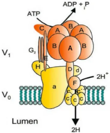
Figura 1. Figura esquemática da H+ ATPase renal com suas subunidades: o dominio
V1 (A-‐H) e o dominio V0 (a, c, c’, c”, d).………...……….……… 15
Figura 2. Mecanismos de transporte tubular no néfron distal: (A) secreção de íons
hidrogênio e acidificação urinária; (B) reabsorção de bicarbonato... 19
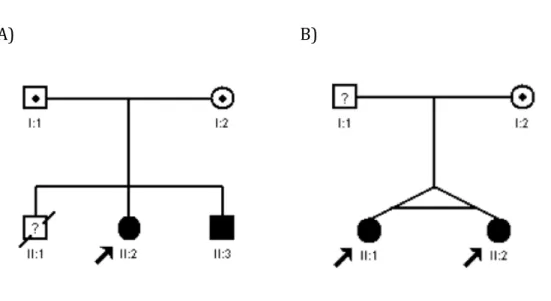
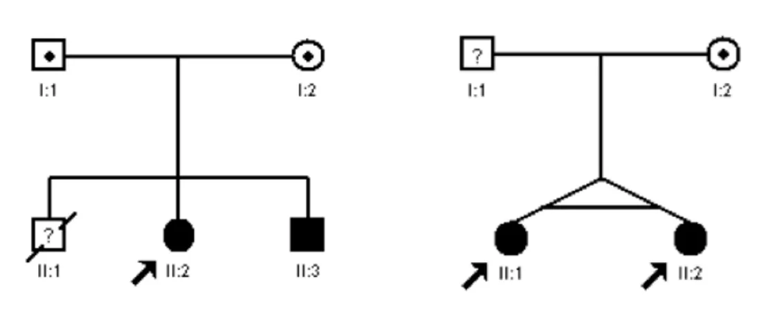
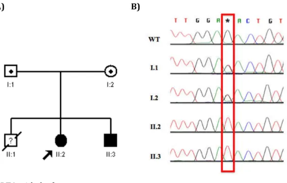
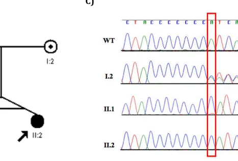
Figura 3. Pedigrees das famílias do estudo. A) Pedigree da família 1. B) Pedigree da
família 2. A seta indica o caso índice... 37
LISTA DE TABELAS
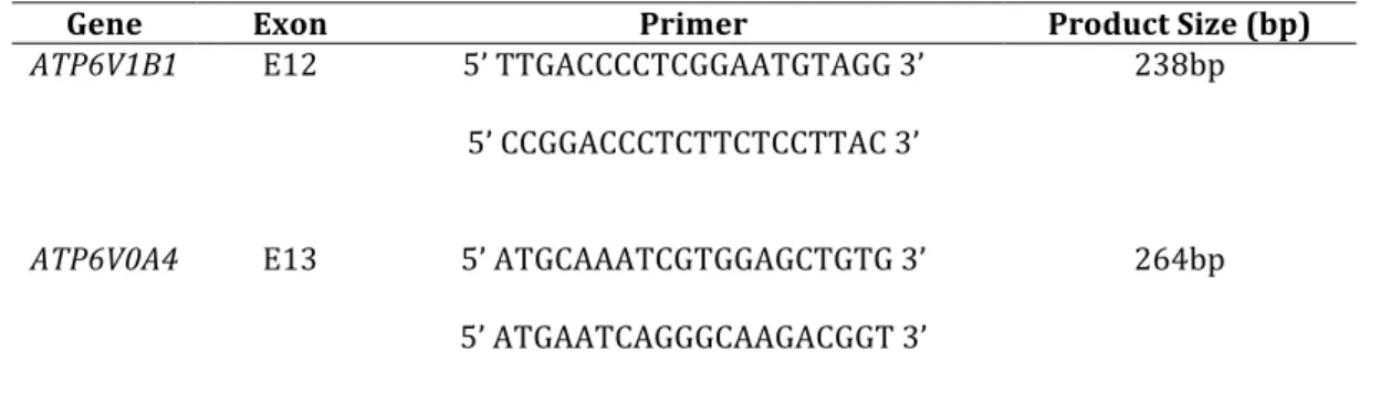
Talela 1. Primers para amplificação genômica e análise de mutações para os genes
ATP6V1B1 e ATP6V0A4………... 41
SUMÁRIO
1 INTRODUÇÃO ... 12
1.1 Acidose Tubular Renal ... 12
1.2 Acidose Tubular Renal Distal ... 13
1.2.1 Etiologia e genética ... 14
1.2.2 Fisiopatologia ... 18
1.2.3 Diagnóstico ... 21
1.2.4 Tratamento ... 23
1.3 Justificativa do estudo ... 24
2 REVISÃO DE LITERATURA (ARTIGO 1) ... 26
3 OBJETIVOS ... 36
3.1 Objetivo Geral ... 36
3.2 Objetivos Específicos ... 36
4 PACIENTES E MÉTODOS ... 37
4.1 Pacientes ... 37
4.2 Metodologia ... 38
4.2.1 Coleta de sangue periferico e extração de DNA ... 38
4.2.2 Sequenciamento por Genoma Inteiro (Whole-‐exome Sequencing -‐ WES) ...38
4.2.3 Análise do Sequenciamento por WES ... 39
4.2.4 Estudo estrutural das alterações encontradas ... 40
4.2.5 Validação dos Resultados: amplificação e sequenciamento das amostras ...40
4.3 Aspectos Éticos ... 42
5 RESULTADOS: ANÁLISE GENÉTICA (ARTIGO 2) ... 43
6 CONCLUSÃO ... 64
7 REFERÊNCIAS BIBLIOGRÁFICAS ... 65
8 ANEXOS ... 70
8.1 Parecer do COEP (comitê de ética em pesquisa)/ UFMG ... 70
8.3 Declaração de aprovação ... 72 8.4 Artigo Original ... 73
1
INTRODUÇÃO
Apesar de não apresentar incidência tão elevada em nosso meio quanto outras nefropatias, como as doenças glomerulares e urológicas, a acidose tubular renal (ATR) assume grande importância, não apenas pela dificuldade diagnóstica, mas também pelo grande impacto sobre o crescimento pôndero-‐estatural das crianças acometidas (1-‐5). O seguimento clínico, os exames complementares e o tratamento dos pacientes portadores de ATR já se encontram bem estabelecidos em nosso meio (6). No entanto, não há nenhum estudo genético com pacientes brasileiros até o momento. Neste contexto, a presente tese pretende extender e ampliar o conhecimento sobre as doenças tubulares renais em crianças e adolescentes brasileiros, focando mais especificamente na caracterização genotípica dos portadores de ATR distal ou tipo 1 e da correlação desses achados com o fenótipo dos pacientes.
1.1 Acidose Tubular Renal
Os rins são responsáveis pela reabsorção do HCO3− filtrado e excreção de H+ numa quantidade igual àquela produzida pelo metabolismo diário de proteínas. A resposta normal a acidemia consiste na reabsorção do HCO3− filtrado e aumento da excreção de ácidos, principalmente através da maior excreção de íons amônio (NH4+) na urina. Assim, para cada H+ excretado há regeneração de um íon HCO3− no plasma (7-‐9).
As ATR são classificadas em quatro categorias: ATR distal ou tipo 1; ATR proximal ou tipo 2 e ATR hipercalêmica ou tipo 4. A ATR mista ou tipo 3 é caracterizada por uma desordem que apresenta características mistas dos tipos 1 e 2 (5).
A seguir, será brevemente descrito a ATR distal ou tipo 1 em relação às características clínicas, fisiopatologia, etiologia e genética, diagnóstco e tratamento.
1.2 Acidose Tubular Renal Distal
A ATR distal caracteriza-‐se por uma inabilidade dos túbulos distal e coletor em promover uma adequada acidificação urinária, resultando numa urina com pH elevado, mesmo em presença de acidose metabólica (5). Inicialmente, a função glomerular encontra-‐se normal ou perto do valor normal em todos os casos (7).
1.2.1 Etiologia e genética
A ATR distal pode ser primária, devido a defeitos genéticos nos mecanismos de transporte, ou secundária a uma variedade de doenças (7,15). Dentre as formas primárias podemos encontrar as seguintes variantes: autossômica dominante e autossômica recessiva com ou sem surdez. Na criança, o defeito é, na maioria das vezes, primário (5). Em algumas famílias, a presença da doença em várias gerações sugere uma forma autossômica dominante. Apesar das manifestações clínicas não serem diferentes das observadas nas formas autossômicas recessivas, estes pacientes podem ter seu diagnóstico mais tardio e evoluírem com sintomatologia mais branda (16). Já pacientes com a forma autossômica recessiva geralmente apresentam manifestações clínicas mais acentuadas com importante déficit de crescimento e nefrocalcinose precoce, podendo evoluir para insuficiência renal (9). Os achados clínicos dos pacientes autossômicos recessivos acompanhados de surdez neurosensorial são idênticos aos dos pacientes portadores de ATR distal autossômica recessiva com audição normal (17). A evolução da surdez é progressiva e não há melhora, mesmo após terapia com álcalis (18). A ATR distal, autossômica recessiva, com função auditiva normal é a forma primária mais comumente encontrada (19).
Dentre as causas secundárias, que são mais comuns em pacientes adultos do que pediátricos, incluem-‐se (20,21):
1 -‐doenças auto-‐imunes: síndrome de Sjögren, hepatite crônica ativa, tireoidite, poliarterite nodosa, hiperparatireoidismo primário, rim esponjoso medular, doença de Wilson, artrite reumatóide e lúpus eritematoso sistêmico;
2 -‐uso de medicamentos: anfotericina B, sulfametoxazol-‐trimetoprim, amilorida, lítio, analgésicos;
3 -‐exposição ao tolueno (cheiradores de cola) e ao mercúrio;
4 -‐doenças túbulo-‐intersticiais: uropatia obstrutiva, pielonefrite crônica, transplante renal;
H+ ATPase
As H+ATPases ou V-‐ATPases (vacuolar) são bombas de prótons essenciais para o funcionamento de diversos compartimentos intracelulares em organismos eucariotas (figura 1). São encontradas em grande quantidade na membrana plasmatica de celulas especializadas como as celulas intercaladas renais, osteoclastos, células do trato genital masculino e da orelha interna (22).
A H+ATPase renal, localizada na membrana apical das células intercaladas tipo α dos tubulos coletor e distal, é a principal bomba de prótons responsável pela acidificação urinária. É uma proteina de membrana com multiplas subunidades divididas em dois dominios funcionais: V1, complexo citoplasmático responsável pela hidrolise do ATP e V0, complexo transmembrana responsável pela translocação do próton. Algumas subunidades possuem isoformas diferentes de acordo com a especie onde é expressa (22-‐24).
O dominio V1 (periférico; 570-‐kDa) é composto por 8 diferentes subunidades (A-‐H), sendo 3 cópias das unidades A e B, 2 cópias da unidade G e uma única cópia das demais. Já o dominio V0 (transmembrana; 260-‐kDa) é composto por 5 subunidades (a, c, c’, c’’, d), sendo seis cópias das unidades c/c’ e uma copia das demais (22).
Gene SLC4A1
O gene SLC4A1 ou AE1 (Solute Carrier Family 4, Anion Exchanger, Member 1) pertence a uma familia de genes trocadores de ânions "Anion Exchanger-‐AE gene family". Era comumente chamado de AE1 (anion exchanger 1 gene), porém, foi recentemente renomeado como SLC4A1, pelo comitê de nomenclatura do mapeamento genético humano (Humam Gene Maping – HGM). É composto de 20
exons e 19 introns estando localizado no cromossomo 17q12-‐q21 e constituído de 17 kb no total (25).
O gene é composto de duas regiões promotoras tecido-‐específicas, uma eritróide localizada acima do exon 1 e uma interna localizada no intron 3, no caso das células renais (26). Codifica a proteína transportadora de íons ou trocador aniônico Cl−/HCO3− (AE1 ou banda 3) nas sua duas isoformas eAE1 (eritrocitária) e rAE1 (renal). Diferem quanto a sua transcrição: o RNA mensageiro (mRNA) da rAE1 não apresenta os três primeiros exons observados na forma eritróide (27). Nos rins, a proteína é expressa na membrana basolateral de células α intercaladas do tubulo coletor, onde é responsável pela reabsorção de HCO3−, compensando a secreção de ácidos pela H+ATPase vacuolar apical, e participando da regulação fina do equilíbrio ácido-‐básico (15).
por acidose metabólica hiperclorêmica, hipocalemia variável, retardo do cresciemento infantil, nefrocalcinose e nefrolitíase, que progride para insuficiência renal. A doença recessiva, frequentemente, se manifesta cedo acompanhada por uma concentração urinária deficiente e suscetibilidade a desidratação grave. Recentemente, foi descrita uma mutação em 3 irmãos indianos causando ATR distal associada a esferocitose heteritária (30). Ressalta-‐se ainda que mutações trocador AE1 já foram descritas em crianças do nosso meio, portadoras de ATR distal e nefrocalcinose (31,32).
Gene ATP6V1B1
O gene ATP6V1B1, localizado no cromossomo 2q13 e composto por 14 exons, codifica a subunidade B1 da H+ATPase, presente na membrana apical das células intercaladas tipo α e também na cóclea e no saco endolinfático (15,33).
Mutações nesse gene já foram detectadas em pacientes portadores de ATR distal autossômica recessiva associada à surdez neurosensorial (34-‐36). Demonstrou-‐se que as células auditivas interdentais e as células do saco endolinfático são muito semelhantes às células intercaladas do tipo α, apresentando tanto a H+ATPase como o trocador AE1. Assim, uma secreção normal de ácidos por estas células é fundamental para a manutenção de um pH reduzido na endolinfa e uma função auditiva normal (15,16). Considerando que a alta concentração de potássio presente nesse compartimento fechado não é normalmente acompanhada por alcalinidade da endolinfa, propõe-‐se que a H+ATPase atue na manutenção do pH em aproximadamente 7,4. A perda de tal função leva ao aumento do pH, promovendo dano celular e perda auditiva (37).
Gene ATP6V0A4
O gene ATP6V0A4 está localizado no cromossomo 7q33-‐34 e é composto por 22 exons, dos quais 20 codificam a subunidade a4 da H+ATPase.
A ATR distal com perda autivida neurossensorial predomina nas mutações do gene ATPV1B1, enquanto a ATR distal com ausência de anormalidade auditiva (pelo menos na infância) se relaciona com alterações no gene ATPV0A4. Contudo, neste último, a surdez pode se desenvolver com idade mais avançada, após a segunda década de vida, embora seja menos grave na maioria dos casos (5). O fato de esse gene ser também expresso em células do ouvido interno pode explicar o desenvolvimento da surdez (15,38).
1.2.2 Fisiopatologia
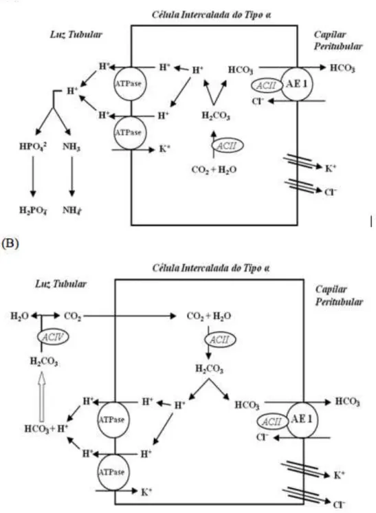
No néfron distal (ND), são definidas as características finais da urina através dos mecanismos de concentração e acidificação urinárias, secreção de K+ e conservação do Na+. O ND é formado pelos seguintes segmentos: túbulo distal (TD), túbulo conector (TCN) e túbulo coletor (TC) (6). O TD é responsável pela reabsorção de 5% do Na+ filtrado enquanto o TC é responsável pela reabsorção de 2% a 3% do cloreto de sódio (NaCl) filtrado.
Esse segmento apresenta dois tipos de células com funções e morfologia
bastante diferentes: células principais ou células claras, onde ocorre reabsorção de
sódio (Na+) e é o principal sítio para secreção de potássio (K+), e células intercaladas ou células escuras, que estão relacionadas ao equilíbrio ácido-básico,
Figura 2. Mecanismos de transporte tubular no néfron distal: (A) secreção de íons hidrogênio e acidificação urinária; (B) reabsorção de bicarbonato.
A ATR distal pode resultar dos seguintes defeitos nos túbulos distais: diminuição da atividade da H+ATPase (ATR distal secretora); aumento da permeabilidade da membrana luminal (ATR distal por difusão retrógrada) ou diminuição da reabsorção distal de Na+ (ATR distal voltagem dependente) (40).
A diminuição, ou mesmo ausência, de atividade da H+ATPase das células intercaladas dos túbulos distais e coletores, geralmente é secundária a um defeito genético. Também já foram detectadas mutações no trocador AE1 que, assim como a H+ATPase, participa do processo de acidificação urinária, reabsorvendo HCO3−. Outra possibilidade para explicar esse sub-‐tipo da ATR distal seria a existência de um defeito na H+/K+ATPase, localizada na membrana apical das células intercaladas. No entanto, alguns autores acreditam que esta bomba esteja mais relacionada à homeostase do K+ que do H+ (5).
como as decorrentes de mutações do trocador AE1, assim como explica a piora da acidose nesses pacientes, induzida por uma carga excessiva de NaCl 0,45% (19).
O exemplo clássico de ATR distal, causada por aumento da permeabilidade da membrana luminal do ND, está associado ao uso de anfotericina B. Postulava-‐se, inicialmente, que esta droga formaria canais aquosos na membrana das células do TD e promoveria o retorno de íons H+ para o interior da célula. Este processo foi chamado de difusão retrógrada. Entretanto, esta teoria tem sido questionada. Alguns autores acreditam que o H2CO3 e/ou o HCO3−, e não o H+, possam retornar para a luz tubular (40). O relato de crianças com quadros de ATR distal associada a doenças auto-‐imunes sugere que o mecanismo de difusão retrógrada como causa ainda não foi totalmente esclarecido (41).
A reabsorção de Na+ no TC cria uma diferença de potencial lúmen-‐ negativa, que é fundamental para a secreção de íons H+ e K+ (8). Os fatores relacionados à diminuição da reabsorção de Na+ ou de seu aporte ao TD, podem reduzir a capacidade secretora deste segmento do néfron, que é voltagem dependente (40). Os fatores mais comumente relacionados a este tipo de ATR são a uropatia obstrutiva, a depleção volumétrica e o uso de diuréticos poupadores de K+ (42). Como a secreção de K+ está igualmente comprometida, também pode evoluir com elevação dos níveis séricos deste cátion (40). Recentemente, entretanto, a teoria de que a ATR distal hipercalêmica seja causada por defeito isolado de uma diferença de potencial transmembrana tem sido questionada. Alguns estudos têm demonstrado que os mecanismos envolvidos são bem mais complexos, envolvendo também defeitos no funcionamento da H+ATPase, H+/K+ATPase e Na+/K+ATPase (42).
1.2.3 Diagnóstico
ou está reduzida, e o paciente é incapaz de reduzir o pH urinário para valores inferiores a 5,5, estabelece-‐se o diagnóstico de ATR distal. A excreção urinária de citrato geralmente está diminuída, devido a sua maior reabsorção proximal, estimulada pela acidose. A excreção urinária aumentada de cálcio (Ca2+) associada a hipocitratúria e ao pH urinário persistentemente elevado, pode contribuir para o desenvolvimento de nefrolitíase e nefrocalcinose. Tais alterações são comuns na ATR distal não tratada, embora existam relatos de nefrocalcinose na ausência de hipercalciúria (43).
O raquitismo e a diminuição da massa óssea também podem ser encontrados, mas sua real incidência ainda é incerta. A acidose metabólica também pode, por si só, alterar o metabolismo da vitamina D, diminuindo sua produção renal, com conseqüente déficit na reabsorção intestinal de Ca2+ e doença óssea secundária.
A hipocalemia está presente em 30 a 50% dos casos. Pode manifestar-‐se como fraqueza muscular, às vezes com episódios agudos de paralisia flácida, que podem evoluir para tetraplegia em até 48h (44).
A apresentação clínica da ATR distal engloba, além do déficit de crescimento nas crianças, um quadro de anorexia, vômitos e poliúria (45). Algumas condições podem mimetizar a ATR distal com pH urinário maior que 5,5. Pacientes portadores de infecção urinária por bactérias urease-‐positivas podem apresentar pH urinário alcalino, porém, em geral, não apresentam acidose sistêmica. Além disso, o exame microbiológico e o sedimento urinário exibem alterações típicas. A fase inicial da ATR proximal, quando ainda há perda urinária de álcalis e hipovolemia, também pode confundir-‐se com a ATR distal. A depleção de K+ e o aumento da excreção urinária de amônia (NH3), que podem ocorrer na acidose metabólica por diarréia aguda, simulam, algumas vezes, o quadro laboratorial desta patologia (5).
horas; e a gasometria seja realizada no início do teste e a cada hora, nas 4 horas subseqüentes à administração do ácido. Se o pH urinário falha em cair abaixo de 5,5 durante a quarta hora após o NH4Cl, é provável que a ATR distal esteja presente, desde que um pH sangüíneo inferior a 7,35 e um bicarbonato menor que 20 mEq/L sejam documentados (6).
1.2.4 Tratamento
O objetivo do tratamento consiste não só na correção das alterações bioquímicas, mas principalmente na retomada do crescimento e na prevenção da nefrocalcinose e da insuficiência renal. Os pacientes adequadamente tratados geralmente são assintomáticos e podem levar uma vida normal, a não ser que já tenha havido lesão renal ou óssea irreversíveis (5). A normalização do pH sérico diminui a perda urinária de K+ e previne a litíase e o desenvolvimento da nefrocalcinose (46). A correção da acidose também reverte as alterações no metabolismo das células ósseas, aumentando, conseqüentemente, a densidade mineral do osso (47).
6,5 e 7,5 e a gasometria revelar equilíbrio ácido básico. A monitoração individual é fundamental para o ajuste das doses (48).
A correção da hipercalciúria é mandatória, mesmo em presença de uma excreção urinária adequada de citrato. O citrato pode melhorar a saturação urinária para o oxalato de cálcio, mas não reverte a tendência para a saturação renal do fosfato de cálcio no osso (48). A monitoração do Ca2+ urinário, através da relação cálcio/creatinina em amostra de urina e/ou dos níveis de cálcio na urina de 24 horas é importante para a avaliação do tratamento (49). O uso de diuréticos tiazídicos é uma opção terapêutica para controlar a hipercalciúria, quando a excreção urinária de cálcio persiste aumentada mesmo após correção do distúrbio ácido-‐básico (50).
Pacientes portadores de ATR distal primária vão requerer tratamento prolongado, possivelmente por toda a vida. Em geral o prognóstico é excelente, sobretudo para as crianças precoce e adequadamente tratadas. O uso adequado da terapia alcalina pode restabelecer o crescimento e prevenir a progressão para nefrocalcinose (5).
1.3 Justificativa do estudo
genótipo e fenótipo dos pacientes. Sendo assim, o presente estudo teve como objetivo principal avaliar a aplicabilidade da técnica de WES no diagnóstico genético desta tubulopatia. Ressalta-‐se ainda que se trata do primeiro estudo em que a técnica de WES foi aplicada para diagnóstico de uma tubulopatia.
2
REVISÃO DE LITERATURA (ARTIGO 1)
Molecular Pathophysiology of Renal Tubular Acidosis
(Current Genomics, 2009, Vol. 10, No. 1, 51-59)
1389-2029/09 $55.00+.00 ©2009 Bentham Science Publishers Ltd. P.C.B. Pereira, D.M. Miranda, E.A. Oliveira* and A.C. Simões e Silva
Pediatric Nephrology Unit, Department of Pediatrics, School of Medicine – Federal University of Minas Gerais (UFMG), Belo Horizonte, MG, Brazil
Abstract: Renal tubular acidosis (RTA) is characterized by metabolic acidosis due to renal impaired acid excretion.
Hy-perchloremic acidosis with normal anion gap and normal or minimally affected glomerular filtration rate defines this dis-order. RTA can also present with hypokalemia, medullary nephrocalcinosis and nephrolitiasis, as well as growth retarda-tion and rickets in children, or short stature and osteomalacia in adults. In the past decade, remarkable progress has been made in our understanding of the molecular pathogenesis of RTA and the fundamental molecular physiology of renal tu-bular transport processes. This review summarizes hereditary diseases caused by mutations in genes encoding transporter or channel proteins operating along the renal tubule. Review of the molecular basis of hereditary tubulopathies reveals various loss-of-function or gain-of-function mutations in genes encoding cotransporter, exchanger, or channel proteins, which are located in the luminal, basolateral, or endosomal membranes of the tubular cell or in paracellular tight junc-tions. These gene mutations result in a variety of functional defects in transporter/channel proteins, including decreased activity, impaired gating, defective trafficking, impaired endocytosis and degradation, or defective assembly of channel subunits. Further molecular studies of inherited tubular transport disorders may shed more light on the molecular patho-physiology of these diseases and may significantly improve our understanding of the mechanisms underlying renal salt homeostasis, urinary mineral excretion, and blood pressure regulation in health and disease. The identification of the mo-lecular defects in inherited tubulopathies may provide a basis for future design of targeted therapeutic interventions and, possibly, strategies for gene therapy of these complex disorders.
Received on: October 9, 2008 - Revised on: November 8, 2008 - Accepted on: November 12, 2008
Key Words: Renal tubular acidosis, acid-base homeostasis, molecular physiology, tubular transport, gene mutations.
INTRODUCTION
The term Renal Tubular Acidosis (RTA) defines many disorders characterized by metabolic acidosis, secondary to defects in renal tubular reabsorption of bicarbonate (HCO3
) and/or in urinary excretion of hydrogen (H+), while glomeru-lar function is little or not affected [1-6]. All forms of RTA present hyperchloremic metabolic acidosis, with normal an-ion gap and are chronic diseases with significant impact on the quality of life of affected patients when left untreated, possibly leading to growth failure, osteoporosis, rickets, nephrolithiasis and even renal insufficiency [1-6].
Defects in proximal bicarbonate reclamation or distal acid secretion give rise to the respective clinical syndromes of proximal or distal RTA [1-6]. These disorders can be pri-mary, originating from genetic defects on tubular transport mechanisms [7], or secondary to systemic diseases and to adverse drug reactions [8-12]. The familial conditions ex-hibit distinct inheritance patterns. Distal RTA can be trans-mitted as either an autosomal dominant or an autosomal re-cessive trait, whereas isolated proximal RTA usually occurs as an autosomal recessive disease [6,7,13]. In the past few years, the molecular genetic strategies of positional cloning and candidate gene analysis have been combined to identify
*Address correspondence to this author at the Rua Engenheiro Amaro Lana-ri, 389 / apt 501, Belo Horizonte-Minas Gerais, 30310-580, Brazil; Tel: +55-31-99797782; Fax: +55-31-32851056;
E-mail: eduolive@medicina.ufmg.br
the genes responsible for these inherited conditions [6,13]. This review will summarize the mechanisms of acid-base regulation by the kidney and the current understanding of the genetic causes of primary inherited RTA. It will, in addition, evaluate the ability of known functional and biochemical properties of these mutant proteins to explain the patho-physiology of associated renal acidification defects.
BRIEF OVERVIEW OF RENAL ACID-BASE HO-MEOSTASIS
bicarbonate exchanger, AE1 [18,20,21].
Proximal Tubular Bicarbonate Reabsorption
HCO3- is freely filtered at the glomerulus and
approxi-mately 80 to 90% of this is reabsorbed in the proximal tubule [6]. In the tubular lumen, HCO3- combines with H+ in a
reac-tion catalyzed by CA IV, which is bound to the luminal membrane of proximal tubular cells [2,14,15]. This reaction produces carbonic acid, which is promptly converted to CO2
and H2O. The resulting CO2 rapidly diffuses into the tubular
cells and is combined with water to produce intracellular H+ and HCO3-. This intracellular reaction is catalyzed by CA II.
HCO3- is then cotransported with Na+ into blood (with a
probable stoichiometry of 3 HCO3– to 1 Na+) [6] via the
NBC-1, located on the basolateral cell membrane. The intra-cellular H+ produced by CA II is secreted into the tubular lumen predominantly via the NHE-3, situated on the luminal membrane [6,15,22]. This transport process is called facili-tated diffusion and depends on the sodium concentration gradient generated by the action of a basolateral membrane Na+-K+-ATPase. It should be mentioned that there is mini-mal net acid excretion in the proximini-mal tubule, since most of the H+ secretion is coupled with HCO3- reabsorption [6,13].
The small amount of remaining H+ will be buffered by phos-phate as titratable acid. HCO3- reabsorption is influenced by
luminal HCO3- concentration and pH, luminal flow rate,
peritubular pCO2, and angiotensin II [2,6,17].
Proximal tubular cells are capable of generating “extra” bicarbonate through the deamination of glutamine to
gluta-This metabolic process produces HCO3 and NH4: the
for-mer reclaimed via the basolateral membrane and the latter secreted into the tubular lumen. This pathway can be upregu-lated in states of chronic acidosis [3,6,15].
The main mechanisms of proximal tubular bicarbonate reabsorption are displayed in Fig. (1).
Distal Tubular Hydrogen Secretion
One of the important roles of the collecting duct segment of the nephron is acid secretion, combined with reclamation of the approximately 10% of filtered HCO3- that is not
reab-sorbed by more proximal nephron segments [18]. The aver-age omnivorous human diet in the `Western' world is rich in protein, and generates 1±1.5 mmol hydrogen/kg body weight each day [23]. Urinary acid excretion is therefore essential, and urine pH can drop as low as 4.5. The -intercalated cell
is the main responsible for hydrogen secretion into the urine. In humans at least, hydrogen pumps, called H+-ATPases, mainly carry out hydrogen secretion [18,19,23]. H+-ATPases are present at high density on the luminal membrane of
-intercalated cells [18]. Studies in nonhuman mammals show that these H+-ATPases are also present within specialized intracellular tubulovesicles close to the membrane, allowing additional pumps to be recruited to the membrane quickly in to response to stimuli, such as systemic acidosis, for example [23]. These cells secrete H+ into the lumen of the distal tu-bule and collecting duct not only via H+-ATPase but possi-bly also by an exchanger, H+/K+-ATPase [7,10]. In addition, the normal function of the luminal H+-ATPase in
-Fig. (1). Schematic model of bicarbonate (HCO3
-) proximal reabsorption. The intracellular carbonic acid (H2CO3
-)dissociates into H+ and HCO3- in a reaction catalysed by a cytoplasmic carbonic anhydrase (CAII). At the luminal membrane, H+ secretion is due to an especific Na+ – H+ exchanger (NHE-3), while, at the basolateral membrane, the 1 Na+ - 3 HCO3
cotransporter (NBC-1) is responsible for HCO3
to the electroneutral transport of HCO3 back across the
baso-lateral surface into the interstitial fluid, and hence to blood. The transporter responsible for this activity in renal
-intercalated cells is the Cl-/HCO3- exchanger AE1 [7,20,21].
The AE1 exchanger is homologous with the red cell anion exchanger known as ‘band 3’ (eAE1) [6,24]. After the red cell, the kidney is the next richest source of this protein (kAE1) [24]. Proton secretion varies with systemic pH and it is also aldosterone-dependent and voltage-dependent [24].
Once secreted, net urinary elimination of H+ depends on its buffering and excretion as titratable acid (mainly phos-phate - HPO42
+ H+ H2PO4
), and excretion as NH4+ [24].
Notably, the production of NH4+ from glutamine by the
proximal tubule, and its subsequent excretion in the urine, also generates ‘new’ bicarbonate, which is added to plasma [24]. Availability of phosphate as a buffer depends on its filtration, whereas NH4+ depends on normal function of the
proximal tubule, as well as a complex process of secretion, reabsorption, and secretion again along the nephron [24]. The final secretory step for NH4+ excretion is ‘diffusion
trapping’ in the collecting duct. Anything that interferes with H+ secretion in the collecting duct will reduce diffusion trap-ping and cause a decrease in excretion of both H+ and NH4+
[6,24]. As previously mentioned,chronic metabolic acidosis stimulates renal NH4+ synthesis and excretion [3,6,15].
Fig.(2)showsrenalacidificationprocessin-intercalated
cells of the distal nephron.
CLASSIFICATION AND CLINICAL FEATURES OF RENAL TUBULAR ACIDOSIS
Clinically, RTA is characterized by a normal anion gap, hyperchloremic metabolic acidosis, and associated failure to
Polyuria and constipation can also be seen, although neither may be apparent in the neonatal period [13]. Hyperchloremic metabolic acidosis in pediatric practice is most often associ-ated with diarrheal disease. Both diarrhea and RTA result in hypokalemia. For this reason, in a young infant with diarrhea and underlying RTA, the true diagnosis may be obscured. Thus, inordinately slow resolution of hyperchloremic meta-bolic acidosis following diarrheal disease should suggest the possibility of an underlying primary RTA [13].
Beyond the difficulties inherent in delineating RTA, RTA can be subcategorized into different disorders with dis-tinctly diverse prognoses [13]. The diagnostic cataloguing of RTA is based on the underlying pathophysiology. The cur-rent model of how the nephron reabsorbs HCO3
and secretes H+ has led to a clinical and functional classification of proximal (tubule) versus distal (tubule and collecting duct) forms of RTA [24]. Thus, the main types of RTA are proxi-mal (or type 2) RTA and distal (or type 1) RTA. Type 3 RTA is a mixed type RTA that exhibits both impaired proximal HCO3– reabsorption and impaired distal
acidifica-tion, and more disturbingly osteopetrosis, cerebral calcifica-tion and mental retardacalcifica-tion [4]. Hyperkalemic (or type 4) RTA is a heterogeneous group of disorders that is character-ized by low urine NH4+, which is probably caused by the
hyperkalemia or by aldosterone deficiency or defective sig-naling [4].
In distal RTA, distal nephron net acid secretion is im-paired. This leads to a high urine pH, even in the presence of systemic acidosis [2,4]. However, there is often no metabolic acidosis and the blood bicarbonate concentration is normal, so-called ‘incomplete’ distal RTA, and a defect in renal acid excretion must be demonstrated by a failure to lower urine
Fig. (2). Schematic model of the -intercalated cell and the H+ secretion in cortical collecting tubule. The -intercalated cell is responsible
for H+ secretion by a vacuolar H+-ATPase (main pump) and also by a H+-K+-ATPase. The luminal ammonia (NH3) buffers H +
to form nondiffusible ammonium (NH4+) and divalent basic phosphate (HPO4-) is converted to the monovalent acid form (H2PO4-) in H+ presence. Intracellularly formed HCO3
leaves the cell via Cl- - HCO3
rosemide test [2,6,24]. Acquired distal RTA is often secon-dary to autoimmune diseases, such as Sjogren’s syndrome [6,24]. Inherited distal RTA can be essentially of three types: autosomal dominant distal RTA (the commonest form) and autosomal recessive distal RTA with and without sen-sorineural deafness [24]. In the complete forms of both dominant and recessive distal RTA bone disease is common (rickets or osteomalacia), as well as nephrocalcinosis (often) complicated by renal stone disease. The occurrence of renal stones is attributed to the combination of hypercalciuria, low urinary citrate excretion (due to systemic and intracellular acidosis) and high urine pH, all favouring calcium phosphate stone formation. Hypokalaemia, another characteristic fea-ture, is less troublesome than in the acquired autoimmune form of distal RTA, but it can become symptomatic, espe-cially if a thiazide diuretic is prescribed to reduce hypercal-ciuria [24]. In recessive distal RTA, some patients suffer from sensorineural deafness, which can be late in onset [24].
Conceptually, the proximal tubule is charged with the task of reclaiming filtered HCO3- (~ 85% of the total) [13].
Failure of this process leads to reduction in systemic base, resulting in metabolic acidosis – proximal RTA [13]. Proxi-mal RTA typically manifests as part of a generalized defect of proximal tubule function, namely the renal Fanconi’s syn-drome (with glycosuria, low molecular weight proteinuria, urinary phosphate wasting, hypophosphataemia and hypouri-caemia) [24]. Isolated proximal RTA occurs rarely and usu-ally presents as growth retardation in childhood. Like distal RTA, it can be divided into three types: autosomal recessive proximal RTA with ocular abnormalities, autosomal reces-sive proximal RTA with osteopetrosis and cerebral calcifica-tion, and autosomal dominant proximal RTA [24]. Autoso-mal recessive proxiAutoso-mal RTA with ocular abnorAutoso-malities is the commonest form of isolated and inherited proximal RTA, but even this is rare. Ocular abnormalities include band kera-topathy, glaucoma and cataracts [24]. Short stature is usual; dental enamel defects, mental retardation, hypothyroidism, abnormal pancreatic function and basal ganglia calcification are also features [24,25]. In inherited CA II deficiency, iso-lated proximal RTA presents with osteopetrosis (due to im-paired osteoclast function), cerebral calcification and vari-able mental retardation [26]. Although this form of inherited RTA is clinically more proximal in type, it can also present with a mixed proximal and distal phenotype, which reflects the presence of CA II in cells all along the renal tubule.
Type 3 RTA can be caused by recessive mutation in the
CA2 gene on chromosome 8q22, which encodes CAII [4] or
NBC1 [27] or Cl/ HCO3 exchanger, SLC26A6 [4,28].
The causes of type 4 RTA include various types of adre-nal failure or pseudohypoaldosteronism type 1 (PHA1) due to defects in the mineralocorticoid receptor or the epithelial Na+ channel, all characterized by salt loss and hypotension [4]. A similar picture may be seen in obstructive uropathy or drug induced interstitial nephritis [4]. Furthermore, a number of drugs may impair signalling in the renin–angiotensin-aldosterone system and cause hyperkalemia and metabolic acidosis (e.g. potassium sparing diuretics, trimethoprim, cy-clo-oxygenase inhibitors, angiotensin converting enzyme inhibitors) [4]. Lately, much interest has been given to a group of rare autosomal dominant diseases characterized by hyperkalaemia and acidosis and age-related hypertension [4]. In spite of hypervolaemia, aldosterone is not low and the disorders have been collectively termed pseudohypoaldos-teronism type 2 (PHA2) [4].
INHERITED FORMS OF DISTAL RENAL TUBULAR ACIDOSIS
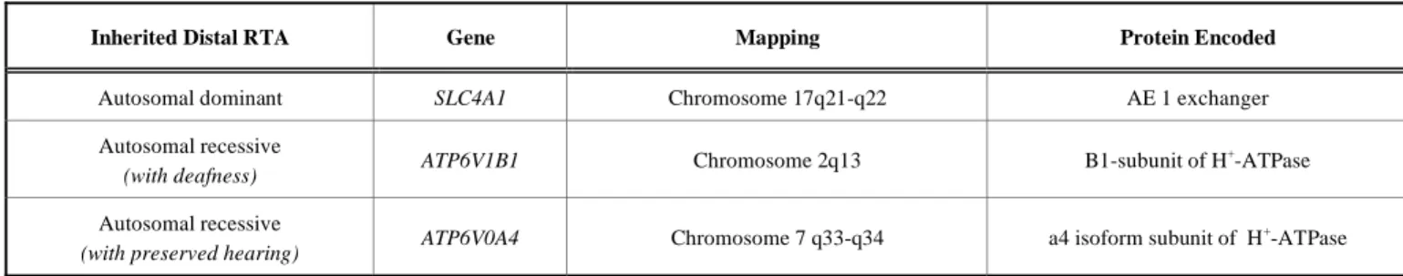
Inherited forms of distal RTA have three variants: auto-somal dominant and autoauto-somal recessive with or without deafness. Dominant disease typically presents more mildly in adolescence or adulthood, and recessive variant occurs in infancy/early childhood, where growth retardation is com-mon [6]. In Table 1 we can see the chromosome mapping of distal RTA.
Autosomal Dominant Distal RTA (Distal RTA Type 1a )
Distal RTA occurs with the greatest frequency as an iso-lated defect, often transmitted as an autosomal dominant trait [13]. In few reported families, the presence of the disorder in several generations suggests an autosomal dominant trans-mission. Although clinical findings are not different from those observed in autosomal recessive or sporadic cases, in these patients the disease may be diagnosed later (in adoles-cence or adulthood) [6] or manifest with milder symptoma-tology.
Autosomal dominant distal RTA has been found to be associated in several kindred with mutations in the SLC4A1 gene encoding the CI-/HCO3- exchanger, AE1 [15].
The Electroneutral Anion Exchanger (AE1)
The Cl-/HCO3- anion exchanger, AE1, is a glycoprotein
encoded by a gene (SLC4A1) present on chromosome 17 q21-22. SCL4A1 gene is a member of the SLC4 family
com-Table 1. Chromosome Mapping of the Inherited Distal Renal Tubular Acidosis
Inherited Distal RTA Gene Mapping Protein Encoded
Autosomal dominant SLC4A1 Chromosome 17q21-q22 AE 1 exchanger
Autosomal recessive
(with deafness) ATP6V1B1 Chromosome 2q13 B1-subunit of H
+
-ATPase
Autosomal recessive
(with preserved hearing) ATP6V0A4 Chromosome 7 q33-q34 a4 isoform subunit of H
+
porters [6,24,29]. AE1 is an integral membrane glycoprotein containing a long cytoplasmic N-terminus (~ 400 amino ac-ids), 12–13 transmembrane domains (responsible for anion transport and dimerization), and a short cytoplasmic C-ter- minus (~ 35 amino acids) [30,31]. It is predominantly ex-pressed in the erythrocytes (eAE1) and in the kidney (kAE1).
kAE1 is a truncated isoform of eAE1 with lacking of 65 amino acids at the N-terminus owing to the use of differen-tial transcription and translation start sites [32]. This extra NH2-terminal sequence confers additional roles for eAE1, including facilitation of red cell metabolism and mainte-nance of erythrocyte structural stability via interaction with a glycolytic enzyme complex and cytoskeletal elements, re-spectively [6]. kAE1 mediates an electroneutral exchange of chloride for bicarbonate at the basolateral membrane of acid secreting -intercalated cells of the distal nephron and
col-lecting duct [32,33]. This ion exchanger promotes the reab-sorption of bicarbonate into the blood. Therefore, eAE1 de-fect results in morphological changes of red blood cells (RBC) while kAE1 abnormality leads to distal RTA [32].
The physiological role of kAE1 in the regulation of distal nephron acid secretion is well established. In the acidifica-tion process of the distal nephron, basolateral kAE1 mediates Na+-independent, electroneutral Cl-/HCO3- exchange,
allow-ing HCO3- to exit the -intercalated cells in concert with
apical H+ secretion via the vacuolar H+-ATPase.
AE1 Gene (SLC4A1) Mutations
Because of the expression of AE1 in two different cells (RBC and -intercalated distal tubular cells) with distinct functions, AE1 mutations show pleiotrophic effects resulting in two distinct and seemingly unrelated phenotypes: heredi-tary spherocytosis (or other forms of erythrocyte abnormali-ties) and distal RTA [31]. The largest group of mutations in human AE1 is associated with autosomal-dominant red cell dysmorphologies (hereditary spherocytosis – HS; and South-east Asian ovalocytosis - SAO), where renal acid-base han-dling is normal [6]. AE1 mutations also result in distal RTA, because the defect in AE1 affects anion Cl-/HCO3- exchanger
at the basolateral membrane of the -intercalated cells in the
distal nephron [31].
SAO, a well-known erythrocyte disorder, is caused by a deletion of 27 bp in codons 400-408 in exon 11 (Ex11D27) of AE1 leading to a lack of 9 amino acids in the protein, which is inactive for anion transport.
How can be explained either the absence of red cell ab-normalities in patients with distal RTA or the rarity of de-fects in distal urinary acidification in patients with hemato-logical disorders, when, in both circumstances, mutations in the same SLC4A1 gene are present? [15]. One exception is the homozygous AE1 mutant V488M (Band 3 Coimbra; GTG → ATG), which presents with severe anemia and renal acidification defect [34,35].
The majority of AE1 mutations apparently cause only erythroid abnormalities without renal phenotype. Most cause autosomal dominant forms of HS and are not encountered in homozygous form, suggesting embryonic lethality [7].
not associated with distal RTA. Conversely, distal RTA-associated AE1 mutations are also not commonly accompa-nied with HS. Whereas HS missense mutations are distrib-uted throughout AE1 cytoplasmic and transmembrane do-mains, distal RTA mutations are restricted to AE1’s trans-membrane domain. Although, the almost complete segrega-tion between mutasegrega-tions associated with HS and with distal RTA is not fully understood [7].
Autosomal dominant distal RTA was first associated with exon 14 nucleotide substitutions encoding missense muta-tions in residue 589 (R589), in which the wild-type Arg is converted to His, Ser, or Cys [30,36]. A single base change alters the identical AE1 residue, R589, in eight of the ten reported kindred with dominant distal RTA, supporting the importance of this residue in the normal acidification proc-ess. R589 lies at the intracellular border of the sixth trans-membrane domain of the protein, adjacent to K590. These basic residues are conserved in all the known vertebrate an-ion exchanger isoforms and are thought to form part of the site of intracellular anion binding. Arginine at this position is conserved in all vertebrate AE proteins, indicating its func-tional importance [37].
Three different mutations at this position (R589C, R589H, and R589S) were found in autosomal dominant dis-tal RTA and two de novo R589H mutations have also been reported [30,32,36]. A high prevalence of AE1 R589 muta-tions and the presence of at least two de novo mutamuta-tions at this position suggest that codon 589 (CGC) is a “mutational hotspot” of AE1. The mechanism of recurrent mutations probably involves methylation and deamination altering cy-tosine (C) to thymine (T) in the CpG dinucleotides [37].
Another missense mutation alters serine to phenylalanine at position 613 [36] within the adjacent transmembrane loop, evidencing the importance of this region of the protein. A further complex mutation results in a C-terminally truncated AE1 protein lacking the last 11 amino acids [29].
AE1 in Autosomal Recessive Distal RTA
Recent gene studies have shown that some of the AE1 mutations are responsible for autosomal recessive distal RTA in several countries in Southeast Asia; these patients may be homozygous for the mutation or be compound het-erozygotes of two different AE1 mutations, one of which is usually the SAO mutation [38,39]. The evaluation of the AE1 G701D mutation has provided the first explanation for how any distal RTA-associated AE1 mutation might cause the disease [40].
sur-hances this movement. Red blood cells contain GPA but GPA is absent from the kidney, hence individuals homozy-gous for the G701D mutation have normal levels of band 3 in their red cells. It is proposed that, in homozygotes, the mutant G701D protein does not reach the basolateral mem-brane of the -intercalated cell, but is turned over within the
cell. In SAO/G701D compound heterozygotes, the SAO pro-tein is presumed to reach the cell surface, but since it is inac-tive in anion transport, it acts as if it were a band 3 null allele [38].
Autosomal Recessive Distal RTA with Deafness (Distal RTA Type 1b)
Recessive forms of distal RTA are related to mutations in the proton pump in -intercalated cells. The gene involved
(ATP6V1B1) is located on chromosome 2q13, and encodes the B1-subunit of H+-ATPase expressed apically on -inter-
calated cells and also in the cochlea and endolymphatic sac [4,23].
In the human cochlea, the H+-ATPase appears to be re-quired to maintain normal endolymph pH [6] given that the very high potassium concentration (approximately 150 mmol/l) in this closed compartment is not normally accom-panied by alkalinity of the endolymph [23]. ATP6V1B1 ex-pression has also been observed in the male genital tract (with acidification requirement for sperm maturation) [29].
Clinical findings, other than deafness, are identical to those present in patients with sporadic or autosomal reces-sive distal RTA and normal hearing. There is great variation in the presentation of deafness, from birth to late childhood, it is progressive and does not respond alkali therapy [15]. The defects in B1 cause irreversible hair cell damage in hu-man cochlea because of ambient electrolyte and pH abnor-malities [29].
Screening for mutations in this gene revealed fifteen dif-ferent mutations in kindred. The majority of these mutations are likely to disrupt the structure, or abrogate the production, of the normal B1 subunit protein [29].
The Human Vacuolar H+-ATPase
The vacuolar-type proton ATPase (V- or H+-ATPase) is a multisubunit pump that is essential for normal acidification of intracellular vesicular structures. In each individual cell, H+-ATPases may function in a variety of distinct but essen-tial cellular processes. However, the mechanisms by which cells regulate the intracellular trafficking, final destination and activity of these proton pumps are unclear [41].
The H+-ATPases are composed of two structural domains (membrane-bound V0 and cytoplasmic or peripheral V1) each
formed of multiple subunits (a–e and A–H, respectively), which are responsible for ATP hydrolysis and proton trans-port, respectively [6,23]. The mammalian H+-ATPase is pre-sumed to be similar to that of yeast (in which most of the structural studies have been performed) [23].
Autosomal Recessive Distal RTA with Preserved Hearing (Distal RTA Type 1c)
Individuals without hearing defects carry mutations at chromossome 7 q33-q34. The defective gene is ATP6V0A4,
ATPase. The involvement of the a4 subunit in distal RTA shows that it must be essential for proper proton pump func-tion in the kidney [29], but its role is not totally clear.
Site-directed mutagenesis studies of the yeast ‘a’ subunit ortholog Vph1p (the ‘a’ subunit in proton pumps localized to the yeast vacuole) have yielded some potential functions [42]. Some mutations showed that this subunit is important for the assembly of the proton pump, whereas other muta-tions had greater effects on ATPase activity and proton transport. These studies suggest that the ‘a’ subunit is impor-tant for both assembly and function of the pump [29,42].
INHERITED FORMS OF PROXIMAL RENAL TUBU-LAR ACIDOSIS
Proximal RTA is caused by a reduction in bicarbonate reabsorption at the proximal tubules, resulting in low renal bicarbonate threshold. The most common proximal RTA in children is secondary to Fanconi Syndrome [2,43]. Rarely, RTA might also be consequence of an inherited or sporadic primary renal disorder.
The acquired proximal RTA follows exposure to drugs or some toxins and the etiopathogenesis is still unknown [2]. Among drugs that cause Fanconi Syndrome are gentamicin, cisplatin, ifosfamide, and sodium valproate [6]. In addition, some hematologic and autoimmune conditions, such as mye-loma and Sjogren syndrome respectively, might also course with proximal RTA.
The proximal RTA resulting from Fanconi Syndrome is frequently part of a systemic syndrome. Among systemic disorders that result in RTA, the inheritance pattern is usu-ally autosomal recessive. Some of these disorders are cysti-nosis, tyrosinaemia, galactosaemia, Fanconi-Bickel syn-drome and others (Table 2) [44]. These synsyn-dromes are a het-erogeneous group of disorders, which genes are mapped in many chromosome regions.
The RTA non-related to Fanconi Syndrome is a rare dis-order and might be sporadic, autosomal dominant or auto-somal recessive. The autoauto-somal recessive disorder is associ-ated with ocular abnormalities, frequently coursing with mental retardation. Other clinical features are short stature, dental enamel defects, pancreatitis, and basal ganglia calcifi-cation [45]. Loss-of-function mutations in the gene that codi-fies the NBC-1, the SLC4A4 gene, were first identified in two Japanese subjects with proximal RTA associated with cataracts, glaucoma and band keratopathy [46]. NBC-1 is formed by 1,035 amino acids; it contains ten transmembrane domains and two cytoplasmic termini, and it is present in kidney, brain, eye, pancreas, heart, prostate, epididymis, stomach, and intestine. In the kidney, NBC-1 is expressed mainly at the basolateral membrane of the proximal tubule. At least two genes encode the NBC proteins. Mutations were identified in the human NBC-1 gene (SLC4A4) mapped at chromosome 4p21 [47,48].