Sugarcane, Which Is Responsive to Biotic and Abiotic
Stresses
Yachun Su1., Jinlong Guo1., Hui Ling1, Shanshan Chen1, Shanshan Wang1, Liping Xu1*, Andrew C. Allan2,
Youxiong Que1*
1Key Laboratory of Sugarcane Biology and Genetic Breeding, Ministry of Agriculture, Fujian Agriculture and Forestry University, Fuzhou, Fujian, China,2The New Zealand Institute for Plant and Food Research, Sandringham, Auckland, New Zealand
Abstract
Catalase is an iron porphyrin enzyme, which serves as an efficient scavenger of reactive oxygen species (ROS) to avoid oxidative damage. In sugarcane, the enzymatic activity of catalase in a variety (Yacheng05–179) resistant to the smut pathogenSporisorium scitamineumwas always higher than that of the susceptible variety (Liucheng03–182), suggesting that catalase activity may have a positive correlation with smut resistance in sugarcane. To understand the function of catalase at the molecular level, a cDNA sequence of ScCAT1 (GenBank Accession No. KF664183), was isolated from sugarcane infected byS. scitamineum.ScCAT1was predicted to encode 492 amino acid residues, and its deduced amino acid sequence shared a high degree of homology with other plant catalases. Enhanced growth of ScCAT1 in recombinant
Escherichia coliRosetta cells under the stresses of CuCl2, CdCl2and NaCl indicated its high tolerance. Q-PCR results showed thatScCAT1was expressed at relatively high levels in the bud, whereas expression was moderate in stem epidermis and stem pith. Different kinds of stresses, includingS. scitamineumchallenge, plant hormones (SA, MeJA and ABA) treatments, oxidative (H2O2) stress, heavy metal (CuCl2) and hyper-osmotic (PEG and NaCl) stresses, triggered a significant induction of
ScCAT1. The ScCAT1 protein appeared to localize in plasma membrane and cytoplasm. Furthermore, histochemical assays using DAB and trypan blue staining, as well as conductivity measurement, indicated thatScCAT1may confer the sugarcane immunity. In conclusion, the positive response ofScCAT1to biotic and abiotic stresses suggests thatScCAT1is involved in protection of sugarcane against reactive oxidant-related environmental stimuli.
Citation:Su Y, Guo J, Ling H, Chen S, Wang S, et al. (2014) Isolation of a Novel Peroxisomal Catalase Gene from Sugarcane, Which Is Responsive to Biotic and Abiotic Stresses. PLoS ONE 9(1): e84426. doi:10.1371/journal.pone.0084426
Editor:Ji-Hong Liu, Key Laboratory of Horticultural Plant Biology (MOE), China
ReceivedAugust 28, 2013;AcceptedNovember 14, 2013;PublishedJanuary 2, 2014
Copyright:ß2014 Su et al. This is an open-access article distributed under the terms of the Creative Commons Attribution License, which permits unrestricted use, distribution, and reproduction in any medium, provided the original author and source are credited.
Funding:This research was supported by National Natural Science Foundation of China (No.31101196), the earmarked fund for the Modern Agriculture Technology of China (CARS-20), Research Funds for Distinguished Young Scientists in Fujian Agriculture and Forestry University (xjq201202), National High Technology Research and Development Program of China (863 Program) Project (2013AA102604). The funders had no role in study design, data collection and analysis, decision to publish, or preparation of the manuscript.
Competing Interests:The authors have declared that no competing interests exist.
* E-mail: [email protected] (LX); [email protected] (YQ)
.These authors contributed equally to this work.
Introduction
Sugarcane smut, a prevalent and worldwide disease of sugarcane, is caused by the basidiomycete Sporisorium scitamineum (S. scitamineum). The characteristic symptom of this disease is the emergence of black whips after three months of exposure to infection with smut [1]. The tainted buds may either produce symptoms, or exist as a latent infection and produce black whips in the following season [2]. The enormous quantity of teliospores as well as the quick spread within the sugarcane-producing area makes it almost impossible to completely eliminate this disease. Smut usually results in poor cane growth with profuse tillering, spindly shoots, and narrow leaves, therefore causing considerable loss in yield and sugar content [3]. The release of smut resistant sugarcane varieties, correct quarantine and integrated field management are three main pathways for the control this disease [1]. It is reported that the rates and patterns of colonization ofS. scitamineumdiffer in resistant and susceptible sugarcane tissues [4]. Solas et al. found that buds of the resistant sugarcane cultivar were
not subjected to intracellular penetration by S. scitamineum compared to that of the susceptible cultivar [5]. Susceptible cultivars produce a large number of sori which develop earlier than that in resistant cultivars [6]. Therefore, breeding for smut resistant sugarcane varieties has proved to be the most effective method [7].
Catalase (E.C.1.11.1.6; H2O2:H2O2oxidoreductase; CAT) is an iron porphyrin enzyme, mostly localized in peroxisomes [11]. It serves as an efficient scavenger of reactive oxygen species (ROS). The main function of catalase is to remove excessive H2O2 (hydrogen peroxide) during developmental process or biotic/ abiotic stress, to avoid oxidative damage [12]. Plant catalases are composed of a multi-gene family and have been reported in many plant species [13]. There are three members identified in Arabidopsis thaliana [14], Nicotiana tabacum and Zea mays [15,16], two inHordeum vulgare[17], one inSolanum lycopersicum[18]. In the catalase gene family, different members encode distinct catalase proteins that exhibit different patterns of subcellular localization and expression regulation [19].
The expression of various plant catalase genes is regulated temporally and spatially and responds to developmental and environmental oxidative stimuli [11,13,20,21]. In Panax ginseng, PgCat1gene was expressed at different levels in leaves, stems, roots of P. ginseng seedlings and was induced by different stresses including heavy metals, osmotic agents, plant hormones and high light irradiances [11]. Kwon and An cloned a Capsicum annuum catalase cDNA, and northern hybridization showed its transcript was more abundant in stems than in leaves and roots, and more in the early stages than that in the mature stage of fruit development [19]. They also found that aluminum, sodium chloride (NaCl) and light treatment could induce its transcript. Previous research also revealed that the expression of three different maize catalase genes was regulated differentially in response to developmental phase or the fungal toxin cercosporin [22], abscisic acid (ABA) and salicylic acid (SA) [16,22]. Wang et al. found increased transcription of a catalase gene (MmeCAT) in resistant clam Meretrix meretrixwhich indicated thatMmeCATcould most probably benefit the immune system of clams to defend against pathogen infection [23]. The positive response of catalase genes to various stimuli suggested that catalase may help to protect the plant against reactive oxidant related environmental stresses. It is therefore interesting to determine the role of sugarcane catalases and their encoding genes in response to biotic and abiotic stresses.
To date, a partial cDNA sequence (GenBank Accession No. CF572408.1) similar to catalase has been cloned from Saccharum hybrid cultivar Q117 [24], while its function remained unclear. In the present research, we analyzed the differences of sugarcane catalase enzyme activity between Yacheng05–179 (resistant) and Liucheng03–182 (susceptible) inoculated with S. scitamineum. A novel full-length catalase geneScCAT1 (GenBank Accession No. KF664183) from sugarcane bud infected by S. scitamineum pathogen was cloned and characterized. Its response inEscherichia coli(E. coli) Rosetta strains, subcellular localization and expression patterns in sugarcane tissues under various biotic/abiotic stresses were reported. Agrobacterium-mediated transient expression of this gene in Nicotiana benthamiana (N. benthamiana) was used to functionally test ScCAT1.
Materials and Methods
Plant Materials and Treatments
Smut whips were collected in the most popular cultivar ‘‘ROC’’22 in the Key Laboratory of Sugarcane Biology and Genetic Breeding, Ministry of Agriculture (Fuzhou, China), and stored at 4uC. Sugarcane varieties of Yacheng05–179 (smut resistant) and Liucheng03–182 (smut susceptible) were cultivated in the Key Laboratory of Sugarcane Biology and Genetic Breeding, Ministry of Agriculture (Fuzhou, China). All of the treatments were repeated independently three times.
For tissue distribution studies, six healthy 10 month old plants were selected. For each plant, the youngest fully expanded leaf viz +1 leaf with a visible dewlap (the collar between the leaf blade and sheath), all the buds, stem epidermis and the stem pith were taken for RNA extraction.
During biotic treatments, two-bud sets of both sugarcane genotypes, Yacheng05–179 and Liucheng03–182, were inoculated with 0.5mL suspension containing 56106spores?mL21in 0.01%
(v/v) Tween-20, while controls were mock inoculated with 0.01% (v/v) Tween-20 in sterile distilled water instead of spores [25,26]. All the inoculated sets were grown at 28uC in condition of 12 h light/12 h dark. Five buds from each of both genotypes were collected at each of the time point of 0 h, 6 h, 12 h, 24 h, 48 h, 72 h and 96 h. Samples were frozen in liquid nitrogen, and stored at280uC.
During abiotic treatments, uniform four-month-old sugarcane tissue cultured plantlets of Yacheng05–179 were grown in water for one week and then transferred to the following seven different treatments in conical tubes at 28uC with 16 h light/8 h dark. The plantlets were treated with 5 mM SA solution, 25mM MeJA (methyl jasmonate) in 0.1% (v/v) ethanol and 0.05% (v/v) Tween-20, 100mM ABA, and 25% PEG (polyethylene glycol), and the plantlets were set to different periods of time (0 h, 6 h, 12 h and 24 h), respectively. In addition, plantlets were separately treated with 250 mM NaCl and 100mM CuCl2(copper chloride) for 0 h, 12 h, 24 h and 48 h [27,28]. For H2O2 stress, the leaves were sprayed with 10 mM H2O2, and the sampling time points were 0 h, 6 h, 12 h and 24 h, respectively. After treatments, three sugarcane plantlets per time point were collected and immediately fixed in liquid nitrogen, and then kept at280uC until used for analysis.
Enzyme Extraction and Activity Assay
To analyze quantitative change in catalase activity in Ya-cheng05–179 and Liucheng03–182 after inoculation with smut pathogen, 0 h, 6 h, 12 h, 24 h, 48 h, 72 h and 96 h buds were sampled as above. Controls were mock inoculated with sterile distilled water. The frozen buds of 0.5 g were homogenized in a mortar and pestle with 3.0 mL of ice-cold phosphoric acid buffer (pH7.8) and a small amount of quartz sand. The supernatant was centrifuged at 4,0006gfor 15 min at 4uC. The supernatant was
used as a crude enzyme solution. After incubation at 25uC (for blank control, incubated in a boiling water for 10 min), 0.2 mL supernatant was mixed with 1.5 mL phosphoric acid buffer (pH7.8) added with polyvinylpyrrolidone (PVP) and 0.3 mL 0.1 mol/L H2O2 in 10 mL tube, which initiated the reaction. The decrease in absorbance was recorded by Lambda 35 UV WinLab software (Perkin Elmer, China) followed by the decom-position of H2O2at 240 nm, and measured for a total of 3 min [29]. One unit of enzyme activity (U) was defined as A240reduced 0.1 enzyme quantity per min per g. The enzyme activity was calculated as follows:
U~ D
A240|VT
0
:1|V1|t|FW
DA240~AS0{
AS1zAS2
2
Among them,AS0means the absorbance of the blank control,
volume of the detected crude enzyme solution (mL),FWmeans the fresh weight of sample (g) and t means the time from adding H2O2 to the last time (min). The activity of catalase was calculated by the activity level of inoculation minus the level of the mock at each corresponding time point.
RNA Extraction
Total RNA of Yacheng05–179 and Liucheng03–182 was extracted with Trizol reagent (Invitrogen, China) according to the manufacturer’s protocol. The quality of the RNA was monitored by measuring the absorbance at 260 nm, 280 nm (NanoVue plus, GE, USA), and the 28 S and 18 S were examinated by electrophoresis. DNase I (Promega, China) was used to remove DNA contamination. The first-strand cDNA synthesis was completed by the Prime-ScriptTMRT Reagent Kit (TaKaLa, China).
Isolation of Sugarcane Catalase Gene
Eighty-six sugarcane expressed sequence tags (ESTs), which share homology to the mRNA sequence of the sugarcane catalase gene (GenBank Accession No. CF572408.1), were obtained from sugarcane sequence database (cultivated sugarcanes (taxid:286192); wild sugarcane (taxid:62335); sugarcane (taxid:128810); sugarcane (taxid:4547)) in GenBank. The CAP3 sequence assembly program (http://pbil.univ-lyon1.fr/cap3.php) was used to construct a putative cDNA sequence of sugarcane catalase gene (ScCAT1). Then the cDNA ofScCAT1was amplified with primers designed on the basis of this assembled sequence.
Amplification of ScCAT1 gene was performed with primers ScCAT1-cDNAF and ScCAT1-cDNAR (Table 1) on first-strand cDNA template of Yacheng05–179 post 48 h S. scitamineum inoculation in a Mastercycler (Eppendorf, Hamburg, Germany). The reaction was performed at 94uC for 5 min, and then subjected to 35 cycles of 94uC for 30 s, 57uC for 45 s and 72uC for 1 min, followed by a final extension at 72uC for 10 min. The expected length of the amplified fragments was 1,658 bp. PCR products were gel-purified, cloned into the pMD18-T vector (TaKaLa, China) and sequenced (Shenggong, China).
Protein Structural Analysis and Phylogenetic Tree Construction
Sequence data were analyzed by ORF (open reading frame) Finder (http://www.ncbi.nlm.nih.gov/gorf/gorf.html), ProtParam (http://web.expasy.org/protparam/), SignalP 4.0 Server (http:// www.cbs.dtu.dk/services/SignalP/), TargetP 1.1 server (http:// www.cbs.dtu.dk/services/TargetP/), SMART (http://smart. embl-heidelberg.de/), PSORT Prediction (http://psort.hgc.jp/ form.html). After blast comparison, the amino acid sequence of ScCAT1 was aligned with published plant catalases, including Sorghum bicolor catalase (XP_002437631.1), Z. mays catalase (NP_001241808.1),Oryza sativacatalase (A2YH64.2),Brachypodium distachyon catalase (XP_003563243.1), Puccinellia chinampoensis catalase (ADN94253.1), H. vulgare catalase (P55307.1), Triticum aestivum catalase (P55313.1) and Setaria italica catalase (XP_004966515.1). Multiple alignment of the amino acid sequences was carried out using the Clustal W software. The phylogenetic tree was constructed following the neighbor-joining (NJ) method (1,000 bootstrap replicates) by using the MEGA 5.05 software [13].
Agrobacterium-mediated Transient Expression and Subcellular Localization Assay
For the studying of subcellular location constructs of pCAMBIA 2300-GFP were generated,ScCAT1gene was PCR amplified from pMD18-T-ScCAT1using primers SublocF and ScCAT1-SublocR (Xba I and Spe I sites) as indicated in Table 1. The fragment was inserted into the vector of pCAMBIA 2300-GFP to construct the fusion protein expression vector of 35S::ScGluA1::GFP (Fig. S1). The recombinant plasmid, was verified by PCR, double digestion and sequencing followed by transfection of the competent cells ofAgrobacterium tumefaciensstrain EHA105.
The assay forAgrobacterium-mediated transformation referred to the method as previously described [30]. Agrobacterium strain EHA105 carrying the indicated construct was grown overnight in LB liquid medium containing 35mg?mL21 rifampicin and 50mg?mL21kanamycin. The suspension at OD600= 0.8 (contain-ing 200mM acetosyringone) was infiltrated into 4–5 weeks oldN. benthamianaleaves and cultured at 24uC for 2 days (16 h light/8 h darkness). The subcellular localization of the fusion protein was
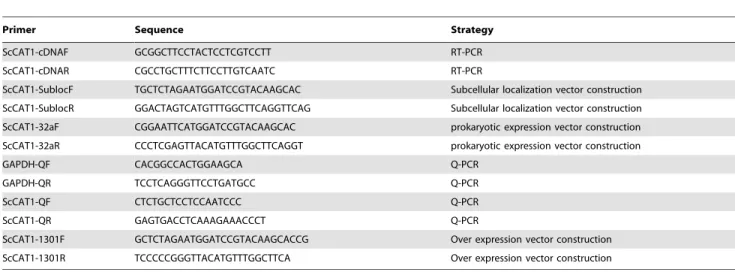
Table 1.Primers used in this study.
Primer Sequence Strategy
ScCAT1-cDNAF GCGGCTTCCTACTCCTCGTCCTT RT-PCR
ScCAT1-cDNAR CGCCTGCTTTCTTCCTTGTCAATC RT-PCR
ScCAT1-SublocF TGCTCTAGAATGGATCCGTACAAGCAC Subcellular localization vector construction
ScCAT1-SublocR GGACTAGTCATGTTTGGCTTCAGGTTCAG Subcellular localization vector construction
ScCAT1-32aF CGGAATTCATGGATCCGTACAAGCAC prokaryotic expression vector construction
ScCAT1-32aR CCCTCGAGTTACATGTTTGGCTTCAGGT prokaryotic expression vector construction
GAPDH-QF CACGGCCACTGGAAGCA Q-PCR
GAPDH-QR TCCTCAGGGTTCCTGATGCC Q-PCR
ScCAT1-QF CTCTGCTCCTCCAATCCC Q-PCR
ScCAT1-QR GAGTGACCTCAAAGAAACCCT Q-PCR
ScCAT1-1301F GCTCTAGAATGGATCCGTACAAGCACCG Over expression vector construction
ScCAT1-1301R TCCCCCGGGTTACATGTTTGGCTTCA Over expression vector construction
visualized using fluorescence microscopy (Axio Scope A1, Germany).
Expression inEscherichia coliRosetta Cells
To study the function ofScCAT1 in prokaryotes, the ScCAT1 ORF was amplified by PCR from the identified cDNA clone using the primers ScCAT1-32aF and ScCAT1-32aR (Table 1) followed by 94uC for 4 min; 94uC for 30 s, 58uC for 30 s, 72uC for 1.5 min, 35 cycles; and 72uC for 10 min. The ScCAT1ORF withEcoR I andXhoI sites was subcloned into pET 32a (+) vector withEco RI-XhoI sites in the E. coli Rosetta strains to generate the putative recombinant (pET 32a-ScCAT1). The desired recombinant plas-mid was identified by PCR amplification, double digestion and sequencing. The prokaryotic expression product was induced in 1.0 mM isopropylb-D-thiogalactoside (IPTG) for 8 h at 37uC and analyzed by sodium dodecyl sulfate-polyacrylamide gel electro-phoresis (SDS-PAGE). Meanwhile, LB medium with blankE. coli Rosetta strains (blank) or Rosetta+pET 32a (control) was each induced in IPTG for 8 h and also analyzed by SDS-PAGE [26,31].
During the response ofE. colicells to various abiotic stresses, the growth ofE. coli Rosetta strains transformed with pET 32a and pET 32a-ScCAT1 was analyzed using spot assay in different treatments of CuCl2, CdCl2 or NaCl. When OD600 of the LB medium (plus 170mg?mL21 chloramphenicol and 80mg?mL21 ampicillin) withE. colicells reached 0.6, IPTG was added to a final concentration of 1.0 mM, and then continued growth for 12 h at 37uC. Thereafter, the cultures were diluted to 0.6 (OD600), and then to two levels (1023 and 1024). Ten microlitres from each dilutions was spotted on LB plates (plus 170mg?mL21 chloram-phenicol and 80mg?mL21ampicillin) containing CuCl2(250, 500 and 750mM), CdCl2(250, 500 and 750mM) or NaCl (250, 500 and 750 mM) [26,31]. All these plates were cultured in 37uC overnight and photographed.
The effect of 750mM CuCl2, 750mM CdCl2 and 250 mM NaCl on the growth of E. coli strains with pET 32a-ScCAT1 or pET 32a was studied in LB medium followed with Su et al. [26]. As above, when cells were grown as earlier described and then diluted to 0.6 (OD600), 400mL of cells were transferred into 10 mL of LB medium containing 170mg?mL21 chloramphenicol and 80mg?mL21 ampicillin, 750mM CuCl2 or 750mM CdCl2 or 250 mM NaCl [32]. Cultures were shaken at 200 rpm at 37uC and growth of the cells was measured at every 2 h by Lambda 35 UV WinLab software (Perkin Elmer, USA).
Real-time Quantitative PCR Analysis
The each time point of 0 h, 6 h, 12 h, 48 h and 72 h during Yacheng05–179-smut incompatible interaction and Liucheng03– 182-smut compatible interaction, as well as mock plants inoculated with sterile distilled water at each corresponding time point, were used to analyze the expression patterns of the ScCAT1. The relative expression of the target gene under certain biotic stress was calculated by the expression level of the inoculated sample minus the level of the mock at each corresponding time point. For tissue-specific expression ofScCAT1, the leaf, bud, stem epidermis and stem pith of sugarcane variety Yacheng05–179 were used as experimental materials. The expression of ScCAT1 under the stresses of SA, MeJA, ABA, H2O2,PEG, CuCl2and NaCl were also performed by real-time quantitative PCR (Q-PCR).
The method of Q-PCR followed the instruction of the SYBR Green Master (ROX) (Roche, China) on a 7500 Q-PCR system (Applied Biosystems, USA). The GAPDH gene (GAPDH-QF/ GAPDH-QR) (Table 1) was chosen as the internal control of the Q-PCR [28]. According to the sequence of ScCAT1, a pair of
specific primers ScCAT1QF/ScCAT1-QR was designed using the Primer Premier 5.0 software. Q-PCR was carried out with FastStart Universal SYBR Green Master (ROX) in a 20mL volume containing 10mL FastStart Universal SYBR Green PCR Master (ROX), 0.5mM of each primer and 2.0mL template (100
6 diluted cDNA). PCR with distilled water as template was performed as control. The Q-PCR reaction condition was held at 50uC for 2 min, 95uC for 10 min, 40 cycles of 95uC for 15 s and 60uC for 1 min. When the reaction was complete, the melting curve was analyzed. Each Q-PCR was repeated three times. The 22ggCtmethod was adopted to analyze the Q-PCR results [33].
Histochemical Assay
For analysis of defense response caused by ScCAT1 over-expression, primers of ScCAT1-1301F/ScCAT1-1301R in Table 1 (XbaI -SmaI sites) were used to construct binary vector expressing ScCAT1 (pCAMBIA 1301-ScCAT1).Agrobacterium strain EHA105 containing recombinant vector and pCAMBIA 1301 vector alone were grown overnight in LB liquid medium (plus 35mg?mL21 rifampicin and 50mg?mL21 kanamycin) at 28uC. Then cultures were pelleted and resuspended in MS liquid medium (plus 200mM acetosyringone) at OD600= 0.8 and infiltrated into N. benthamianaleaves at eight-leaf stage [34]. Plants were incubated at 24uC for 1–2 days (16 h light/8 h darkness), which were employed to following different tests.
DAB (3,39-diaminobenzidinesolution) staining. Agroinfiltrated leaves were incubated in 1.0 mg?mL21DAB-HCl solution in the dark overnight. Then the leaves were destained by boiling in 95% ethanol for 5 min. The bronzing color of the leaves for H2O2 detection which generated in leaves after treatments was photographed [35].
Trypan blue staining. The infiltrated leaves were boiled for 5 min in lactophenol-ethanol trypan blue solution (10 mL glycerol, 10 mL lactic acid, 10 g phenol, 10 mg trypan blue, 30 mL absolute ethanol and 10 mL distilled water). Then the leaves were destained in 2.5 g?mL21choloral hydrate in distilled water and the blue color indicates the cell death [30].
Measurement of ion conductivity. It was performed as previously described with some modifications [36]. Six leaf discs (11 mm in diameter per leaf) were cut and washed in distilled water and then incubated in 20 mL of distilled water and shaken slowly at room temperature for 60 min. After that, electrolyte leakage was measured using a conductivity meter (SevenEasy, METTLER TOLEDO, Switzerland).
Results
Enzyme Activity of Catalase
correlations between catalase activity and smut resistance in sugarcane.
Cloning and Sequence Analysis ofScCAT1
To study the sugarcane catalase at the molecular level, a 1,658 bp full-length catalase gene ScCAT1 (GenBank Accession No. KF664183) was cloned using RT-PCR method combined within silicocloning technique. There were 1,479 nucleotides in its ORF (Fig. S2). ScCAT1encoded a predicted polypeptide of 492 amino acids with no signal peptide. The predicted protein had a molecular mass of 56.81 kDa with a pI of 6.72. A search at the NCBI for conserved protein domains indicated that ScCAT1 belonged to a member of catalase-like superfamily. The catalytic active site and heme binding motifs of ScCAT1 were detected by Motif Scan Online program. 17 amino acids at the position of 54– 70 (FDRERIPERVVHARGAS) were reported to be a catalase active site signature, and the heme-ligand signature was detected at the position of 344–352 (RIFSYADTQ). These data suggested clearly that sugarcane ScCAT1 encoded a putative peroxisomal catalase. Furthermore, it also predicted that ScCAT1 contains no transmembrane helix domain, implying that ScCAT1 is not a membrane located or secretory protein.
A GenBank Blastp comparison showed that ScCAT1 exhibited high identity with other plant catalases, includingS. bicolorcatalase (XP_002437631.1) (97.97% identity), Z. mays catalase (NP_001241808.1) (97.56% identity), O. sativa catalase (A2YH64.2) (94.92% identity), B. distachyon catalase (XP_003563243.1) (93.29% identity), P. chinampoensis catalase (ADN94253.1) (92.07% identity), H. vulgare catalase (P55307.1) (91.67% identity),T. aestivumcatalase (P55313.1) (91.26% identity) and S. italica catalase (XP_004966515.1) (87.02% identity). Phylogenetic analysis (Fig. 2) revealed that ScCAT1 was closely related to S. bicolor catalase (XP_002437631.1), Z. mays catalase (NP_001241808.1) andS. italicacatalase (XP_004966515.1), with the homologies of 97.97%, 97.56% and 87.02%, respectively.
Subcellular Localization of ScCAT1
To further understand the function of ScCAT1 gene, its subcellular localization was conducted.ScCAT1 was recombined into plant expression vector pCAMBIA 2300 between the sites of
the35Spromoter andGFP(Figs. S1 and S3), and its location was characterized by transient expression of the target gene and GFP in N. benthamiana leaves with Agrobacterium-mediated transforma-tion. After 2 days of cultivation, the infiltrated leaves were harvested and the reporter protein GFP was observed under a fluorescence microscope. The results revealed that 35S::ScCAT1::GFP was located in plasma membrane and cytoplasm (Fig. 3). In contrast, GFP was shown in the nucleus, cytoplasm and plasma membrane cells transiently transfected with 35S::GFP.
Expressions of ScCAT1 inE. coli
As reported before, different stresses, such as copper (Cu), cadmium (Cd), high temperature, wounding, ethylene (ET), H2O2, SA, jasmonic acid (JA), ABA and other inducers, could trigger an induction of plant catalases [11,13,21,37,38]. To study the function of ScCAT1 in response to different kinds of adverse environmentsin vivo, pET 32a-ScCAT1(Fig. S4A) was transformed intoE. coliRosetta cell. The recombinant protein of 62 kDa was specifically induced and accumulated approximately after 8 h IPTG induction on the SDS-PAGE (Fig. S4B).
The growth of gene-expressed cells (Rosetta+pET 32a-ScCAT1) and mock (Rosetta+pET 32a) was analyzed on LB plates with different supplements (Figs. 4A, B, C, and D). After one day culture, Rosetta+pET 32a-ScCAT1showed an increased number of colonies as compared to the control cells on LB plates containing CuCl2, CdCl2 and NaCl. The growth was also analyzed in the LB liquid medium containing 750mM CuCl2, 750mM CdCl2and 250 mM NaCl (Figs. 4E, F, G and H). All the Rosetta+pET 32a-ScCAT1cells showed faster growth as compared to that of the mock which revealed that ScCAT1 had an effect on increasing the tolerance to CuCl2, CdCl2and NaCl. These results demonstrat that the recombinant protein of ScCAT1 enhanced growth ability of prokaryotic E. coli Rosetta strains in stress conditions.
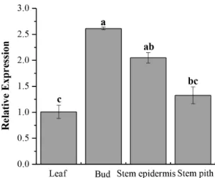
Tissue-specific Expression Analysis ofScCAT1
The relative expression ofScCAT1was detected in four kinds of sugarcane tissues, including leaf, bud, stem epidermis and stem pith. As showed in Fig. 5, the bud exhibited the highest mRNA expression, while the mRNA expression of stem epidermis and stem pith was at a moderate level. The leaf showed a relatively low level in comparison with the other three kinds of tissues.
ScCAT1Expression in Response to Different Stress Treatments
Smut challenged sugarcane (Yacheng05–179 and Liucheng03– 182) buds were detected by Q-PCR for examination whether the expression ofScCAT1was induced or inhibited (Fig. 6). In order to eliminate the influence of wounding, the relative expression of the target gene was calculated by the expression level of the inoculated sample minus the level of the mock at each corresponding time point. As indicated in Fig. 6, after the inoculation of smut pathogen, the mRNA expression of ScCAT1 in resistant variety Yacheng05–179 was higher than that in susceptible variety Liucheng03–182. During the sugarcane-smut incompatible inter-action, the transcript of ScCAT1 in Yacheng05–179 began was elevated as early as 6 h post-inoculation (6 hpi), while that of ScCAT1in Liucheng03–182 appeared delayed (12 hpi). Further-more, the transcript of ScCAT1 in Yacheng05–179 and Liu-cheng03–182 reached the maximum at 48 h, but the expression in incompatible interaction was 1.55 times that of the compatible one, and then decreased in both. During the whole process of
Figure 1. The catalase activity in smut resistant (Yacheng05– 179) and smut susceptible (Liucheng03–182) sugarcane varie-ties inoculated with Sporisorium scitamineum. All data points (deduction its mock) are means6SE (n = 3).
interaction, the transcript ofScCAT1in the incompatible cultivar almost always higher than that of the compatible, except at 12 h. These data reveal that the up-regulation ofScCAT1expression was most probably associated with smut resistance in sugarcane.
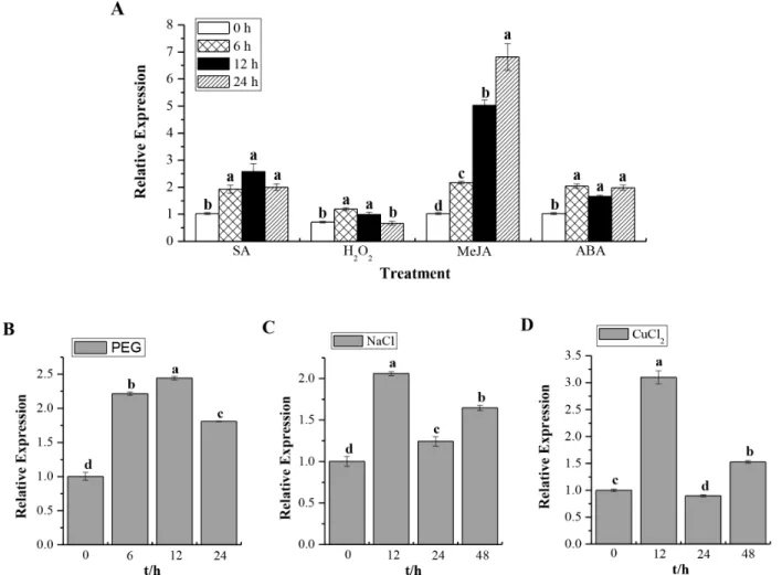
Expression ofScCAT1in response to various abiotic stimuli in Yacheng05–179 plantlets was checked after treatment with 5 mM SA, 10 mM H2O2, 25mM MeJA, 100mM ABA, 25% PEG, 250 mM NaCl and 100mM CuCl2, and the results shown in Fig. 7. Interestingly, ScCAT1 showed a positive response to exogenous stresses, including plant hormones stresses of SA, MeJA and ABA, oxidative stress of H2O2, hyper-osmotic stresses of PEG and NaCl, as well as mental stress of CuCl2.ScCAT1transcription was always up-regulated and the expression level usually increased steadily from 0 h to 24 h or 48 h after post-treatment with these seven exogenous inducers. These results suggest thatScCAT1may be a positive responsive component of abiotic stress in sugarcane.
Transient Over-expression ofScCAT1inN. benthamiana
Leaves Induces Hypersensitive Reaction Response To test whetherScCAT1can induce HR and immunity in plant, ScCAT1was transient over-expressed inN. benthamiana leaves by infiltration withAgrobacterium EHA105 carrying pCAMBIA 1301 (mock) and pCAMBIA 1301-ScCAT1. The results showed that at the time point of 48 h after infiltration, a typical HR symptom, darker DAB staining and enhanced electrolyte leakage, was found in the leaves expressing the target gene (Figs. 8A and C). Furthermore, injected leaves 5 d after agroinfiltrated by 35S::ScCAT1presented yellow symptoms (Fig. 8B). What is more, cell death measured by qualitative trypan blue staining showed a darker color than that in mock (Fig. 8B). These results indicate the involvement ofScCAT1in cell death responses.
Discussion
Fungal disease is a major concern worldwide for sugarcane production and most other crops. During plant-pathogen
interac-Figure 2. Phylogenetic trees based on catalase amino acid sequences, showing the phylogenetic relationships between ScCAT1 (KF664183) and the catalases from other plant species.Neighbor-joining method was used.
doi:10.1371/journal.pone.0084426.g002
Figure 3. Subcellular localizations of ScCAT1 and empty vector inNicotiana benthamianaleaves 48 h after infiltration.The epidermal cells were used for taking images of green fluorescence, visible light and merged light. Read arrows 1, 2 and 3 indicated plasma membrane, nucleus and cytoplasm, respectively. Bar = 50mm.
tions, many antifungal components have been identified [39]. Peroxidase (POD) activity increased in resistant sugarcane varieties
(and not in susceptible) implies that it may be related to smut resistance [40]. Our previous report showed thatb-1,3-glucanase activity in the
Figure 4. Spot assays of Rosetta+pET 32a-ScCAT1(a) and Rosetta+pET 32a (mock) (b) on LB plates with CuCl2, CdCl2and NaCl (A–D).
And liquid culture assay in LB liquid medium with 750mM CuCl2, 750mM CdCl2and 250 mM NaCl (E–H). IPTG (isopropylb-D-thiogalactoside) was
added to the cultures of Rosetta+pET 32a-ScCAT1and Rosetta+pET 32a to induce the expression of recombinant protein. The cultures were adjusted to OD600= 0.6. Ten microliters from 1023(left side of red line on plate) to 1024(right side of red line on plate) dilutions were spotted onto LB basal (A)
plates or with CuCl2(250, 500 and 750mM) (B), CdCl2(250, 500 and 750mM) (C), NaCl (250, 500 and 750 mM) (D). For studying the growth analysis of ScCAT1, Rosetta+pET 32a-ScCAT1and Rosetta+pET 32a were grown in LB liquid medium with LB basal medium (E) or with 750mM CuCl2(F), 750mM
CdCl2(G), and 250 mM NaCl (H). All data points are means6SE (n = 3). CuCl2:copper chloride; CdCl2: cadmium chloride; NaCl: sodium chloride.
resistant variety increased faster and lasted longer than that of the susceptible one after challenged byS. scitamineum, which showed a positive relationship between the activity of the sugarcaneb -1,3-glucanase and smut resistance [26]. Plant catalase, one of the scavenger enzymes, has been also shown to be involved in plant defense and development [41]. Wang et al. found that catalase could be induced by pathogen infection in resistant clamMeretrix meretrix [23]. As showed in Fig. 1, the activity of catalase increased in the resistant genotype (Yacheng05–179) after challenged byS. scitami-neumin comparison to the susceptible cultivar (Liucheng03–182). There appears to be a positive correlation between catalase activity and sugarcane smut resistance. This observation should be repeated in different resistant genotypes.
The capacity of a plant to scavenge H2O2 may result from increased activities of scavenger enzymes or up-regulated expression of genes increasing of the levels of the corresponding proteins [42].
Multiple catalase isozymes in plants have been observed. Previous research demonstrated that there were at least six catalase isozymes existing inA. thalianaencoded by a multi-gene family including three genes (cat1,cat2andcat3) [14]. InZ. mays, three catalase isoenzymes encoded by three different structural genes were observed [38]. A sweet potato catalaseSPCAT1was cloned from mature leaves treated with ethephon and found that it could alleviate ethephon-mediated leaf senescence and H2O2elevation [13]. Until now, there has been no report on sugarcane catalase genes involved in the sugarcane-smut interaction. In this study, we isolated and characterized a full-length sugarcane catalase geneScCAT1which encoded a polypeptide of 492 amino acids and had high identities with several other plant catalases. Using the method ofAgrobacterium-mediated transformation inN. benthamiana leaves, 35S::ScCAT1::GFP was located in plasma membrane and cytoplasm in cells (Fig. 3) which is consistent with a previous report that catalase mostly localized in peroxisomes, glyoxysome and cytoplasm [11,43].
Recent publications have reported that E. coli cells can be enhanced or inhibited under stress expressing recombinant proteins [27,31,32,44]. Some of the protective mechanisms were similar in both eukaryotes and prokaryotes under stress stimuli [45]. Gupta et al. studied an A-2 type DREB transcription factor from extreme halophyteSalicorniabrachiata and found it conferred abiotic stress tolerance in E. coli cells under NaCl, PEG and mannitol treatments, which may be due to the stress regulated function by this transcription factor [32]. Guo et al. tested a sugarcane dirigent protein geneScDirand a metallothionein gene ScMT2-1-3in theE. colisystem, which indicated that they offered different tolerance against PEG, NaCl and mental stresses [27,31]. Chaurasia et al. studied that phytochelatin synthase gene PCS, when expressed inE. coli, provided better protection against the stresses of heat, salt, carbofuron, cadmium, copper and UV [44]. In the present study, the ScCAT1 recombinant protein expressed inE. coliRosetta cells leads to a better growth under the stresses CuCl2, CdCl2and NaCl. In eukaryote, the previous studies found that the increased tolerance to stress maybe due to the activity and expression of scavenging enzymes which increased in plants placed in different conditions [12]. It has been proposed that catalase, one
Figure 5. Tissue-specific expression analysis of theScCAT1in sugarcane variety Yacheng05–179. Data are normalized to the
GAPDHexpression level. All data points are means6SE (n = 3). doi:10.1371/journal.pone.0084426.g005
Figure 6. Q-PCR analysis of theScCAT1expression patterns in biosystem of sugarcane-smut (Sporisorium scitamineum) interaction.
Data was normalized to theGAPDHexpression level. All data points (deduction its mock) are means6SE (n = 3). Resistant: Yacheng05–179 variety; Susceptible: Liucheng03–182 variety.
Figure 7. Q-PCR analysis of theScCAT1expression patterns in Yacheng05–179 plantlets with abiotic elicitors.Data are normalized to theGAPDHexpression level. (A) The relative expression ofScCAT1under the stresses of 5 mM SA, 10 mM H2O2, 25mM MeJA and 100mM ABA. (B)
The relative expression ofScCAT1under 25% PEG stress. (C) The relative expression ofScCAT1under 250 mM NaCl stress. (D) The relative expression ofScCAT1under 100mM CuCl2stress. All data points are means6SE (n = 3). SA: salicylic acid; H2O2: hydrogen peroxide; MeJA: methyl jasmonate; ABA:
abscisic acid; PEG: polyethylene glycol; NaCl: sodium chloride; CuCl2:copper chloride.
doi:10.1371/journal.pone.0084426.g007
Figure 8. The effect of transient over-expression ofScCAT1on immunity induction inNicotiana Benthamianaleaves.(A) DAB staining withN. benthamianaleaves 48 h after35S::ScCAT1-containingAgrobacteriumstrain infiltration to assess the H2O2production; a: images taken by
SONY camera; b: images taken by microscope. (B) Cell death measured by trypan blue staining of transient expression leaves 5 d after agroinfiltration; a: phenotypes ofN. Benthamiana5 d after infiltration taken by SONY camera; b: images of trypan blue staining taken by microscope. (C) Conductivity measurement ofN. Benthamianaleaves infiltrated with35S::ScCAT1-containingAgrobacteriumstrain after 48 h. Mock:Agrobacteriumstrain carrying
of the antioxidant enzymes, can be modulated and controlled in response to excessive iron stress, due to alterations in the electron transport chain and damages to the thylakoidal membranes [46]. Therefore, it is plausible to predict that the ScCAT1 encoded by ScCAT1 gene cloned in this study could be helpful for the tolerance/stresses of sugarcane to CuCl2, CdCl2and NaCl.
The plant faces variable environmental stresses like soil salinity, temperature, drought and cold, and may often present a series of physiological and biochemical changes which are a highly complex and disturb plant growth and yield. To examine the accumulation of sugarcane catalase gene in different developmental processes and environmental conditions, the expression of ScCAT1 gene in sugarcane was analyzed by Q-PCR method (Figs. 5, 6 and 7). Results indicated that while expressed at moderate levels in stem epidermis and stem pith,ScCAT1was expressed at a relatively high level in the bud (Fig. 5). Similar to other species ofUstilago, theS. scitamineumis a parasite of young meristematic tissues and gains entry into the host, exclusively through the bud scales [47]. From above, high expression ofScCAT1in sugarcane bud may help to defend against the smut pathogen. In our study, the target transcript ofScCAT1was found to be higher in the incompatible interaction than that in the compatible one during sugarcane-S. scitamineuminteraction (Fig. 6). After the smut pathogen challenge in Yacheng05–179, the expression ofScCAT1 increased at 6 h and reached the maximum level at 48 h (1.5 times that in Liucheng03–182). As previous reported, the phenomenon of smut hypha entry into the sugarcane bud meristem occurs between 6 h and 36 h after the teliospore deposition [48]. It should be also noted thatScCAT1expression decreased gradually after 48 h, but the expression level still maintained at a higher level than that at 0 h, and the gene expression pattern of ScCAT1 was coincident with the activity change of catalase in this study. So we assume thatScCAT1 may have a protective effect on smut penetration in sugarcane.
Q-PCR analysis of the expression of ScCAT1 in response to hydrogen peroxide and plant hormones showed that from 0 h to 24 h its levels increased under the stresses of 10 mM H2O2, 5 mM SA, 25mM MeJA and 100mM ABA (Fig. 7A). In Panax ginseng, PgCat1 transcript accumulated during 1–12 h of 10 mM H2O2 treatment [11]. MaizeCat1gene transcript increased in developing embryos by the treatments of 1.5 mM SA, 50 mM H2O2, 100mM JA and 1 mM ABA [38,49]. In the present study, for hyper-osmotic stress, ScCAT1 mRNA levels increased until 12 h then slightly decreased at 24 h and induced at 48 h under 250 mM NaCl treatment. ScCAT1transcript was also stimulated till 24 h after 25% PEG stress (Figs. 7B and C). 500 mM NaCl stress induced the expression ofCat1inAvicennia marinaseedlings till 12 h then subsequently decreased [50]. In Panax ginseng, PgCat1 transcripts accumulated till 24 h then decreased till 72 h after 100 mM NaCl treatment [11]. Plants suffering from NaCl stress not only because of increased osmolarity but also oxidative stress caused by ionic character [51]. In our study, theScCAT1transcript increased 1.5 fold until 48 h under the stress of 100mM CuCl2. The maximum expression was observed to be 3.0 fold at 12 h after treatment. Previous study revealed that copper toxicity caused ultra structural damage which resulting in the increasing production of ROS [46]. ThePrunus cerasifera Cat1gene expression and enzyme activity were high for 10 days under 100 mM copper stress [52]. These results lead us to conclude thatScCAT1may be a positive responsive component of abiotic stresses in sugarcane.
N. benthamianahas been widely employed in functional character-ization of the target genes by over-expression [30]. Cell death presented at the infected site is the most efficient strategy to restrict pathogen growth and development [30]. The induction of R gene expression, ion fluxes, stimulation of ROS and defense-related hormones, are the common response of cell death [53,54]. Here,
DAB staining showed deep brown in the presence of H2O2inN. benthamianaleaves after 48 h infiltration and resulted in an increase of electrolyte leakage (Figs. 8A and C). Trypan blue staining exhibited a darker color post 5 d injection than that in mock (Fig. 8B). Previous studies have shown that there is a close relationship between HR and H2O2accumulation [55]. It can be deduced from this study that H2O2accumulation by transient over-expression ofScCAT1may confer the HR cell death in sugarcane.
In conclusion, after inoculation withS. scitamineum, sugarcane catalase was found to significantly increase in the resistant variety and maintain at much higher level than that of the susceptible one which suggested a positive correlation between the activity of the catalase and the smut resistance in sugarcane.ScCAT1was isolated from sugarcane buds and the recombinant protein resulted in a better growth ofE. coliRosetta cells under certain stresses. The expression of ScCAT1was up-regulated by smut infection and by different stresses such as plant hormones (SA, MeJA and ABA) treatments, oxidative (H2O2) stress, heavy metal (CuCl2) and hyper-osmotic (PEG and NaCl) stresses. ScCAT1 was located in plasma membrane and cytoplasm in cells. Histochemical assays indicated thatScCAT1acted positively in sugarcane immunity. From these observations, we can conclude thatScCAT1should be a positive responsive component of biotic and abiotic stresses in sugarcane.
Supporting Information
Figure S1 Construction of subcellular localization vec-tor35S::ScCAT1::GFP.
(TIF)
Figure S2 Nucleotide acid sequences and deduced amino acid sequences ofScCAT1 obtained by RT-PCR.
The deduced amino acid sequences were shown in one-letter code under the cDNA sequences. The underlines showed the catalase active site signature (FARERIPERVVHARGAS) and the heme-ligand signature (RVFAYADTQ) of ScCAT1.
(TIF)
Figure S3 The enzyme digestion to identify the insert-integrated subcellular localization expression vector 35S::ScCAT1::GFP. 1, 15,000+2,000 bp DNA marker; 2,
35S::GFP/Xba I; 3, ScCAT1 ORF PCR product; 4, 35S::ScCAT1::GFP/Xba I; 5, 35S::ScCAT1::GFP/Xba I+Spe I; 6, 100 bp ladder DNA marker.
(TIF)
Figure S4 The enzyme digesting identification of insert-integrated prokaryotic expression vector pET 32a-ScCAT1 (A) and corresponding protein expressions in
Escherichia coli Rosetta strains (B). (A)1, 100 bp ladder
DNA marker; 2, pET 32a/EcoR I; 3,ScCAT1ORF PCR product; 4, pET 32a-ScCAT1/EcoR I; 5, pET 32a-ScCAT1/EcoR I+XhoI; 6, 15,000+2,000 bp DNA Marker.(B) 1, Protein marker; 2, blank without induction; 3, blank induction for 8 h; 4, control without induction; 5, control induction for 8 h; 6, pET 32a-ScCAT1 without induction; 7 and 8, pET 32a-ScCAT1induction for 4 h and 8 h, respectively. The induced protein was shown by arrow. (TIF)
Author Contributions
References
1. Sundar AR, Barnabas EL, Malathi P, Viswanathan R (2012) A mini-review on smut disease of sugarcane caused bySporisorium scitamineum. In: Mworia J, editor. Botany. Croatia: InTech. 109–128.
2. Agnihotri VP (1990) Diseases of sugarcane and sugarbeet Oxford & IBH Publishing Co Pvt Ltd. New Delhi: 72–103.
3. Hoy JW, Hollier CA, Fontenot DB, Grelen LB (1986) Incidence of sugarcane smut in Louisiana and its effects on yield. Plant Dis 70: 59–60.
4. Singh N, Somai BM, Pillay D (2004) Smut disease assessment by PCR and microscopy in inoculated tissue cultured sugarcane cultivars. Plant Sci 167: 987– 994.
5. Solas MT, Pinon D, Vicent C, Legaz ME (1999) Ultrastructural aspects of sugarcane bud infection byUstilago scitamineateliospores. Sugar Cane 2: 14–18. 6. Waller JM (1970) Sugarcane smut (Ustilago scitaminea) in Kenya: II. Infection and
resistance. T British Mycol Soc 54: 405–414.
7. Xu LP, Chen RK, Chen PH (2004) Analysis on infection index of smut caused byUstilago scitamineain sugarcane segregated population. Chin J Trop Crop 25: 33–36.
8. Lin YQ, Chen RK, Gong DM (1996) Analysis of quantitative inheritance for smut resistance in sugarcane. J Fujian Agr U China 25: 271–275.
9. Dussle CM, Quint M, Melchinger AE, Xu ML, Lubberstedt T (2003) Saturation of two chromosome regions conferring resistance to SCMV with SSR and AFLP markers by targeted BSA. Theor Appl Genet 106: 485–493.
10. Lakshmanan P, Geijskes RJ, Aitken KS, Grof CLP, Bonnett GD, et al. (2005) Sugarcane biotechnology: the challenges and opportunities. In Vitro Cell Dev-Pl 41: 345–363.
11. Purev M, Kim YJ, Kim MK, Pulla RK, Yang DC (2010) Isolation of a novel catalase (Cat1) gene fromPanax ginsengand analysis of the response of this gene to various stresses. Plant Physiol Bioch 48: 451–460.
12. Mhamdi A, Queval G, Chaouch S, Vanderauwera S, Van-Breusegem F, et al. (2010) Catalase function in plants: a focus onArabidopsismutants as stress-mimic models. J Exp Bot 61: 4197–4220.
13. Chen HJ, Wu SD, Huang GJ, Shen CY, Afiyanti M, et al. (2012) Expression of a cloned sweet potato catalase SPCAT1 alleviates ethephon-mediated leaf senescence and H2O2elevation. J Plant Physiol 169: 86–97.
14. Frugoli JA, Zhong HH, Nuccio ML, McCourt P, McPeek MA, et al. (1996) Catalase is encoded by a multigene family inArabidopsis thaliana(L.) Heynh. Plant physiol 112: 327–336.
15. Willekens H, Villarroel R, Van-Montagu M, Inze D, Van-Camp W (1994) Molecular identification of catalases fromNicotiana plumbaginifolia(L.). FEBS Lett 352: 79–83.
16. Guan L, Scandalios JG (1995) Developmentally related responses of maize catalase genes to salicylic acid. P Natl A Sci 92: 5930–5934.
17. Skadsen RW, Schulze-Lefert P, Herbst JM (1995) Molecular cloning, characterization and expression analysis of two catalase isozyme genes in barley. Plant Mol Biol 29: 1005–1014.
18. Drory A, Woodson WR (1992) Molecular cloning and nucleotide sequence of a cDNA encoding catalase from tomato. Plant Physiol 100: 1605.
19. Kwon SI, An CS (2001) Molecular cloning, characterization and expression analysis of a catalase cDNA from hot pepper (Capsicum annuumL.). Plant Sci 160: 961–969.
20. Du YY, Wang PC, Chen J, Song CP (2008) Comprehensive functional analysis of the catalase gene family inArabidopsis thaliana. J Integr Plant Biol 50: 1318– 1326.
21. Guan ZQ, Chai TY, Zhang YX, Xu J, Wei W (2009) Enhancement of Cd tolerance in transgenic tobacco plants overexpressing a Cd-induced catalase cDNA. Chemosphere 76: 623–630.
22. Williamson JD, Scandalios JG (1992) Differential response of maize catalases and superoxide dismutases to the photoactivated fungal toxin cercosporin. The Plant J 2: 351–358.
23. Wang C, Yue X, Lu X, Liu BZ (2013) The role of catalase in the immune response to oxidative stress and pathogen challenge in the clamMeretrix meretrix. Fish Shellfish Immun 34: 91–99.
24. Casu RE, Dimmock CM, Chapman SC, Grof CP, McIntyre CL, et al. (2004) Identification of differentially expressed transcripts from maturing stem of sugarcane by in silico analysis of stem expressed sequence tags and gene expression profiling. Plant Mol Biol 54: 503–517.
25. Moosawi-Jorf SA, Mahin BI (2007) In vitro detection of yeast-like and mycelial colonies ofUstilago scitaminea in tissue-cultured plantlets of sugarcane using polymerase chain reaction. J Appl Sci 7: 3768–3773.
26. Su YC, Xu LP, Xue BT, Wu QB, Guo JL, et al. (2013) Molecular cloning and characterization of two pathogenesis-relatedb-1,3-glucanase genesScGluA1and
ScGluD1from sugarcane infected bySporisorium scitamineum.Plant Cell Rep 32: 1503–1519.
27. Guo JL, Xu LP, Su YC, Wang HB, Gao SW, et al. (2013)ScMT2-1-3, a metallothionein gene of sugarcane, plays an important role in the regulation of heavy metal tolerance/accumulation. BioMed Res Int: doi.10.1155/2013/ 904769.
28. Que YX, Xu LP, Xu JS, Zhang JS, Zhang MQ, et al. (2009) Selection of control genes in real-time qPCR analysis of gene expression in sugarcane. Chin J Trop Crop 30: 274–278.
29. Hao JJ, Kang ZL, Yu Y (2001) Plant physiology experiment technology. In: Hao JJ, editor. China: Liaoning Sci Technol Press. 53–55.
30. Hwang IS, Hwang BK (2012) Requirement of the cytosolic interaction between pathogenesis-related protein10 and leucine-rich repeat protein1 for cell death and defense signaling in pepper. Plant Cell 24: 1675–1690.
31. Guo JL, Xu LP, Fang JP, Su YC, Fu HY, et al. (2012) A novel dirigent protein gene with highly stem-specific expression from sugarcane, response to drought, salt and oxidative stresses. Plant Cell Rep 31: 1801–1812.
32. Gupta K, Agarwal PK, Reddy MK, Jha B (2010) SbDREB2A, an A-2 type DREB transcription factor from extreme halophyteSalicornia brachiataconfers abiotic stress tolerance inEscherichia coli. Plant Cell Rep 29: 1131–1137. 33. Livak KJ, Schmittgen TD (2001) Analysis of relative gene expression data using
real-time quantitative PCR and the 22ggCtmethod. Methods 25: 402–408. 34. Choi HW, Kim YJ, Hwang BK (2011) The hypersensitive induced reaction and
leucine-rich repeat proteins regulate plant cell death associated with disease and plant immunity. Mol Plant Microbe In 24: 68–78.
35. Huckelhoven R, Fodor J, Trujillo M, Kogel KH (2000) Barley Mla and Rar mutants compromised in the hypersensitive cell death response againstBlumeria graminisf. sp.hordeiare modified in their ability to accumulate reactive oxygen intermediates at sites of fungal invasion. Planta 212: 16–24.
36. Hwang IS, Hwang BK (2011) The pepper mannose-binding lectin gene
CaMBL1is required to regulate cell death and defense responses to microbial pathogens. Plant Physiol 155: 447–463.
37. Scandalios JG, Acevedo A, Ruzsa S (2000) Catalase gene expression in response to chronic high temperature stress in maize. Plant Sci 156: 103–110. 38. Guan LM, Zhao J, Scandalios JG (2000) Cis-elements and trans-factors that
regulate expression of the maizeCat1antioxidant gene in response to ABA and osmotic stress: H2O2 is the likely intermediary signaling molecule for the
response. The Plant J 22: 87–95.
39. Takken FLW, Joosten MHAJ (2000) Plant resistance genes: their structure, function and evolution. Eur J Plant Pathol 106: 699–713.
40. Xu LP, Wang JN, Chen RK (1994) Biochemical reaction of sugarcane to smut and its relation to resistance. Sugarcane 1: 13–16.
41. Wan LL, Zha WJ, Cheng XY, Liu C, Lv L, et al. (2011) A riceb-1,3-glucanase geneOsg1is required for callose degradation in pollen development. Planta 233: 309–323.
42. Rezaee F, Ghanati F, Behmanesh M (2013) Antioxidant activity and expression of catalase gene of (Eustoma grandiflorumL.) in response to boron and aluminum. S Afr J Bot 84: 13–18.
43. Song XX, Zhao FY (2007) Research progress on catalase in plants. J Anhui Agric Sci, 35: 9824–9827.
44. Chaurasia N, Mishra Y, Ai LC (2008) Cloning expression and analysis of phytochelatin synthase (pcs) gene fromAnabaenasp. PCC 7120 offering multiple stress tolerance inEscherichia coli. Biochem Bioph Res Co 376: 225–230. 45. Liu Y, Zheng Y (2005) PM2, a group 3 LEA protein from soybean, and its
22-mer repeating region confer salt tolerance inEscherichia coli. Biochem Bioph Res Co 331: 325–332.
46. Sandmann G, Boger P (1980) Copper-mediated lipid peroxidation processes in photosynthetic membranes. Plant Physiol 66: 797–800.
47. Fawcett GL (1942) Circular, Estacion Experimental Agricola. In: Fawcett GL, editor. Tucuman. 114.
48. Alexander KC, Ramakrishnan K (1980) Infection of the bud, establishment in the host and production of whips in sugarcane smut (Ustilago scitaminea) of sugarcane. Proc Int Soc Sugar Cane Technol 17: 1452–1455.
49. Guan LM, Scandalios JG (2000) Hydrogen peroxide-mediated catalase gene expression in response to wounding. Free Radical Bio Med 28: 1182–1190. 50. Jithesh MN, Prashanth SR, Sivaprakash KR, Parida A (2006) Monitoring
expression profiles of antioxidant genes to salinity, iron, oxidative, light and hyperosmotic stresses in the highly salt tolerant grey mangrove,Avicennia marina
(Forsk.) Vierh. by mRNA analysis. Plant Cell Rep 25: 865–876.
51. Munns R (2002) Comparative physiology of salt and water stress. Plant Cell Environ 25: 239–250.
52. Lombardi L, Sebastiani L (2005) Copper toxicity inPrunus cerasifera: growth and antioxidant enzymes responses of in vitro grown plants. Plant Sci 168: 797–802. 53. Melech-Bonfil S, Sessa G (2010) Tomato MAPKKKeis a positive regulator of cell-death signaling networks associated with plant immunity. The Plant J 64: 379–391.
54. Li Y, Tessaro MJ, Li X, Zhang Y (2010) Regulation of the expression of plant resistance geneSNC1by a protein with a conserved BAT2 domain. Plant physiol 153: 1425–1434.
55. Levine A, Tenhaken R, Dixon R, Lamb C (1994) H2O2from the oxidative burst