S E
Biotechnol. Agron. Soc. Environ.2004 8(4), 229–234
1. INTRODUCTION
Methods are required that enable diff e r e n t i a t i o n between muscle and non-muscle tissues such as liver, kidney and heart in meat samples. This is because only muscle tissue is considered meat from the point of view of labelling (Food Labelling (Amendment) (England) Regulations 2003) and Quantitative Ingredient Declaration (QUID), and also certain parts of the carcass are prohibited to be used in raw meat products (Meat Products (England) Regulations 2003). Included in the prohibited offal is brain and spinal cord. The described methodology has therefore been developed primarily to enforce labelling rules and also to contribute to the enforcement of BSE legislation requiring the detection of Central Nervous System (CNS) tissue. This requires the removal of central nervous system tissue from carcasses prior to processing (Government UK, 1989). To permit eff e c t i v e enforcement there is a need for the development of robust CNS specific detection methods that are applicable to a wide range of processed meat products. Methods for the detection of CNS tissue in meat
include direct tissue examination (Bauer et al., 1996), histological preparation and microscopic examination (FSIS/USDA, 1998) and the analysis of cholesterol content (Lücker et al., 1999; Schmidt et al., 1999). The latter method has used antibodies directed against CNS specific proteins to enable the immunogenic detection of CNS tissue. One of these (Schmidt et al., 1999) utilises an ELISA method based upon the detection of Glial Fibrillary Acidic Protein (GFAP), a protein whose expression is restricted to the CNS (Kaneko, Sueoka, 1993). Our studies have also utilised detection of GFAP but at the DNA rather than the protein level. Such an assay may be more suitable for processed products where DNAdegradation can be overcome by designing a PCR based assay that generates small amplicons and also has the potential to be utilised on a range of assay formats.
Methylation of cytosine occurring in CpG dinucleotides in the promoter of a gene is one of several ways of controlling tissue specific regulation (Condorelli et al., 1994; Condorelli et al., 1997; Teter
et al., 1994). Several tissue specific genes have been shown to possess unmethylated promoter sequences in
Detection of neuronal tissue in meat using tissue specific
DNA modifications
Sally Gout, Hernan Valdivia, David Mc Dowell, Neil Harris
LGC Limited. Queens Road. Teddington. Middlesex TW11 0LY (United Kingdom). E-mail: [email protected]
A method has been developed to differentiate between non-muscle tissues such as liver, kidney and heart and that of muscle in meat samples using tissue specific DNA detection. Only muscle tissue is considered meat from the point of view of labelling (Food Labelling [Amendment] (England) Regulations 2003) and Quantitative Ingredient Declaration (QUID), and also certain parts of the carcass are prohibited to be used in raw meat products (Meat Products [England] Regulations 2003). Included in the prohibited offal are brain and spinal cord. The described methodology has therefore been developed primarily to enforce labelling rules but also to contribute to the enforcement of BSE legislation on the detection of Central Nervous System (CNS) tissue. The latter requires the removal of Specified Risk Material (SRM), such as bovine and ovine brain and spinal cord, from the food chain. Current methodologies for detection of CNS tissue include histological examination, analysis of cholesterol content and immunodetection. These can potentially be time consuming, less applicable to processed samples and may not be readily adapted to high throughput sample analysis. The objective of this work was therefore to develop a DNAbased detection assay that exploits the sensitivity and specificity of PCR and is potentially applicable to more highly processed food samples. For neuronal tissue, the DNA target selected was the promoter for Glial Fibrillary Acidic Protein (GFAP), a gene whose expression is restricted to astroglial cells within CNS tissue. The promoter fragments from both cattle and sheep have been isolated and key differences in the methylation patterns of certain CpG dinucleotides in the sequences from bovine and sheep brain and spinal cord and the corresponding skeletal muscle identified. These have been used to design a PCR assay exploiting Methylation Specific PCR (MSP) to specifically amplify the neuronal tissue derived sequence and therefore identify the presence of CNS tissue in an assay sample.
expressing tissue but be heavily methylated in those where little or no expression occurs (Yeivin et al., 1993). In several cases, methylation of specific CpG dinucleotides within the promoter has been shown to correlate with transcriptional repression and hence control gene expression (Li et al., 1993; Yoder et al., 1997).
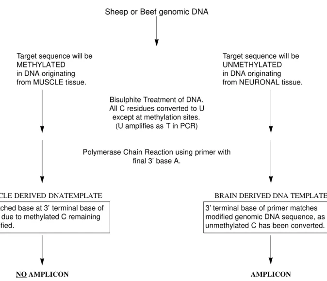
One technique that can be used to study promoter methylation is the chemical modification of cytosine to uracil by bisulphite treatment. In this reaction all cytosines are converted to uracil but those that are methylated (5-methylcytosine) are resistant to modification (Wang e t a l ., 1980). Following amplification and sequencing, the converted bases will appear as thymine compared to cytosine in the native sequence. The sequence differences resulting from bisulphite modification of specific bases can also be used to design PCR primers specific to the methylation status of a target template, so called Methylation Specific PCR (MSP) (Herman e t a l ., 1996), allowing discrimination of methylated and non-methylated sequences. By utilising differences in the methylation patterns of the GFAP promoter sequence in bovine and ovine brain and muscle tissue, we have been able to use MSP to design an assay that enables the specific detection of brain tissue in both raw and processed mixed meat samples.
2. DETERMINATION OF METHYLATION STATUS
Using the high degree of conservation that exists in the published promoter sequences for human (Besnard
et al., 1991) and rat GFAP (Condorelli et al., 1994), it was possible to design PCR primers to conserved regions to amplify ca. 1700 bp of the bovine and sheep forms of the promoter. Each target was then sequenced to obtain the native form of the promoter sequence prior to methylation analysis (GenBank Accession numbers AF251845 and AF251846).
Genomic DNA was isolated from samples of bovine and ovine brain and spinal cord. Bisulphite treatment of the DNA was then carried out essentially as described by Frommer et al. (1992) with some modifications (Paulin et al., 1998; Woodcock et al., 1997). This procedure chemically converts unmethylated cytosine residues to uracil and leaves methylated cytosine residues unconverted giving a permanent record of the methylation status of the gene in each tissue type. Subsequent PCR amplification then records the uracil residues as thymine. PCR primers were designed to amplify only from bisulphite modified DNAs and not any native (unmodified) strands. Care was taken to exclude any CpG residues from the primers where possible. For each tissue type
the target gene was amplified and cloned into a plasmid vector and replicate plasmids were sequenced to give a representation of the percentage methylation in each tissue. Each tissue specific set of sequences was then compared to that of the native sequence to determine any methylation differences (as the presence of thymine or cytosine at a CpG target site) between sequences resulting from muscle or neuronal derived amplicons. Three cytosine residues contained between 90 and 100% methylation in muscle derived DNA and 10% or less in neuronal tissue (based upon replicate analysis), making them suitable for assay design.
3. ASSAY DESIGN
PCR primers were designed to discriminate between methylated (unmodified by bisulphite treatment) and unmethylated (modified by bisulphite treatment) sequences to enable discrimination between skeletal muscle and neuronal tissue derived DNA. Primers were designed to encompass a terminal nucleotide at the 3’ end corresponding to the unmethylated form (i.e. that derived from neuronal tissue) of cytosine in a discriminatory CpG site identified within the promoter sequence. This would be converted to uracil by bisulphite modification and hybridise effectively with the terminal adenosine of the primer. This ensures that Taq Polymerase cannot efficiently amplify a promoter sequence containing the methylated form of the gene (i.e. that derived from muscle tissue) because the cytosine residue is unchanged, leading to a 3’ mismatch (Figure 1).
F i g u re 2 illustrates an example of the tissue specificity of the neuronal tissue detection assay for ovine neuronal tissue. An amplicon is visible for DNA derived from brain tissue but absent for that from muscle, heart and liver.
4. TRANSFER OF ASSAYS TO OTHER FORMATS
4.1. Use of a reference gene
levels of target gene in each reaction were the same. It also enabled the ratio of the amount of target tissue specific signal to that of the total promoter signal (methylated plus unmethylated) to be calculated to normalise for any differences.
4.2. Real time PCR detection of neuronal tissue
One technique that has been applied for sensitive detection of methylated alleles using real time PCR detection is the Methylight assay (Eads et al., 2000). This utilises methylation specific amplification primers as with MSP but also allows the use of methylation state specific TaqMan probes to add an extra level of discrimination. It also enables the accumulation of fluorescence resulting from the increase in amplicon concentration during amplification to be measured, potentially enabling quantitative PCR measurement. Using this principal, the assays were further refined to incorporate a
Sheep or Beef genomic DNA
Target sequence will be Target sequence will be
METHYLATED UNMETHYLATED
in DNA originating in DNA originating
from MUSCLE tissue. from NEURONAL tissue.
Bisulphite Treatment of DNA. All C residues converted to U except at methylation sites.
(U amplifies as T in PCR)
Polymerase Chain Reaction using primer with final 3’ base A.
MUSCLE DERIVED DNATEMPLATE BRAIN DERIVED DNA TEMPLATE
NO AMPLICON AMPLICON
Mismatched base at 3’ terminal base of primer, due to methylated C remaining unmodified.
3’ terminal base of primer matches modified genomic DNA sequence, as unmethylated C has been converted.
Figure 1.Diagram illustrating how it is possible to use the methylation sites identified in target promoters to design a PCR assay using Methylation Specific PCR (MSP) for the detection of neuronal tissue.
M 1 2 3 4 5 M
fluorescent TaqMan probe containing a methylation site in the GFAP promoter to enable the transfer of the method to the ABI 7700 SDS Prism system. This enables discrimination of DNA derived from bovine spinal cord, brain or muscle, by measuring the cycle at which the accumulated fluorescence signal for each amplicon crosses a given threshold (Figure 3).
This reaction can be run in duplex with the reference gene to enable the ratio of the neuronal specific form to that of the total gene concentration to be measured as the difference in threshold cycle (∆Ct). Using a serie of admixtures of neuronal tissue in muscle we obtain a linear relationship between ∆Ct and target tissue concentration demonstrating the quantitative potential of the assay (Figure 4).
4.3. Capillary electrophoresis (CE) based detection of neuronal tissue
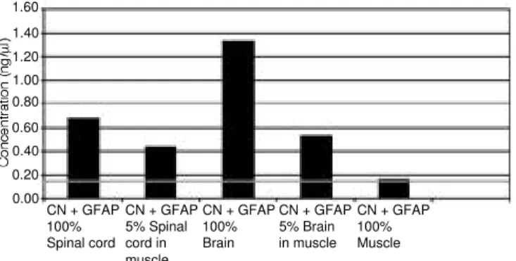
The Agilent CE based detection system relies on the use of conventional PCR followed by CE based separation of amplicons in a polyacrylamide matrix using a 12 well Labchip. This gives a high level of product separation and the potential for quantitative measurement by being able to measure the net fluorescent signal obtained for each PCR product and is a relatively low cost alternative to that of real time PCR. Reactions were carried out as a duplex utilising the ratio of the total GFAP amplicon signal obtained compared to that of the neuronal specific amplicon signal, to normalise and attempt to quantitate the level of non-muscle specific amplicon. This method has been proven to work in a similar fashion for the quantitative detection of Roundup Ready soya (Burns
et al., 2003) using the ratio of the lectin gene signal (detecting GM plus non-GM soya) to that of the Roundup Ready soya specific amplicon (GM specific). It enables a cut off limit to be set, that is designated as the background signal resulting from any residual non-methylated GFAP sequence normally present in muscle tissue which will be linked to the possible presence of GFA P protein in peripheral nervous system tissue. Any signal designated as resulting from neuronal tissue would therefore have to be above this as compared to a 100% muscle control run on the chip.
By using admixtures of neuronal tissue in muscle, a linear relationship was demonstrated between target concentration and signal obtained and we were able to set a threshold for any background non-methylation present in the muscle sample, giving the basis for quantitative measurement (F i g u re 5). T h i s demonstrates the clear potential of the CE system as an alternative platform for this type of assay.
5. SUMMARY
C u r r e n t l y, visual or histological examination, cholesterol content or more recently developed immunoassays can be used to detect neuronal tissue in meat products. Some of these methods are subjective and time consuming and may not be applicable to highly processed samples. We report here a new technique for the detection of CNS tissue in meat products that exploits the robustness of DNA, a molecule that is relatively resistant to the rigours of food processing and may give greater applicability to both raw, processed and cooked meat products.
To enable the use of DNA as the target molecule we focused on GFA P, a gene that has been characterized as a marker for CNS tissue and has previously been utilised at the protein level for the 1.000
0.800
0.600
0.400
0.200
0.000
-0.200
25 27 29 30 32 3435 37 39 40 42 4445 Cycle
Spinal Cord
Brain
Muscle
F i g u re 3 .TaqMan based differentiation of bovine brain, spinal cord and muscle using the ABI SDS Prism 7700 platform.
16.00
14.00
12.00
10.00
8.00
6.00
4.00
2.00
0.00
0.00 0.50 1.00 1.50 2.00 2.50
Brain in muscle Spinal cord in muscle
R2= 0.9509
R2= 0.9386
Log % concentration
immunogenic detection of CNS tissue in meat (Schmidt et al., 1999). Tissue specific methylation patterns for the promoter of this gene have been reported in several species (Condorelli et al., 1994, 1997; Teter et al., 1994) and this has enabled us to design a PCR based assay to discriminate between differentially methylated forms of the same sequence and hence identify the tissue from which the template was derived. By utilising the technique of methylation sensitive PCR (MSP) it was possible to design a PCR based assay specific to the methylation status of the GFAP promoter in the target tissue and hence be able to detect bovine and sheep brain and spinal cord tissue in a background of non-target muscle tissue.
In conclusion, we have demonstrated the first use of a PCR based assay for the specific detection of D N A derived from neuronal tissue in muscle admixtures. This has utilised methylation specific PCR (MSP), a technique previously only used to distinguish between tumour and non-tumour tissue in carcinogenesis. Further work is now underway on the use of one non-gel based detection platform, the Agilent CE system, in order to fully validate the method for the detection of neuronal and other non-muscle targets in a non-muscle background.
Acknowledgements
This work was supported by the United Kingdom Food Standards Agency.
Bibliography
Bauer NE., Garland T., Edwards JF. (1996). Brain emboli in slaughtered cattle. Vet. Pathol.33, p. 600.
Besnard F., Brenner M., Nakatani Y., Chao R., Purohit HJ., F r e e s e E. (1991) Multiple interacting sites regulate astrocyte-specific transcription of the human gene for glial fibrillary acidic protein. J. Biol. Chem. 266(28), p. 18877–18883.
Burns M., Shanahan D., Valdivia H., Harris N. (2003). Quantitative event-specific multiplex PCR detection of Roundup Ready soya using LabChipTMtechnology.Eur.
Food Res. Technol.216(5), p. 428–433.
C o n d o r e l l i D F., Nicoletti VG., Barresi V., Caruso A . , Conticello S., de Vellis J., Giuffrida Stella AM. (1994). Tissue-specific DNA methylation patterns of the rat glial fibrillary acidic protein gene. J. Neurosci. Res.39, p. 694–707.
Condorelli DF., Dell’Albni P., Conticello SG., Barresi V., N i c o l e t t i VG., Caruso A., Kahn M., Va c a n t i M . , Albanese V., de Vellis J., Giuffrida Stella AM. (1997). A neural-specific hypomethylated domain in the 5’ flanking region of the glial fibrillary acidic protein gene. Dev. Neurosci.19, p. 446–456.
E a d s CA., Danenberg KD., Kawakami K., Saltz L B . , Blake C., Shibata D., Danenberg PV., Laird PW. (2000). Methylight: a high-throughput assay to measure DNA methylation. Nucl. Acids Res.28(8), E32.
FSIS/USDA. (1998). Proposed rules: meat produced by advanced meat/bone separation machinery and recovery systems. Fed. Reg.63, p. 17959–17965.
F r o m m e r M., McDonald LE., Millar DS., Collis C M . , Watt F., Grigg GW., Molloy PL., Paul CL. (1992). A genomic sequencing protocol that yields a positive display of 5-methylcytosine residues in individual DNA strands. Proc. Natl. Acad. Sci. USA89, p. 1827–1831. Government UK. (1989). The bovine offal (prohibition)
regulations. United Kingdom SI No. 2061.
H e r m a n JG., Graff JR., Myohanen S., Nelkin B D . , Baylin S. (1996). Methylation-specific PCR: a novel PCR assay for methylation status of CpG islands. Proc. Natl. Acad. Sci. USA93, p. 9821–9826.
Kaneko R., Sueoka N. (1993). Tissue-specific versus cell type-specific expression of the glial fibrillary acidic protein. Proc. Natl. Acad. Sci. USA90, p. 4698–4702. L i E., Beard C., Jaenisch R. (1993). Role for DNA
methylation in genomic imprinting. N a t u re 3 6 6, p. 362–365.
Lücker E., Eigenbrodt E., Wenisch S., Failing K., Leiser R., B u l t e M. (1999). Development of an integrated procedure for the detection of central nervous tissue in meat products using cholesterol and neuron-specific enolase as markers. J. Food. Prot.62, p. 268–276. Paulin R., Grigg GW., Davey MW., Piper AA. (1998). Urea
improves efficiency of bisulphite-mediated sequencing of 5’-methylcytosine in genomic DNA. Nucl. Acids Res.
CN + GFAP 100% Spinal cord
CN + GFAP 5% Spinal cord in muscle
CN + GFAP 100% Brain
CN + GFAP 5% Brain in muscle
CN + GFAP 100% Muscle 1.60 1.40 1.20 1.00 0.80 0.60 0.40 0.20 0.00
26, p. 5009–5010.
S c h m i d t GR., Hossner KL., Ye m m RS., Gould D H . , O ’ C a l l a g h a n J P. (1999). An enzyme-linked immunosorbent assay for Glial Fibrillary Acidic Protein as an indicator of the presence of brain or spinal cord in meat. J. Food Prot.62, p. 394–397.
Te t e r B., Finch CE., Condorelli D F. (1994). DNA methylation in the Glial Fibrillary Acidic Protein gene: map of CpG methylation sites and summary of analysis by restriction enzymes and by LMPCR. J. Neurosci. Res.39, p. 708–709.
Wang RY.-H., Gerke CW., Ehrlich M. (1980). Comparison of bisulphite modification of 5-methyldeoxycytidine and deoxycytidine residues. Nucl. Acids Res. 8, p. 4777–4790.
Wo o d c o c k DM., Lawler CB., Linsenmeyer M E . , D o h e r t y J P., Wa r r e n WD. (1997). A s y m m e t r i c methylation in the hypermethylated CpG promoter region of the human L1 retrotransposon. J. Biol. Chem. 272, p. 7810–7816.
Yeivin A., Razin A. (1993). Gene methylation patterns and expression. In J o s t J P., Saluz H P. (Eds). D N A methylation: Molecular biology and biological significance, 8 ed. Basel: Birkhauser, p. 523–568. Yo d e r JA., Wa l s h C P., Bestor TH. (1997). Cytosine
methylation and the ecology of intragenomic parasites. Trends Genet. 13, p. 335–340.