Submitted17 November 2015 Accepted 2 February 2016 Published25 February 2016
Corresponding authors Wouter Boomsma, [email protected] Lars Ellgaard, [email protected]
Academic editor Vladimir Uversky
Additional Information and Declarations can be found on page 13
DOI10.7717/peerj.1725
Copyright 2016 Boomsma et al.
Distributed under
Creative Commons CC-BY 4.0
OPEN ACCESS
Bioinformatics analysis identifies several
intrinsically disordered human E3
ubiquitin-protein ligases
Wouter Boomsma, Sofie V. Nielsen, Kresten Lindorff-Larsen, Rasmus Hartmann-Petersen and Lars Ellgaard
Linderstrøm-Lang Centre for Protein Science, Department of Biology, University of Copenhagen, Copenhagen, Denmark
ABSTRACT
The ubiquitin-proteasome system targets misfolded proteins for degradation. Since the accumulation of such proteins is potentially harmful for the cell, their prompt removal is important. E3 ubiquitin-protein ligases mediate substrate ubiquitination by bringing together the substrate with an E2 ubiquitin-conjugating enzyme, which transfers ubiquitin to the substrate. For misfolded proteins, substrate recognition is generally delegated to molecular chaperones that subsequently interact with specific E3 ligases. An important exception is San1, a yeast E3 ligase. San1 harbors extensive regions of intrinsic disorder, which provide both conformational flexibility and sites for direct recognition of misfolded targets of vastly different conformations. So far, no mammalian ortholog of San1 is known, nor is it clear whether other E3 ligases utilize disordered regions for substrate recognition. Here, we conduct a bioinformatics analysis to examine >600 human andS. cerevisiaeE3 ligases to identify enzymes that are similar to San1 in terms of function and/or mechanism of substrate recognition. An initial sequence-based database search was found to detect candidates primarily based on the homology of their ordered regions, and did not capture the unique disorder patterns that encode the functional mechanism of San1. However, by searching specifically for key features of the San1 sequence, such as long regions of intrinsic disorder embedded with short stretches predicted to be suitable for substrate interaction, we identified several E3 ligases with these characteristics. Our initial analysis revealed that another remarkable trait of San1 is shared with several candidate E3 ligases: long stretches of complete lysine suppression, which in San1 limits auto-ubiquitination. We encode these characteristic features into a San1 similarity-score, and present a set of proteins that are plausible candidates as San1 counterparts in humans. In conclusion, our work indicates that San1 is not a unique case, and that several other yeast and human E3 ligases have sequence properties that may allow them to recognize substrates by a similar mechanism as San1.
SubjectsBioinformatics, Cell Biology, Biochemistry
Keywords E3 ligase, Hrd1, San1, Intrinsic disorder, STUbL, Ubiquitin, Protein folding, Protein degradation, Quality control
INTRODUCTION
homeostasis. In addition to its role in controlling protein levels for regulatory purposes, the proteasome also degrades misfolded proteins that may otherwise accumulate as toxic protein aggregates.
Cellular proteins are targeted for proteasomal degradation by the attachment of multiple moieties of the small regulatory protein ubiquitin, most often to lysine residues, but in some cases also to serines, threonines or cysteines (Wang, Herr & Hansen, 2012). This process oc-curs through the sequential action of a ubiquitin-activating enzyme (a so-called E1 enzyme), a ubiquitin-conjugating enzyme (E2) and a ubiquitin-protein ligase (E3). First, an E1 enzyme gets activated with a ubiquitin moiety in an ATP-dependent process, and subsequently passes on ubiquitin to an E2 enzyme. The E2 enzyme then interacts with an E3 ubiquitin-protein ligase. The most common class of E3 ligases contains a Really Interesting New Gene (RING) domain, which acts to bind ubiquitin-conjugating E2 enzymes (Metzger et al., 2014). RING E3 ligases thus bridge ubiquitin-charged E2s with substrates, which then become ubiquitinated. Multiple rounds of this process ensure substrate poly-ubiquitination, a requirement for degradation of most proteasomal substrates (for a recent review of the ubiquitin-proteasome system seeKleiger & Mayor, 2014).
Although most components and the basic overall mechanisms of this system are well understood, we still lack a comprehensive molecular understanding of how proteins are recognized and marked for degradation. In some cases, E3 enzymes bind ancillary protein factors that aid in the recognition of the substrate. The E3 enzyme CHIP, for example, ubiquitinates misfolded proteins that are recognized and delivered to CHIP via Hsp70 chaperones (Arndt, Rogon & Hohfeld, 2007). In this case, recognition of partially folded or misfolded substrates is offloaded to a chaperone, and the E3 enzyme then recognizes the substrate-loaded Hsp70.
Intrinsic disorder in proteins is now recognized to be broadly distributed across all kingdoms of life, and to play an important role in a large number of biological processes (Dyson & Wright, 2005). The large flexibility of fully intrinsically disordered proteins, or long stretches of intrinsic disorder in conjunction with well-folded domains, is believed to provide a number of particular properties to proteins (Babu, Kriwacki & Pappu, 2012). Importantly, the heterogeneous structures attained by these proteins allow them to bind a broad range of target proteins, either through a process called folding-upon-binding in which the disordered regions gain a specific structure upon binding to a target, or through the formation of ‘‘fuzzy complexes’’ in which substantial disorder is retained (Cumberworth et al., 2013).
The finding that San1 can recognize its protein substrates directly via its intrinsically disordered regions prompted us to explore whether this mechanism might be employed by other E3 enzymes. Using sequence conservation as a search criterion proved to be insufficient in this respect, presumably due to the low levels of sequence conservation that characterize intrinsically disordered protein regions. As an alternative, site-specific disorder profiles have been shown to display substantial preservation through evolution (Brown, Johnson & Daughdrill, 2010; Brown et al., 2011;Chen et al., 2006a;Chen et al., 2006b;Daughdrill et al., 2007), and have recently been used as the basis for detecting similar disorder functionalities across species (Mahani, Henriksson & Wright, 2013;Petrovich et al., 2015). We employ such an approach here, by constructing a procedure that searches for disorder properties similar to those of San1, combined with information about the unique pattern of binding regions known from the San1 protein. A previous study of structural disorder in the human ubiquitin-proteasome system found evidence for disorder in E1, E2 and—to the greatest extent—E3 enzymes, and provided examples for how disordered regions in E3 enzymes can function either as flexible linker regions or sometimes as sites for interaction with folded domains in substrates (Bhowmick et al., 2013). Our work extends this analysis by focusing on the extent to which E3 enzymes may use their intrinsic disorder in direct substrate recognition. The method was applied on datasets of 563 human and 80 yeast E3 enzymes. We demonstrate that a number of human E3 enzymes, in addition to displaying pervasive protein disorder, show two other hallmarks of the San1 sequence: short stretches of higher order potentially involved in substrate binding and a remarkably low content of lysine residues. Overall, our data indicate that San1 is not a unique case among E3 ligases and that a number of these enzymes could display a substrate recognition mode similar to San1.
RESULTS
Sequence-conservation based analyses reveal no obvious human orthologs of San1
As yet, no human ortholog of yeast San1 is known. In a first attempt to identify such a protein, we focused on the unusual RING domain of San1. Whereas a classical RING domain has a consensus motif of Cys-X2-Cys-X9–39-Cys-X1−3-His-X2–3
Nakatani & Day, 2012), the distance between the first Cys-X2-Cys sequence and the
following cysteine in the San1 RING domain is 88 residues (inS. cerevisiae), making it unusually long. However, using MOTIF search (http://www.genome.jp/tools/motif/), an algorithm designed to detect a specific sequence motif, we did not find obvious San1 candidates among the human proteins identified. Moreover, using theS. cerevisiae San1 RING domain in PSI-BLAST and DELTA-BLAST (Altschul et al., 1997) searches we did also not detect any human E3 ligase comprising the same characteristic RING domain as San1. To increase the sensitivity of the sequence-based search, we proceeded with an HMMSearch protocol, where the San1 sequence was encoded into a hidden Markov model using the jackhmmer program (both part of the HMMer software package;http://hmmer.org/). This procedure detected a number of potential matches, but the hits were predominantly defined by similarities in the ordered regions of San1—primarily the RING domain, and the detected homologues did not generally display the disorder and binding patterns characteristic of San1 (Fig. S1). In light of these results, we decided to search for human E3 ligases that contain characteristic sequence features of San1 other than those associated with the RING domain.
Initial San1-similarity analysis
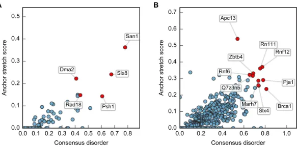
Although San1 fromS. cerevisiae is largely intrinsically disordered outside of the RING domain, the flexible parts are predicted to contain short interspersed regions of higher order that coincide with sequence conservation among orthologs in other yeast species (Rosenbaum et al., 2011). Experimentally, it has been verified that these more ordered regions confer substrate binding (Rosenbaum et al., 2011). The overall model that emerges is therefore one where multiple binding sites (independently or in conjunction) can engage the substrates, whereas the flexibility provided by the intrinsic disorder allows San1 to adopt its structure to a wide variety of misfolded substrates (Rosenbaum & Gardner, 2011). We therefore analyzed datasets of S. cerevisiae and human E3s to identify E3 enzymes containing the same overall disorder/order signature as San1, in addition to a high overall content of intrinsic disorder. Specifically, we employed the consensus disorder annotation from the MobiDB database (Potenza et al., 2015) to identify regions of disorder in E3 enzymes (Fig. 1A,Table S1). For the San1 sequence, 78% was predicted to be disordered (Fig. 1B,Table S1).
Figure 1 San1 is highly disordered and contains a characteristic pattern of interspersed ANCHOR positive regions for substrate interaction.(A) Disorder annotation of theS. cerevisiaeSan1 protein, as provided by the MobiDB database (Potenza et al., 2015). Nine individual predictors were used by MobiDB as the basis for a consensus annotation, which we used for all analyses in this paper. The darkness of the shade of blue indicates how many of the nine predictors agree about the disorder annotation at a particu-lar site. (B) Combined plot of the MobiDB consensus disorder annotation (blue) and the ANCHOR pre-diction (red) for San1. The position of the RING domain is indicated. This compact view is used as the ba-sis for the comparative analyses throughout the paper.
regions of length 15–40 in each protein of the datasets, and normalized it to the length of the protein.
Plotting this ANCHOR score against the consensus disorder, and considering the candidates with highest scores in both dimensions, we indeed found San1 at the top of the yeast dataset, accompanied by a few other high-scoring candidates (e.g., Slx8;Figs. 2and3). The disorder and ANCHOR profiles for several of the top candidates in the human dataset (e.g., Rnf6, Rnf12, Pja1, Rn111;Figs. 2and3) showed clear similarities to the San1 profile, with general high levels of disorder and the distinct ANCHOR propensity pattern. This initial approach, based on a simple combination of disorder and anchor score, however, had a few shortcomings. First of all, it relies on global high values of disorder, thereby excluding proteins containing one or more ordered domains. Secondly, it fails to take into account another remarkable feature of San1: long segments without a single occurrence of lysine residues. Both these limitations are addressed below.
San1-like E3s show substantial lysine suppression
Figure 2 Characterization of the E3 dataset in terms of disorder and protein binding sites in disor-dered regions.Plots of ANCHOR score versus fraction consensus disorder for allS. cerevisiae(A) and hu-man (B) proteins in the datasets. Protein names are given for the top-scoring candidates, for which disor-der and ANCHOR profiles are depicted inFig. 3.
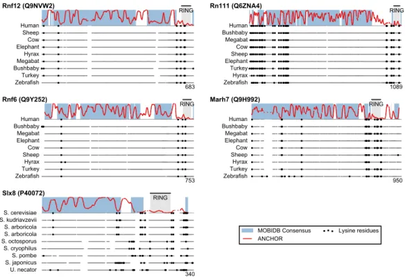
Figure 4 Low lysine content in San1 candidates is conserved broadly across species.The lysine distri-butions of the candidates with strongest lysine suppression inFig. 3are compared to select orthologs from representative species of evolutionary divergence, suggesting lysine suppression to be a strongly conserved feature in these candidates. Note that the number below the alignment denotes the length of the alignment (including gaps), rather than the query sequence length.
domain or in a short sequence stretch of relatively low disorder immediately adjacent to this domain (Fig. 3, black dots), a feature which seems to be shared with several of the highest scoring candidates (Fig. 3), in some cases to a remarkable extent (e.g., >600 consecutive residues were devoid of lysines in the case of RN111). The most significant cases are highlighted in Fig. 4, demonstrating that the lysine suppression is strongly conserved among orthologs in a range of different species. The lack of lysines is particularly remarkable in light of the finding that disordered proteins are typically enriched in lysines compared to folded proteins (Tompa, 2002). Taken together, these results suggest that the San1-like E3 ligases identified here could employ the same strategy as San1 itself to avoid auto-ubiquitination and degradation (but see also ‘Discussion’).
Definition of a San1 similarity score
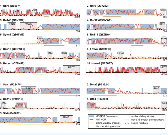
Figure 5 San1-like candidates can be detected using a feature-based similarity score.Top-scoring can-didates retrieved when applying the sliding-window San1-similarity score (seeTable S1). Three comple-mentary quantities are calculated over a window of 150 residues: (1) the percentage of disorder, (2) the percentage of residues within an ANCHOR positive stretch of length 15–40 and (3) the percentage of residues occurring in the maximum lysine-free stretch within the window. The San1-similarity score is the maximum value of the product of these numbers, with the corresponding window depicted as an orange line under each plot. The window size is chosen to be of fixed size, and the score is therefore not defined for the first and last 75 positions. Note that certain of the top-scoring candidates found using the San1 similarity score overlap with our initial, simpler approach that considered only disorder and ANCHOR score over the entire sequence (seeFig. 3).
residues, normalized by the length of the window (chosen here as 150 residues). For each position in the sequence, a product of these three quantities is calculated, and each protein is represented with the maximum value (‘‘the San1 similarity score’’) observed among its residues. Thus, proteins with a high San1 similarity score contain a particular region that simultaneously has a high level of disorder, many potential sites for molecular interaction and strong lysine suppression.
a further discussion of the choice of parameters and how they affect the San1-similarity score we refer to the ‘Methods’ section.
Analysis of potential functional orthologs of San1
Based on the bioinformatics investigation described above, we next analyzed specific proteins with high San1 similarity in more detail founded on the current literature. Specifically, among the top scoring hits inFig. 5, we consider those candidates with known links to protein quality control, i.e., the human E3s Syvn1/Hrd1 and Hecw1 and the yeast E3s Doa10, Slx8, and Ufd4.
A well-characterized protein appearing in our analysis is human Syvn1/Hrd1, a multispanning transmembrane protein of the ER. The low overall disorder displayed by this protein is due to the transmembrane region ending at around residue 240, followed by a disordered C-terminal cytosolic region, which is also low in lysine content (Fig. 5). Curiously, the cytosolic tail of the budding yeast protein contains more lysines than in its orthologs. Whether this difference reflects a basic difference in the respective functional mechanisms is presently unknown.
Syvn1/Hrd1 is of particular interest in the current context, because it is known to interact with misfolded proteins. Thus, Syvn1/Hrd1 is involved in targeting misfolded ER and secretory proteins for proteasomal degradation in the cytosol through a process known as ER-associated degradation (ERAD) (Christianson & Ye, 2014). In this process, misfolded proteins are first recognized by ER lumenal chaperones and retrotranslocated across the ER membrane. When emerging in the cytosol, substrate proteins are ubiquitinated by ER-membrane localized E3 ligases, such as Syvn1/Hrd1, before they are finally degraded by the 26S proteasome (Christianson & Ye, 2014). Along with the RING domain, the predicted intrinsically disordered area in HRD1 faces the cytosol, and has so far not been ascribed any particular function. Presumably, Syvn1/Hrd1 transiently engages the at least partially extended ERAD substrates during ubiquitination, and the disordered region could therefore play a role, similar to the situation for San1, in this function.
Intriguingly, another ERAD E3, Doa10, was one of the best scoring yeast E3s. Like Syvn1/Hrd1, Doa10 is a multispanning transmembrane protein localized to the ER and inner nuclear membrane where it targets a variety of misfolded proteins, e.g., squalene monooxygenase, an enzyme involved in sterol homeostasis (Foresti et al., 2013), for proteasomal degradation (Habeck et al., 2015and references therein). As with Syvn1/Hrd1 the disordered region of Doa10 faces the cytosol, which fits well with its established role in targeting also soluble nuclear and cytosolic proteins for degradation (Ravid, Kreft & Hochstrasser, 2006).
SLX8were identified as suppressors of a temperature sensitive mutant in the DNA binding protein Mot1 (Wang, Jones & Prelich, 2006). At high temperatures, the mutant Mot1 protein is highly unstable, but stabilized inSLX5andSLX8null mutants (Wang, Jones & Prelich, 2006). This is similar to how San1 was first connected to protein quality control of the mutant proteins Sir4-9 and Cdc68-1 (Gardner, Nelson & Gottschling, 2005), and suggests that the mutant Mot1 protein is still functional at the restrictive temperature, but targeted for degradation. However, the degradation of Mot1 was also shown to depend on the SUMO-ligases Siz1 and Siz2 (Wang, Jones & Prelich, 2006), revealing that sumoylation of Mot1 was required for Slx5-Slx8 dependent degradation. Although in sequence, they do not appear highly conserved, the mammalian STUbL Rnf4/Snurf is a functional ortholog of budding yeast Slx5-Slx8 (Prudden et al., 2007;Sriramachandran & Dohmen, 2014). However, as opposed to the situation with Slx5-Slx8 in yeast, Rnf4/Snurf forms a homodimer (Plechanovova et al., 2011), which has primarily been connected with the SUMO-dependent degradation of the Pml-Rarα oncoprotein (Tatham et al., 2008). Of note, Rnf4/Snurf did not display a high San1-similarity score. However, another less characterized human STUbL, Rn111 (Fig. 5), which is also involved in targeting Pml-Rarα for SUMO-dependent degradation (Erker et al., 2013), did show similarity to San1. In contrast to Rnf4/Snurf, Rn111 has also been shown to promote non-proteolytic, K63-linked ubiquitination of SUMO-modified substrate proteins (Poulsen et al., 2013).
In addition to Syvn1/Hrd1, Doa10 and Slx8 described above, also Hecw1 and Ufd4 have been connected with protein quality control. Specifically, Hecw1, also known as NEDL1, was found to ubiquitinate amyotrophic lateral sclerosis (ALS)-linked SOD1 variants, but not wild type SOD1 (Miyazaki et al., 2004). However, it is not clear that Hecw1 catalyzed SOD1 ubiquitination leads to proteasomal degradation. Genetic experiments have also linked the yeast E3 Ufd4 to protein quality control (Theodoraki et al., 2012), and showed that Ufd4 as a proteasome-associated E3 (Xie & Varshavsky, 2002) targets an orphan ER-membrane protein for degradation (Ravid & Hochstrasser, 2007).
DISCUSSION
In the case of San1, it has been shown that one reason for its unusually low content of lysine residues is to protect it from auto-ubiquitination and untimely proteasomal degradation (Fredrickson et al., 2013). Many of the disordered San1-like E3s that we identify here also display long stretches devoid of lysine residues. For instance, Rnf6 and Rnf12 both contain a remarkably long stretch of around 500 residues devoid of lysines, a feature conserved among their orthologs (Fig. 4). It is therefore possible that these proteins employ the same strategy as San1 to avoid auto-ubiquitination and degradation. Indeed, recent work shows a clear propensity for ubiquitinated lysines in proteasomal substrates to occur in regions of intrinsic disorder, and moreover these lysines are often located in close proximity to segments of >19 consecutive disordered residues (Guharoy et al., 2016). However, since lysine residues can undergo numerous post-translational modifications, including for instance sumoylation and acetylation, the low lysine content need not necessarily be connected with ubiquitination and degradation. Indeed, for Dma2, auto-ubiquitination followed by degradation was shown to occur despite the presence of a region with low lysine content (Loring et al., 2008).
It is noteworthy that both Rn111 and Slx8 belong to the rather small family of STUbL-type E3s. Since data suggest that several proteins are conjugated to SUMO in response to heat shock (Tammsalu et al., 2014;Tatham et al., 2011), such STUbL E3s are likely to be important for protein quality control. Indeed, like San1, Slx8 has been linked to protein quality control (Wang, Jones & Prelich, 2006). However, since the SUMO interacting motifs that are found in STUbLs are generally located in intrinsically disordered regions (Vogt & Hofmann, 2012), it cannot be ruled out that our approach is biased to detect STUbLs.
further examples could be added in the near future, opening up for more detailed functional and biophysical studies of the mechanisms by which intrinsic disorder in E3 enzymes is used to regulate protein homeostasis.
METHODS
HMM sequence-based search procedure
The hidden Markov model based sequence searches were conducted using the HMMER software package (http://hmmer.org/) by constructing a model using the jackhmmer tool, and subsequently using the hmmsearch program to search through the entire Uniprot databases ofSaccharomyces cerevisiae and human, respectively. Default values were used for these programs. The top-10 matches for human and top-5 hits for yeast are included asFig. S1.
Datasets
For the remaining analyses, we searched directly within a set of 80 yeast E3 ligases (Li et al., 2008) and a set of 563 human E3 ligases recently published by Tompa and coworkers (Bhowmick et al., 2013). This set was constructed as a merge of an extract from the KEGG BRITE database (Kanehisa & Goto, 2000) and well-characterized E3 ligases from a previ-ously published dataset (Li et al., 2008), filtered for uncertain annotations, identical gene names, and internal sequence homology (>85%), leading to an unbiased, representative set of the currently known E3 proteins. It should be noted, however, that additional human E3 ligases probably exist (Bhowmick et al., 2013;Li et al., 2008), and more than 600 RING-type E3 ligases are likely expressed in human cells (Deshaies & Joazeiro, 2009).
All results obtained in the analysis for this paper are made available as an annotation of this original data set, presented as an Excel file inTable S1. This dataset contains all position-specific prediction results, it provides the resulting San1 similarity score, and allows for easy sorting of candidates based on the criteria outlined in the paper. The spreadsheet also permits reanalyses of the data with different parameter values (e.g., the lengths of the sliding window and the ANCHOR stretch).
Prediction of protein binding regions
We used the ANCHOR (Meszaros, Simon & Dosztanyi, 2009) program to assign position-specific scores for protein binding within disordered regions. For the purpose of the San1 similarity score, we considered stretches of between 15 and 40 residues (see below) for which the ANCHOR scores are all higher than 0.5. The total number of residues occurring within such regions was normalized by the length of the window to give an ANCHOR stretch score. No requirements were imposed on the disorder status of such regions.
Choice of parameters
than the window size will be assigned a score of zero), and choosing smaller values makes the score less sensitive to long lysine-free stretches. The 15 and 40 residue ANCHOR stretch boundaries were based directly on the lengths observed in San1 (Rosenbaum et al., 2011). We probed the sensitivity of our approach to these choices by considering the top candidates with varying values for both of these parameters and found the presented results to be quite robust, as long as the chosen values were not directly at odds with the characteristics observed for San1. For instance, when changing the ANCHOR stretch length to 5–35, we find that 17 out of 20 of the Top 20 candidates inTable S1are preserved.
ADDITIONAL INFORMATION AND DECLARATIONS
Funding
This work was supported by grants from the Lundbeck Foundation (SVN, RHP: R108-A10121; LE: R83-A7446), Danish Council for Independent Research/Natural Sciences (RHP: 18490), the Villum Foundation (WB: VKR023445), the Novo Nordisk Foundation (KLL, RHP) and the Danish Cancer Society (RHP, KLL). The funders had no role in study design, data collection and analysis, decision to publish, or preparation of the manuscript.
Grant Disclosures
The following grant information was disclosed by the authors: Lundbeck Foundation: R108-A10121, R83-A7446.
Danish Council for Independent Research/Natural Sciences: 18490. Villum Foundation: VKR023445.
Novo Nordisk Foundation. Danish Cancer Society.
Competing Interests
The authors declare there are no competing interests.
Author Contributions
• Wouter Boomsma conceived and designed the experiments, performed the experiments, analyzed the data, wrote the paper, prepared figures and/or tables, reviewed drafts of the paper.
• Sofie V. Nielsen performed the experiments, reviewed drafts of the paper.
• Kresten Lindorff-Larsen and Rasmus Hartmann-Petersen conceived and designed the experiments, analyzed the data, wrote the paper, reviewed drafts of the paper.
• Lars Ellgaard conceived and designed the experiments, analyzed the data, wrote the paper, prepared figures and/or tables, reviewed drafts of the paper.
Data Availability
Supplemental Information
Supplemental information for this article can be found online athttp://dx.doi.org/10.7717/ peerj.1725#supplemental-information.
REFERENCES
Altschul SF, Madden TL, Schaffer AA, Zhang J, Zhang Z, Miller W, Lipman DJ. 1997.
Gapped BLAST and PSI-BLAST: a new generation of protein database search programs.Nucleic Acids Research25:3389–3402DOI 10.1093/nar/25.17.3389.
Arndt V, Rogon C, Hohfeld J. 2007.To be, or not to be–molecular chaperones in protein degradation.Cellular and Molecular Life Science64:2525–2541
DOI 10.1007/s00018-007-7188-6.
Babu MM, Kriwacki RW, Pappu RV. 2012.Structural biology. Versatility from protein disorder.Science337:1460–1461DOI 10.1126/science.1228775.
Bhowmick P, Pancsa R, Guharoy M, Tompa P. 2013.Functional diversity and structural disorder in the human ubiquitination pathway.PLoS ONE8:e65443
DOI 10.1371/journal.pone.0065443.
Brown CJ, Johnson AK, Daughdrill GW. 2010.Comparing models of evolution for ordered and disordered proteins.Molecular Biology and Evolution27:609–621
DOI 10.1093/molbev/msp277.
Brown CJ, Johnson AK, Dunker AK, Daughdrill GW. 2011.Evolution and disorder.
Current Opinion in Structural Biology21:441–446DOI 10.1016/j.sbi.2011.02.005.
Budhidarmo R, Nakatani Y, Day CL. 2012.RINGs hold the key to ubiquitin transfer.
Trends in Biochemical Sciences37:58–65DOI 10.1016/j.tibs.2011.11.001.
Chen JW, Romero P, Uversky VN, Dunker AK. 2006a.Conservation of intrinsic disorder in protein domains and families: I. A database of conserved predicted disordered regions.Journal of Proteome Research5:879–887DOI 10.1021/pr060048x.
Chen JW, Romero P, Uversky VN, Dunker AK. 2006b.Conservation of intrinsic
disorder in protein domains and families: II. functions of conserved disorder.Journal of Proteome Research5:888–898 DOI 10.1021/pr060049p.
Christianson JC, Ye Y. 2014.Cleaning up in the endoplasmic reticulum: ubiquitin in charge.Nature Structural & Molecular Biology21:325–335DOI 10.1038/nsmb.2793.
Cumberworth A, Lamour G, Babu MM, Gsponer J. 2013.Promiscuity as a functional trait: intrinsically disordered regions as central players of interactomes.Biochemical Journal 454:361–369DOI 10.1042/BJ20130545.
Daughdrill GW, Narayanaswami P, Gilmore SH, Belczyk A, Brown CJ. 2007.Dynamic behavior of an intrinsically unstructured linker domain is conserved in the face of negligible amino acid sequence conservation.Journal of Molecular Evolution 65:277–288DOI 10.1007/s00239-007-9011-2.
Deshaies RJ, Joazeiro CA. 2009.RING domain E3 ubiquitin ligases.Annual Review of Biochemistry78:399–434DOI 10.1146/annurev.biochem.78.101807.093809.
Dyson HJ, Wright PE. 2005.Intrinsically unstructured proteins and their functions.
Erker Y, Neyret-Kahn H, Seeler JS, Dejean A, Atfi A, Levy L. 2013.Arkadia, a novel SUMO-targeted ubiquitin ligase involved in PML degradation.Molecular and Cellular Biology 33:2163–2177DOI 10.1128/MCB.01019-12.
Foresti O, Ruggiano A, Hannibal-Bach HK, Ejsing CS, Carvalho P. 2013.Sterol homeostasis requires regulated degradation of squalene monooxygenase by the ubiquitin ligase Doa10/Teb4.Elife2:e00953DOI 10.7554/eLife.00953.
Fredrickson EK, Clowes Candadai SV, Tam CH, Gardner RG. 2013.Means of preservation: how an intrinsically disordered ubiquitin-protein ligase averts self-destruction.Molecular Biology of the Cell24:1041–1052
DOI 10.1091/mbc.E12-11-0811.
Gallagher PS, Clowes Candadai SV, Gardner RG. 2014.The requirement for Cdc48/p97 in nuclear protein quality control degradation depends on the substrate and
correlates with substrate insolubility.Journal of Cell Science127:1980–1991
DOI 10.1242/jcs.141838.
Gardner RG, Nelson ZW, Gottschling DE. 2005.Degradation-mediated protein quality control in the nucleus.Cell120:803–815DOI 10.1016/j.cell.2005.01.016.
Guerriero CJ, Weiberth KF, Brodsky JL. 2013.Hsp70 targets a cytoplasmic quality control substrate to the San1p ubiquitin ligase.Journal of Biological Chemistry 288:18506–18520DOI 10.1074/jbc.M113.475905.
Guharoy M, Bhowmick P, Sallam M, Tompa P. 2016.Tripartite degrons confer diversity and specificity on regulated protein degradation in the ubiquitin-proteasome system.
Nature Communications7:10239DOI 10.1038/ncomms10239.
Habeck G, Ebner FA, Shimada-Kreft H, Kreft SG. 2015.The yeast ERAD-C ubiq-uitin ligase Doa10 recognizes an intramembrane degron.Journal of Cell Biology 209:261–273DOI 10.1083/jcb.201408088.
Her YR, Chung IK. 2009.Ubiquitin Ligase RLIM Modulates Telomere Length Home-ostasis through a Proteolysis of TRF1.Journal of Biological Chemistry284:8557–8566
DOI 10.1074/jbc.M806702200.
Kanehisa M, Goto S. 2000.KEGG: kyoto encyclopedia of genes and genomes.Nucleic Acids Research28:27–30DOI 10.1093/nar/28.1.27.
Kleiger G, Mayor T. 2014.Perilous journey: a tour of the ubiquitin-proteasome system.
Trends in Cell Biology24:352–359DOI 10.1016/j.tcb.2013.12.003.
Kriegenburg F, Jakopec V, Poulsen EG, Nielsen SV, Roguev A, Krogan N, Gordon C, Fleig U, Hartmann-Petersen R. 2014.A chaperone-assisted degradation pathway targets kinetochore proteins to ensure genome stability.PLoS Genetics10:e1004140
DOI 10.1371/journal.pgen.1004140.
Li W, Bengtson MH, Ulbrich A, Matsuda A, Reddy VA, Orth A, Chanda SK, Batalov S, Joazeiro CA. 2008.Genome-wide and functional annotation of human E3 ubiquitin ligases identifies MULAN, a mitochondrial E3 that regulates the organelle’s dynamics and signaling.PLoS ONE3:e1487DOI 10.1371/journal.pone.0001487.
promotes degradation of Smad7 by Arkadia.EMBO Journal25:1646–1658
DOI 10.1038/sj.emboj.7601057.
Loring GL, Christensen KC, Gerber SA, Brenner C. 2008.Yeast Chfr homologs retard cell cycle at G1 and G2/M via Ubc4 and Ubc13/Mms2-dependent ubiquitination.
Cell Cycle7:96–105DOI 10.4161/cc.7.1.5113.
Mahani A, Henriksson J, Wright AP. 2013.Origins of Myc proteins–using intrinsic protein disorder to trace distant relatives.PLoS ONE8:e75057
DOI 10.1371/journal.pone.0075057.
Meszaros B, Simon I, Dosztanyi Z. 2009.Prediction of protein binding regions in disordered proteins.PLoS Computational Biology5:e1000376
DOI 10.1371/journal.pcbi.1000376.
Metzger MB, Pruneda JN, Klevit RE, Weissman AM. 2014.RING-type E3 ligases: master manipulators of E2 ubiquitin-conjugating enzymes and ubiquitination.Biochimica et Biophysica ACTA/General Subjects1843:47–60DOI 10.1016/j.bbamcr.2013.05.026.
Miyazaki K, Fujita T, Ozaki T, Kato C, Kurose Y, Sakamoto M, Kato S, Goto T, Itoyama Y, Aoki M, Nakagawara A. 2004.NEDL1, a novel ubiquitin-protein isopeptide ligase for dishevelled-1, targets mutant superoxide dismutase-1.Journal of Biological Chemistry279:11327–11335DOI 10.1074/jbc.M312389200.
Nielsen SV, Poulsen EG, Rebula CA, Hartmann-Petersen R. 2014.Protein quality control in the nucleus.Biomolecules4:646–661DOI 10.3390/biom4030646.
Park SH, Kukushkin Y, Gupta R, Chen T, Konagai A, Hipp MS, Hayer-Hartl M, Hartl FU. 2013.PolyQ proteins interfere with nuclear degradation of cytosolic proteins by sequestering the Sis1p chaperone.Cell154:134–145DOI 10.1016/j.cell.2013.06.003.
Petrovich A, Borne A, Uversky VN, Xue B. 2015.Identifying similar patterns of struc-tural flexibility in proteins by disorder prediction and dynamic programming. Inter-national Journal of Molecular Sciences16:13829–13849DOI 10.3390/ijms160613829.
Plechanovova A, Jaffray EG, McMahon SA, Johnson KA, Navratilova I, Naismith JH, Hay RT. 2011.Mechanism of ubiquitylation by dimeric RING ligase RNF4.Nature Structural & Molecular Biology18:1052–1059DOI 10.1038/nsmb.2108.
Potenza E, Di Domenico T, Walsh I, Tosatto SC. 2015.MobiDB 2.0: an improved database of intrinsically disordered and mobile proteins.Nucleic Acids Research 43:D315–D320DOI 10.1093/nar/gku982.
Poulsen SL, Hansen RK, Wagner SA, Van Cuijk L, Van Belle GJ, Streicher W, Wikstrom M, Choudhary C, Houtsmuller AB, Marteijn JA, Bekker-Jensen S, Mailand N. 2013.RNF111/Arkadia is a SUMO-targeted ubiquitin ligase that facilitates the DNA damage response.Journal of Cell Biology201:797–807
DOI 10.1083/jcb.201212075.
Prudden J, Pebernard S, Raffa G, Slavin DA, Perry JJ, Tainer JA, McGowan CH, Boddy MN. 2007.SUMO-targeted ubiquitin ligases in genome stability.EMBO Journal 26:4089–4101DOI 10.1038/sj.emboj.7601838.
Ravid T, Kreft SG, Hochstrasser M. 2006.Membrane and soluble substrates of the Doa10 ubiquitin ligase are degraded by distinct pathways.EMBO Journal 25:533–543DOI 10.1038/sj.emboj.7600946.
Rodrigo-Brenni MC, Gutierrez E, Hegde RS. 2014.Cytosolic quality control of mislo-calized proteins requires RNF126 recruitment to Bag6.Molecular Cell55:227–237
DOI 10.1016/j.molcel.2014.05.025.
Rosenbaum JC, Fredrickson EK, Oeser ML, Garrett-Engele CM, Locke MN, Richardson LA, Nelson ZW, Hetrick ED, Milac TI, Gottschling DE, Gardner RG. 2011.
Disorder targets misorder in nuclear quality control degradation: a disordered ubiq-uitin ligase directly recognizes its misfolded substrates.Molecular Cell41:93–106
DOI 10.1016/j.molcel.2010.12.004.
Rosenbaum JC, Gardner RG. 2011.How a disordered ubiquitin ligase maintains order in nuclear protein homeostasis.Nucleus2:264–270DOI 10.4161/nucl.2.4.16118.
Sriramachandran AM, Dohmen RJ. 2014.SUMO-targeted ubiquitin ligases.Biochimica et Biophysica ACTA/General Subjects1843:75–85DOI 10.1016/j.bbamcr.2013.08.022.
Tammsalu T, Matic I, Jaffray EG, Ibrahim AF, Tatham MH, Hay RT. 2014. Proteome-wide identification of SUMO2 modification sites.Science Signaling 7:rs2. 1–10
DOI 10.1126/scisignal.2005146.
Tanaka H, Takahashi T, Xie Y, Minami R, Yanagi Y, Hayashishita M, Suzuki R, Yokota N, Shimada M, Mizushima T, Kuwabara N, Kato R, Kawahara H. 2016.A conserved island of BAG6/Scythe is related to ubiquitin domains and participates in short hydrophobicity recognition.FEBS Journal283:662–677
DOI 10.1111/febs.13618.
Tatham MH, Geoffroy MC, Shen L, Plechanovova A, Hattersley N, Jaffray EG, Palvimo JJ, Hay RT. 2008.RNF4 is a poly-SUMO-specific E3 ubiquitin ligase required for arsenic-induced PML degradation.Nature Cell Biology 10:538–546
DOI 10.1038/ncb1716.
Tatham MH, Matic I, Mann M, Hay RT. 2011.Comparative proteomic analysis identifies a role for SUMO in protein quality control.Science Signaling 4:rs4. 1–12
DOI 10.1126/scisignal.2001484.
Theodoraki MA, Nillegoda NB, Saini J, Caplan AJ. 2012.A network of ubiquitin ligases is important for the dynamics of misfolded protein aggregates in yeast.Journal of Biological Chemistry 287:23911–23922DOI 10.1074/jbc.M112.341164.
Tompa P. 2002.Intrinsically unstructured proteins.Trends in Biochemical Sciences 27:527–533DOI 10.1016/S0968-0004(02)02169-2.
Uzunova K, Gottsche K, Miteva M, Weisshaar SR, Glanemann C, Schnellhardt M, Niessen M, Scheel H, Hofmann K, Johnson ES, Praefcke GJ, Dohmen RJ. 2007.
Ubiquitin-dependent proteolytic control of SUMO conjugates.Journal of Biological Chemistry282:34167–34175DOI 10.1074/jbc.M706505200.
Vittal V, Shi L, Wenzel DM, Scaglione KM, Duncan ED, Basrur V,
Elenitoba-Johnson KS, Baker D, Paulson HL, Brzovic PS, Klevit RE. 2015.Intrinsic disorder drives N-terminal ubiquitination by Ube2w.Nature Chemical Biology11:83–89
Vogt B, Hofmann K. 2012.Bioinformatical detection of recognition factors for ubiquitin and SUMO.Methods in Molecular Biology832:249–261
DOI 10.1007/978-1-61779-474-2_18.
Wang X, Herr RA, Hansen TH. 2012.Ubiquitination of substrates by esterification.
Traffic 13:19–24DOI 10.1111/j.1600-0854.2011.01269.x.
Wang Z, Jones GM, Prelich G. 2006.Genetic analysis connects SLX5 and SLX8 to the SUMO pathway in Saccharomyces cerevisiae.Genetics172:1499–1509
DOI 10.1534/genetics.105.052811.
Wang Q, Liu Y, Soetandyo N, Baek K, Hegde R, Ye Y. 2011.A ubiquitin ligase-associated chaperone holdase maintains polypeptides in soluble states for proteasome degrada-tion.Molecular Cell42:758–770 DOI 10.1016/j.molcel.2011.05.010.
Ward JJ, Sodhi JS, McGuffin LJ, Buxton BF, Jones DT. 2004.Prediction and functional analysis of native disorder in proteins from the three kingdoms of life.Journal of Molecular Biology 337:635–645DOI 10.1016/j.jmb.2004.02.002.
Westerbeck JW, Pasupala N, Guillotte M, Szymanski E, Matson BC, Esteban C, Kerscher O. 2014.A SUMO-targeted ubiquitin ligase is involved in the degradation of the nuclear pool of the SUMO E3 ligase Siz1.Molecular Biology of the Cell25:1–16
DOI 10.1091/mbc.E13-05-0291.
Xie Y, Varshavsky A. 2002.UFD4 lacking the proteasome-binding region catalyses ubiquitination but is impaired in proteolysis.Nature Cell Biology 4:1003–1007
DOI 10.1038/ncb889.
Xu Y, Liu Y, Lee JG, Ye Y. 2013.A ubiquitin-like domain recruits an oligomeric chap-erone to a retrotranslocation complex in endoplasmic reticulum-associated degrada-tion.Journal of Biological Chemistry288:18068–18076DOI 10.1074/jbc.M112.449199.
Xu K, Shimelis H, Linn DE, Jiang R, Yang X, Sun F, Guo Z, Chen H, Li W, Chen H, Kong X, Melamed J, Fang S, Xiao Z, Veenstra TD, Qiu Y. 2009.Regulation of androgen receptor transcriptional activity and specificity by RNF6-induced ubiquitination.Cancer Cell 15:270–282DOI 10.1016/j.ccr.2009.02.021.
Yang L, Mullen JR, Brill SJ. 2006.Purification of the yeast Slx5-Slx8 protein complex and characterization of its DNA-binding activity.Nucleic Acids Research34:5541–5551