ABSTRACT
Bone regenerat ion in surgically creat ed defect s
illed with autogenous bone: an epiluorescence
m icroscopy analysis in rat s
Marcos Heidy GUSKUMA1, Eduardo HOCHULI-VIEIRA2, Flávia Priscila PEREIRA1, Idelmo RANGEL-GARCIA JUNIOR2,
Roberta OKAMOTO2, Tetuo OKAMOTO2, Osvaldo MAGRO FILHO2
1- DDS, MSc, Discipline of Oral and Maxillofacial Surgery, Department of Surgery and Integrated Clinics, Araçatuba Dental School, São Paulo State University (UNESP) Araçatuba, SP, Brazil.
2- DDS, MSc, PhD, Discipline of Oral and Maxillofacial Surgery, Department of Surgery and Integrated Clinics, Araçatuba Dental School, São Paulo State University (UNESP) Araçatuba, SP, Brazil.
Corresponding address: Marcos Heidy Guskuma - Rua Dulcídio Pereira, 138 - Jd Higienópolis - Londrina, PR - Brazil - 86015-170 - Phone: (43) 91013448 - e-mail: [email protected]
Received: January 11, 2009 - Modiication: July 20. 2009 - Accepted: September 16, 2009
A
lt hough t he search for t he ideal bone subst it ut e has been t he focus of a large num berof studies, autogenous bone is still the gold standard for the illing of defects caused by
pat hologies and t raum as, and m ainly, for alveolar ridge reconst ruct ion, allowing t he t it anium im plant s inst allat ion. Obj ect ives: The aim of t his st udy was t o evaluat e t he dynam ics of aut ogenous bone graft incorporat ion process t o surgically creat ed defect s in rat calvaria,
using epiluorescence microscopy. Material and methods: Five adult male rats weighing
200- 300 g were used. The anim als received t wo 5- m m - diam et er bone defect s bilat erally in each pariet al bone wit h a t rephine bur under general anest hesia. Two groups of defect s
were formed: a control group (n=5), in which the defects were illed with blood clot, and a graft group (n=5), in which the defects were illed with autogenous bone block, removed from the contralateral defect. The luorochromes calcein and alizarin were applied at the
7t h and 30t h post operat ive days, respect ively. The anim als were killed at 35 days. Result s: The m ineralizat ion process was m ore int ense in t he graft group ( 32.09% ) and occurred m ainly bet ween 7 and 30 days, t he period labeled by calcein ( 24.66% ) . Conclusions:
The luorochromes showed to be appropriate to label mineralization areas. The interfacial areas between luorochrome labels are important sources of information about the bone
regenerat ion dynam ics.
Ke y w or ds: Fluorescent dyes. Bone t ransplant at ion. Bone regenerat ion.
I N TROD UCTI ON
The rehabilit at ion of t he st om at ognat hic syst em is of great im port ance as it associat ed w it h t he recovery of est het ic, funct ional and psychosocial r ecover y of t he pat ient s. Ot her w ise, t he lack of bone in t he alveolar ridge is a great challenge for t he rehabilit at ion success. Alt hough t he search for t he ideal bone subst it ut e has been t he focus of a large num ber of st udies5,9,24, aut ogenous bone is
st ill t he gold st andard6,14 for the illing of defects
caused by pat hologies and t raum as23 and m ainly,
for t he alveolar ridges reconst ruct ion, allowing t he t it anium im plant s inst allat ion2. The ost eoinduct ion
and ost eoconduct ion propert ies, biocom pat ibilit y, an d im p ossib ilit y of d isease t r an sm ission ar e
different ial charact erist ics of t he high success rat es of aut ogenous bone26,28.
St udies on bone reconst ruct ion aim at im proving t h e qu alit y r espon se an d in cr easin g n ew bon e form at ion, as well as accelerat ing t his process19.
The knowledge of bone regenerat ion dynam ics in aut ogenous graft s and t heir cellular and m olecular m echanism s collaborat es t o t hese purposes, and allows for t he exchange bet ween biology and clinics,
thus relecting in the success of rehabilitation.
Burchardt4 ( 1983) int roduced t he t erm “ creeping
subst it ut ion” t o describe t he process of aut ogenous
bone graft incorporation. More recently, Paciici,
et al.1 6 ( 2 0 0 2 ) pr oposed a sim plif ied m odel of
being progressively replaced by newly form ed bone. From t he 4t h t o 6t h week, m ineralizat ion occurs, result ing in an im m at ure and disorganized bone. Finally, t he im m at ure bone ( woven bone) will be gradually rem odeled and resorbed, being replaced by a lam ellar bone, according t o t he funct ional and m ast icat ory st im ulus.
Ma n y m e t h o d o l o g i e s h a v e b e e n u s e d t o obser ve t he bone r egenerat ion pr ocess13,25. The
m ineralizat ion of t he ost eoid m at rix const it ut es an im port ant st ep of t his process, and t he observat ion
of this phenomenon by epiluorescence microscopy using luorochromes may contribute to the evolution
of bone regenerat ion dynam ics concept .
Fl u o r o ch r o m es ar e f l u o r escen t l ab el s w i t h calciu m af f in it y, t h e m ost u sed bein g alizar in , calcein and oxyt et racycline. When different t ypes
of luorochromes are injected in the organism at different moments of ossiication, they bind to
t he available calcium t hat is precipit at ing in t he
mineralization areas. With the aid of ilters that catch speciic wavelengths for each luorochrome,
it is possible t o visualize t he m ineralized areas in different colors for each period.
Methodologies using luorochromes have been
m ore frequent ly used in bone biology researches7,12, and studies using luorochromes to evaluate the
dynam ics of aut ogenous bone graft s incorporat ion are needed. Therefore, aim of t his st udy was t o evaluat e t he aut ogenous bone graft regenerat ion d y n am ics in su r g ically cr eat ed d ef ect s in r at
calvaria, using epiluorescence microscopy. The
possible cont ribut ions of aut ogenous bone t o bone defect regenerat ion were also invest igat ed.
M ATERI AL AN D M ETH OD S
This st udy was conduct ed in accordance wit h t he Et hical Principles for Anim al Experim ent at ion a d o p t e d b y t h e Br a zi l i a n Co l l e g e o f An i m a l Ex per im ent at ion ( COBEA) , have been appr oved by t he Anim al Experim ent at ion Et hics Com m it t ee of t he Vet erinary Medicine Facult y of Araçat uba/
(n=5), in which the defects were illed with blood
clot , and a graft group ( n= 5) , in which t he defect s
were illed with autogenous bone block, removed
from t he cont ralat eral defect ( Figures 1 and 2) .
The luorochromes calcein and alizarin (Sigma
Ch em i cal , St . Lo u i s, MO, USA) w er e ap p l i ed int ram uscularly at t he 7t h and 30t h post operat ive days, respect ively, at a dose of 20 m g/ kg body weight17, as present ed schem at ically in Figure 3.
The anim als were killed by anest het ic overdose at t he 35t h post operat ive day.
The pieces w er e pr ocessed accor ding t o t he prot ocol described by Maniat opoulos, et al.11 ( 1986) .
Aft er inclusion in resin, blocks wit h t he pieces were ground t o a t hickness of 150 µm ( Figure 4) .
An epiluorescence microscope (Leica Aristoplan,
Lei ca Mi cr o sy st em s, Wet zl a r, Ger m a n y ) w i t h
speciic ilters for each luorochrome was used
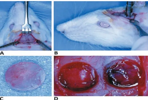
Figure 1- Rat calvaria scheme in an upper view. The 5-mm-diameter surgical bone defects were localized bilaterally at
for evaluat ion of t he r esult s. Phot om ult iplicat or
ilters of 488 nm wavelength for calcein and 594
nm wavelengt h for alizarin were used. For im age acquisit ion, a digit al cam era was coupled t o t he
epiluorescence microscope (Leica DFC 300 FX,
Leica Microsyst em s, Wet zlar, Germ any) , whoch was
connect ed t o a com put er ( Pent ium I V, Program I M 50 – Leica Microsyst em s, Wet zlar, Germ any) . For st andar dizat ion of t he analysis, only t he cent ral region of t he defect s was acquired ( Figure 5) . This
criterion was established considering the dificulty
t o visualize t he whole ext ension of defect wit h t he
alizarin ilter in 4x increase and considering the low quality of image in this magniication.
I n each slide, t wo im ages were acquired, one labeled for calcein and one for alizarin. The im ages
were saved in a digital ile. The acquired images
w er e super im posed using t he Adobe Phot oshop 7 . 0 . 1 ( Adobe Sy st em s I ncor porat ed, San Jose, CA, USA) ( Figure 6) t o illust rat e t he dynam ics of bone repair.
I n t he quant it at iv e analy sis, I m agelab 2000 so f t w ar e ( Can b o r o u g h , Wel l an d p o r t , On t ar i o, Canada) was used. Considering t hat t he t ot al area ( TA) of t he defect in t he analy zed r egions m ay correspond t o t he area occupied by calvaria bone in t he sam e region before defect preparat ion, t he TA w as det er m ined as follow s: t he defect t hat pr esen t ed t h e gr eat er h eigh of labelin g in t h e lat eral side of t he im age was est ablished as t he pat t er n. Fr om t he m ost super ior and t he m ost inferior point s of t he labels in t he lat eral board t hat present ed a great er height , t wo parallel lines were t raced t owar ds t he ot her lat eral boar d, for m ing a rect angle delim it at ing t he TA ( Figure 7a) . The program calculat ion spreadsheet resource was used t o obt ain t he TA value ( Figure 7b) , considering t hat t he calvaria bone t hickness was sim ilar t o all t he anim als included in t he experim ent . The calcein-labeled regions ( green) ( Figure 8a) and alizarin-labeled regions ( red) ( Figure 8b) were delim it ed and t he areas were calculat ed in pixels. Only t he
Figure 2- Surgical procedure. A) Surgical access. A linear incision in an anterior-posterior direction was made in the median region of calvaria and the dermoperiostic was detached. B) Trephine bur used to prepare the bone defects. C) The bone
block removed from the left side was positioned in the right side defect. D) An approximate view of the defects to be illed
Figure 3- Schematic presentation of the application of calcein (7th postoperative day) and Alizarin (30th postoperative day) application (20 mg/kg body weight, IM)
m ost int ensively labeled ar eas w er e consider ed ( Figure 8) . The TA was considered as 100% and t he percent age of each labeled area was calculat ed for each group. Five percent values were obt ained for
each group (n=5) and averaged to obtain the inal
value of m ineralized area in each analyzed period. The m ean values were analyzed st at ist ically by t he t - t est and Mann- Whit ney t est , using t he Sigm a St at soft ware, version 3.1. ( Syst at I nc., Chicago, I L, USA)
RESULTS
The obt ained im ages showed areas labeled in green and red t hat represent regions of calcium
precipitation, labeled by luorochromes in different
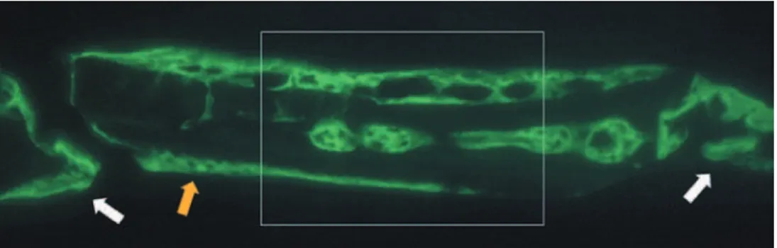
m om en t s of t issu e m in er alizat ion . Th e calcein labelin g ( gr een ) r epr esen t s t h e r egion s w h er e calcium precipit at ed from t he 7t h t o 30t h day. The alizarin labeling ( red) represent s t he regions where calcium precipit at ed from t he 30t h t o 35t h days ( Figures 9 and 10) .
I n t he Cont rol Group, t he t wo anim als present ed im ages of isolat ed sit es of m ineralizat ion in t he cent er of t he defect . I n t he ot her 3 anim als, labeling
by luorochromes was not observed. The sites of
m in eralizat ion pr esen t ed sim ilar ch aract er ist ics wit h areas int ensely labeled m ainly by calcein, in cont rast wit h areas of less int ense labeling. Alizarin show ed int ense labeling, r evealing a pat t er n of lam inar form at ion, covering t he calcein labels and locat ed m ainly bet w een t his and t he per iost eal surface ( Figure 9) .
I n t he Graft Group, all t he slices of t his group sh ow ed sign if ican t ly m or e calcein an d alizar in labeling t han t he Cont rol Group. Ot herwise, calcein ( in gr een) pr esent ed labeling sit es w it h gr eat er densit y and a t endency of bone form at ion in blocks. Alizarin ( in red) was charact erized by less int ense labeling wit h m ore lam inar form at ion, prim arily in t he periost eal surfaces, covering t he calcein labels ( Figure 10) . The im ages revealed t hat one of t he surfaces ( periost eal or dural) of t he defect s always
Figure 6- Image superposition scheme. The calcein and the alizarin images were superimposed in a computer program
to obtain the inal studied images
Figure 7- Total area (TA) standardization. A) The dashed lines delimit the area; B) Imagelab 2000® program
showed m ore ext ensive and dense labels t han t he ot her, prim arily by calcein. The alizarin labels in t hese surfaces were always locat ed ext ernally t o t hose of calcein ( Figure 10) .
Large areas with no luorochrome label or with
w eak labels ( w hich w er e not consider ed) in t he m edullar region charact erized t his group ( Figure 10) , except for one slice, where t he labels seem s t o be fused, form ing a single block in t he periost eal surface ( Figure 10E) . Ot herwise, in t hese areas,
som e spread m ineralizat ion sit es w ere observed ( Figure 10) .
The t hickest areas labeled wit h calcein seem ed t o be excavat ed wit h cavit at ions apparent ly lined by alizarin labels. I n som e slices, proj ect ions seem ed t o be ext ending from t he alizarin labels, j oining t he cavit at ions ( Figures 10C and 10D) .
Figure 9- Fluorochrome-labeled areas in the Control Group. Three of ive animals did not show labeled areas (green =
calcein ; red = alizarin)
Figure 10- Fluorochrome-labeled areas in the Graft Group. The ive animals showed strong labeled areas (green = calcein;
D I SCUSSI ON
Bone is a specialized m ineralized connect ive t i ssu e co m p o se d b y 3 3 % o f o r g a n i c m a t r i x ( prim arily collagen t ype I ) and 67% of inorganic m at rix ( hydroxyapat it e cryst als)27.
The incorporat ion of aut ogenous graft process involves t he st eps of induct ion, revascularizat ion, resorption, osteoid m atrix production, m ineralization and rem odeling4. Mineralizat ion involves num erous
com plex ev en t s t h at ar e n ot w ell u n der st ood. Aft er ost eoid m at r ix deposit ion by ost eoblast s,
the collagen ibers of the matrix presents areas of
cracks in t heir m olecules, where t he calcium ions are linked t o prot eoglycans. Under enzym at ic act ion, phosphoprot eins occupy t he place of prot eoglycans, init iat ing t he pr ecipit at ion of calcium phosphat e com plex. This way, t he form at ion of hydroxyapat it e st ar t s and t he hydr oxyapat it e cr yst als gradually occupies t he areas of cracks, expanding bet ween
the ibers and completely mineralizing the tissue10. The action mechanism of luorochromes is not
w ell under st ood. I m ages obt ained in t he Graft Group ( Figure 10) suggest t hat t hese labels link t o
calcium while it st ays available in t he ost eoid m at rix, befor e t he for m at ion of hydr oxyapat it e cr yst als.
Therefore, the luorochromes would act only on
t he t issues undergoing m ineralizat ion, not labeling already m ineralized t issues. The absence of labels over t he graft surfaces reinforces t his hypot hesis.
Acco r d i n g t o t h i s a u t o g e n o u s b o n e g r a f t incorporat ion process described, we m ay consider t he labeled areas as new bone form at ion areas, w h i ch a r e u n d e r g o i n g m i n e r a l i za t i o n o f t h e ost eoid m at r ix . Ot her w ise, car e m ust be t ak en w h en t h e m in er alizat ion ar eas ar e r elat ed t o
bone formation. Paritt, et al.18 ( 1990) concluded
t h at t h e bon e for m at ion in dex calcu lat ed fr om
the labels of tetracycline luorochrome is under
est im at ed in 10% appr ox im at ely. Som e fact or s m ay cont ribut e t o t his discrepancy: t he decline of t he ost eoblast act ivit y during t he short period of life18 may weaken tetracycline ixation in the inal
of m ineralizat ion7; t he areas of ost eoid m at rix t hat are not mineralized do not retain suficient amount
of t et racycline t o achieve t he t hreshold of det ect ion
in the epiluorescence microscopy technique18; and
m at r ix sy nt hesis and m ineralizat ion t em porar ily cease during bone rem odeling7,12. Alt hough t here
is no st udy t est ing t he discrepancy bet ween calcein and alizarin, it is suggest ed t hat t hese fact ors m ust be considered.
According t o Frost7 ( 1983) and considering t he periods of application of labels and sacriice, the
Figure 11- Percentage reached by calcein and alizarin in relation to the total area (TA). (G = Graft Group; C = Control Group). Results are expressed as mean ± standard deviation
*Differs signiicantly from the Control group labeled by
calcein (P = 0.008) (Mann-Whitney Test)
**Differs signiicantly from the Graft Group labeled by
calcein. (P = 0.008) (Mann-Whitney Test)
*** Differs signiicantly from the Graft Group labeled by
alizarin. (P = <0.001) (T Test)
Figure 12- Sum of the areas labeled by calcein and alizarin in the Graft (G) and Control (C) groups. Results are expressed as mean ± standard deviation
* Differs signiicantly from the Control Group. (P < 0.001)
calcein labels m ust represent t he areas t hat suffered m ineralizat ion bet ween t he 7t h and 30t h days, and
t h e alizar in labels, t h e ar eas t h at m in er alized bet ween t he 30t h and 35t h days, wit h t he occurrence
of areas of superposit ioning bet ween t he t wo labels. The result s of quant it at ive analysis seem s, at least pr opor t ionally, in accor dance t o m any published st u dies2 0 . 2 2. Th e qu an t it y of labels in t h e Gr aft
Group ( 32.09% ) showed a regenerat ive pot ent ial of aut ogenous bone4,28,and t he weaker labels in
t he Cont rol Group ( 2.68% ) suggest a crit ical size defect1,3,8,21,29 ( Figures 11 and 12) .
Wh en t h e lab els of t w o f lu or och r om es ar e com p ar ed , som e f act or s m u st b e con sid er ed . O’Brien, et al.15 ( 2002)classiied the luorochromes
accor d in g t o t h eir lev el of af f in it y t o calciu m . Alizar in w as consider ed as hav ing t he gr eat est
afinity, followed by xylenol, blue calcein, calcein
and oxyt et racycline. The predom inance of calcein labels ( 24.66% ) on alizarin labels ( 7.43% ) in t he
Graft Group possibly relects the time of action for
each label ( calcein = 23 days, alizarin = 5 days) . ( Figures 11 and 12) I f t he m ineralizat ion occurred in t he sam e int ensit y during t he evaluat ed period,
and considering the greater afinity of calcium to
alizarin, we should have had a m ore int ense label
for this luorochrome. As alizarin caused more
discret e labels, we can suppose t hat t he great est m ineralizat ion rat e occur s bet w een t he 7t h and
t he 30t h day, which is t he period of calcein act ion.
However, it is not possible t o det erm ine t he m om ent w h en m i n er a l i za t i o n st a r t ed a n d u n t i l w h i ch m om ent it occurred, since t his process m ight have st art ed before t he 7t h day and would cont inue aft er the 35th day, if the rats had not been sacriiced,.
The areas wit h m ore int ense bright in calcein l a b e l s a r e t h e r e g i o n s w i t h g r e a t e r ca l ci u m pr ecipit at ion. I t is supposed t hat t hese ar e t he a r ea s w h er e t h e m i n er a l i za t i o n st a r t ed . Th e lim it s regions where t he calcein labels t ouch t he alizar in labels, r epr esent s t he r egion w her e t he m ineralizat ion occurred in t he 30t h day aft er t he
graft im plant at ion. Aft er t hese considerat ions, it is possible t o clearly see in som e im ages, nuclei of cent rifugal m ineralizat ion in m any sit es of t he graft ( Figure 10A, 10B and 10D) .
The feat ures observed in t he im ages and t he m ore int ense and dense label in one of t he surfaces of t he defect ( periost eal or dural) m ight be due t o t he posit ion t hat t he bone block was placed in t he defect , wit h t he ext ernal cort ical in direct ion t o t he periost eal or dural surface. As no st andardizat ion of t his sense was m ade at t he m om ent of surgery,
and also by the dificulty imposed by the size of the
obt ained blocks, we believe t hat t he surface wit h great er label corresponds t o t he ext ernal cort ical of t he bone block ( Figure 10) . The im ages indicat e t hat t he m ineralizat ion init iat es by t he sur faces
( periost eal or dural) of t he graft s and by sit es of m ineralizat ion locat ed in t he m edullar r egions. These nuclei suggest an ost eoinduct ion act ivit y of t he aut ogenous graft .
Th e r eg ion s of alizar in lab els seem s t o b e lining t he cavit ies form ed by t he calcein labels and sending proj ect ions linking t he cavit at ions ( Figures 10C and 10D) , possibly represent ing t he form at ion of Haver s and Volk m ann channels, t hat shelt er blood capillaries. These st ruct ures t hat had t heir form at ion init iat ed in t he calcein period of act ion ( bet ween days 7 and 30) seem t o becom e m ore m at ure aft er t he 30t h day.
CON CLUSI ON
The luorochromes used in the present study
appeared adequat e t o label m ineralizat ion areas, and t he int er facial ar eas bet w een t he labels of
two luorochromes revealed important information
about t he dynam ics of bone regenerat ion in regions gr aft ed w it h au t ogen ou s t r an splan t s. How ev er, t he obt ained result s cannot be ext rapolat ed t o t he clinical condit ions, considering t he differences in ev olut ion, m et abolism and dim ensions bet w een m an and rat . More st udies are needed using m ore
than two types of luorochromes with experimental
m odels of m et abolism m or e sim ilar t o t h at of hum ans, and wit h a longer post operat ive evaluat ion period.
REFEREN CES
1- Bosch C, Melsen B, Vargervik K. I m port ance of t he crit ical- size bone defect in t est ing bone- regenerat ion m at erials. J Craniofac Surg. 1998; 9( 4) : 310- 6.
2- Brånem ark PI , Lindst röm J, Hallén O, Breine U, Jeppson PH, Ohm an A. Reconst ruct ion of t he defect ive m andible. Scand J Plast ic Reconst Surg. 1975; 9: 116- 28.
3 - Bu sch O, Solh eim E, Ban g G, Tor n es K. Gu id ed t issu e regenerat ion and local delivery of insulinlike growt h fact or I by bioerodible polyort hoest er m em branes in rat calvarial defect s. I nt J Oral Maxillofac I m plant s. 1996; 11( 4) : 498- 505.
4- Burchardt H. The biology of bone graft repair. Clin Ort hop Relat Res. 1983; 174: 28- 42.
5- Const ant ino PD, Hilt zik D, Govindaraj S, Moche J. Bone healing and bone subst it ut es. Facial Plast Surg. 2002; 18( 1) : 13- 26. 6- Est eves JC, Aranega AM, Borrasca AG, Fat t ah CM, Garcia-Júnior IR. Repair process of surgical defects illed with autogenous bone graft s in t ibiae of diabet ic rat s. J Appl Oral Sci. 2008; 16( 5) : 316- 20. 7- Frost HM. Bone hist om orphom et ry: correct ion of t he labeling “ escape er r or ”. I n: Recker PR, ed. Bone hist om or phom et r y : t echniques and int erpret at ion. Boca Rat on: CRC Press; 1983. p 133- 42.
8- Hollinger JO, Kleinschm idt JC. The crit ical size defect as an ex per im ent al t o t est bone r epair m at er ials. J Craniofac Sur g. 1990; 1( 1) : 60- 8.
9- Leit e FR, Ram alho LT. Bone regenerat ion aft er dem ineralized bone m at r ix and cast or oil (Ricinus com m unis) poly ur et hane im plant at ion. J Appl Oral Sci. 2008; 16( 2) : 122- 6.
17- Papalexiou V, Novaes AB Jr, Grisi MFM, Souza SSLS, Taba M Jr, Kajiwara JK. Inluence of implant microstructure on the dynam ics of bone healing around im m ediat e im plant s placed int o periodont ally infect ed sit es. A confocal laser scanning m icroscopic st udy. Clin Oral I m plant s Res. 2004; 15( 1) : 44- 53.
18- Paritt AM. Bone forming cells in clinical disorders. In: Hall BK, ed. Bone: a t reat ise. Caldwell: Teford Press; 1990. v. 1
19- Raghoebar GM, Schort inghuis J, Liem RS, Ruben JL, van der Wal JE, Vissink A. Does plat elet- rich plasm a prom ot e rem odeling of aut ologous bone graft s used for augm ent at ion of t he m axillary sinus loor? Clin Oral Implant Res. 2005;16(3):349-56.
2 0 - Sch leg el KA, Lan g FJ, Don at h K, Ku low JT, Wilt f an g J. Th e m on ocor t ical cr it ical size bon e def ect as an alt er n at iv e experim ent al m odel in t est ing bone subst it ut e m at erials. Oral Surg Oral Med Oral Pat hol Oral Radiol Endod. 2006; 102( 1) : 7- 13.
2006; 26( 2) : 113- 25.
26- Triplet t RG, Schow SR. Aut ologous bone graft s and endosseous im plant s: com plem ent ar y t echniques. J Oral Max illofac Sur g. 1996; 54( 11) : 486- 94.
27- Whit son SW. Bone. I n: Ten Cat e R, ed. Oral hist ology - developm ent , st ruct ure, and funct ion. 5t h ed. St Louis: Mosby; 1998. p.104- 27.
28- Wood RM, Moore DL. Graft ing of t he m axillary sinus wit h int raorally harvest ed aut ogenous bone prior t o im plant placem ent . I nt J Oral Maxillofac I m plant s. 1988; 3( 3) : 209- 14.