Beef Carcass Contamination by Shiga Toxin–Producing
Escherichia coli
Strains in an Abattoir in Brazil:
Characterization and Resistance to Antimicrobial Drugs
Everlon Cid Rigobelo,1Edilene Santo,2 and Jose Moacir Marin3
Abstract
A survey was performed to estimate the frequency of Escherichia coliand Shiga toxin–producing E. coli (STEC) in carcasses obtained from an abattoir in Brazil between February 2006 and June 2007. A total of 216 beef carcasses were sampled at three stages of the slaughter process—preevisceration, postevisceration, and postprocessing—during the rain and dry seasons, respectively. Of the carcasses sampled, 58% were preeviscerationE. coli positive, 38% were postevisceration positive, and 32% postprocessing positive. At the postprocessing stage, the isolation ofE. coliwas twice as high in the rain season. E. coliwas isolated from 85 carcasses of which only 3 (1.4%) were positive forstx-encoding genes. NoE. coliO157 serogroup isolates were detected. No antimicrobial resistance was found in nine of the isolates (10% of the total). The most frequent resistances were seen against cephalothin (78%), streptomycin (38%), nalidixic acid (36%), and tetracycline (30%). Multidrug resistance (MDR) to three or more antimicrobial agents was determined in 28 (33%)E. coliisolates. The presence of STEC and MDR strains among the isolates in the beef carcasses emphasizes the importance of proper handling to prevent carcass contamination.
Introduction
E
scherichia coliformpart of the bacterial population of cattle’s gastrointestinal tract. During beef carcass processing, the presence ofE. coli is an indicator of fecal contamination. Levels ofE. coliassociated with cattle carcasses may increase or decrease during processing ac-cording to the extent of such contamination of the living cattle, efficiency of evisceration, and hygienic practices in the abattoir (Bell, 1997). Increased consumer’s concern about beef safety started in 1983 (Rileyet al., 1983) and continued to rise in recent years due to the large number of reported outbreaks and sporadic cases of human infections with Shiga toxin–producing
E. coli (STEC) (Hussein and Bollinger, 2005). STEC strains most frequently associated with diseases in the United States and Europe are of the O157:H7 serotype (Nataro and Kaper, 1998; Caprioli et al., 2005). However, several other serotypes (O26, O103, O111, O113, and O121) are also commonly found in association with severe disease outbreaks; in some countries they are isolated more often from clinical cases than O157 (Bettelheim, 2007).
Cattle are considered primary reservoirs of both O157 and non-O157 STEC bacteria (Bettel-heim, 2000), and frequently carry STEC without showing pathological symptoms (Blanco et al., 1997). The complete list of bacterial virulence determinants required for STEC’s pathological
1Faculdade de Zootecnia de Dracena, Universidade Estadual Paulista, Ribeira˜o Preto, Brazil. 2
Faculdade de Cieˆncias Agra´rias e Veterina´rias de Jaboticabal, Universidade Estadual Paulista, Ribeira˜o Preto, Brazil. 3Departamento de Morfologia, Estomatologia e Fisiologia, FORP, Universidade de Sa˜o Paulo-Campus, Ribeira˜o Preto, Brazil.
ªMary Ann Liebert, Inc. DOI: 10.1089=fpd.2008.0138
effects is not known, although stx appears to be a key factor in pathogenesis (Acheson, 2000). Other virulence factors such as intimin (eae) and hemolysin (hlyA) are thought to enhance path-ogenicity, but are not required for strains to produce severe disease, including hemolytic uremic syndrome (HUS) (Acheson, 2000; Ca-prioli et al., 2005). STEC occurrence in feces of healthy or diarrheic cattle in Brazil has been re-ported with a high prevalence of strains and a great diversity of serotypes (Irino et al., 2005; Rigobelo et al., 2006; Aidar-Ugrinovich et al., 2007; Farahet al., 2007).
Although antimicrobial therapy is an impor-tant tool for infection treatment, antimicrobial resistance may become a major problem in veterinary medicine as a consequence of the intensive use or misuse of antimicrobial drugs (Monroe and Polk, 2000). Susceptibility patterns of indicator bacteria obtained from healthy an-imals have been suggested as good predictors of resistance situation in a bacterial population as a whole (Van den Bogaard and Stobberingh, 2000). During the processing of carcasses, fecal contamination or transfer of bacteria from the animal’s hide to the carcass can promote trans-mission of pathogenic E. coli to food supplies (Bell, 1997; Barkocy-Gallagher et al., 2001). An-timicrobial drug resistance data of fecal E. coli
strains from animals were difficult to find in the literature from Brazil, most of them showing high levels of resistance against several antimi-crobial agents from commensal E. coli isolated from diarrheic calves (Rigobelo et al., 2006) as well as from STEC strains isolated from meat (Rodolpho and Marin, 2007).
The aim of the present study was to deter-mine the incidence of E. coli on beef carcass at three stages of the slaughter process, during the rain and dry seasons; the survey also included assessment of the prevalence of virulence genes and antimicrobial drug resistance in the isolates obtained at a chosen abattoir in Brazil.
Materials and Methods
Carcass samples
Two hundred and sixteen samples from bo-vine carcasses of pasture-raised cattle were col-lected between February 2006 and June 2007, at a small abattoir in Sa˜o Paulo State (Dracena
city), in southwestern Brazil. The abattoir had a slaughtering capacity of 100 cows per day; after antemortem inspection, healthy cows selected for slaughter rested in the bairage for a day prior to slaughter. Food was withdrawn, but water was given. Carcass sampling was performed according to the abattoir processing plan and permission. Sampling of the feedlot cattle was done on five different occasions, three in the dry and two in the rainy seasons, respectively, dur-ing three stages of the carcass handldur-ing process, namely, preevisceration, postevisceration, and postprocessing. Preevisceration samples were taken immediately after complete hide removal; postevisceration samples after splitting and trimming; postprocessing samples were taken after washing carcasses hanging in the cooler. Due to an abattoir ruling of its processing plan, all samples were taken from one carcass at only one stage of the process; it was therefore not possible to take samples of the same carcass at different stages of the processing. Each sample was obtained using a Specie-Sponge (3M-Brazil) moistened with sterile 0.1% peptone water (Ba-singstoke, Oxoid, UK) in a bag. Sponges were wrung out as much as possible within the bag, withdrawn, and used to swab the rump of each carcass, near the anus, over an area of 1030 cm, delineated by a sterile metal template placed on the same half of each carcass. Each sponge was immersed in a stomacher bag with 25 mL of sterile-modified tryptone soy broth (Oxoid) supplemented with 2% novobiocin (Sigma, St. Louis, MO) (mTSB) and mixed by handling for 2 min. All samples were then taken to the labo-ratory in an ice-cooled bag and kept for 12 h at room temperature.
Bacterial isolates
considered non-O157 strains. E. coli EDL 933 strain was used as a positive control for O157 serogroup. All isolates were confirmed as being
E. coli by their biochemical analysis and sub-mitted to PCR for the detection ofstx,eae, and
ehlygenes. From each MacConkey agar plate, a loopful from a confluent bacterial growth was collected and analyzed. From each plate posi-tive forE. coli, reisolation for individual colonies was done and the isolated colonies were used for polymerase chain reaction (PCR) and sus-ceptibility testing.
PCR screening of samples
Bacterial strains grown overnight in nutrient broth (Sigma) at 378C were pelleted by centri-fugation at 12,000g for 1 min, resuspended in 200mL of sterile distilled water, and lysed by boiling for 10 min. Lysates were centrifuged as described above, and 150mL of the supernatants was used as DNA template for the PCR (Wani
et al., 2003). A total of 85 E. coli isolates were subjected to PCR;stx1,stx2, andeaegenes were detected using the primers and PCR conditions described by Chinaet al. (1996). Control refer-ence strains wereE. coliEDL 933 (O157:H7,stx1,
stx2,eae) andE. coliK12 (negative control).
Expression ofehly
Expression of enterohemolysin was deter-mined as described by Beutinet al.(1989). Plates were incubated at 378C for 24 h and observed for hemolysis after 3 h (for expression of a -hemolysin) and 24 h (forehly), respectively, and the genotype was confirmed by PCR using the primers hlyA1 and hlyA4 described by Schmidtet al.(1995). The reference strains used
in this assay wereE. coliU4-41 (positive control fora-hemolysin),E. coli32511 (STEC O157:H7) (positive control forehly), and E. coliK12 (neg-ative control).
Susceptibility testing
Antimicrobial disk susceptibility tests were performed using the disk diffusion method, as recommend by the National Committee for Clinical Laboratory Standards (NCCLS, 2002). From each E. coli–positive plate, one isolated colony was tested against 11 antimicrobial agents: ampicillin, amoxicillin=clavulanic acid, cephalothin, ceftriaxone, tetracycline, gentami-cin, streptomygentami-cin, amikagentami-cin, cotrimoxazole, nal-idixic acid, and ciprofloxacin. E. coli reference strains ATCC 25922 and ATCC 35218 were used for strain quality control.
Results
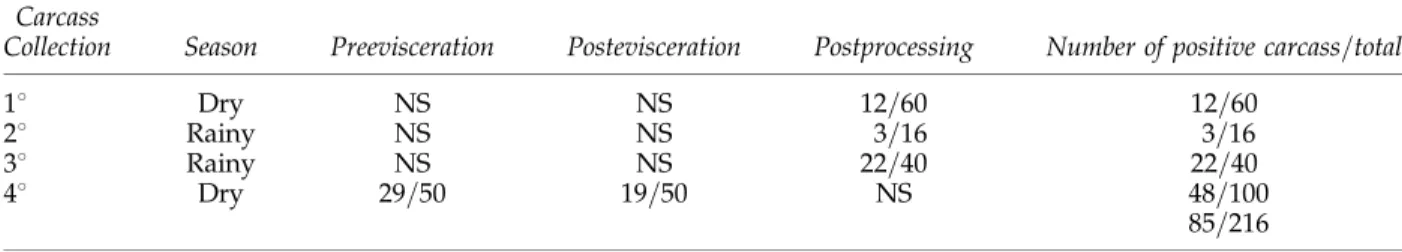
The distribution of positive carcass responses forE. coli corresponding to each sampling sea-son is shown in Table 1. At the postprocessing stage, 32% (37=116) of the carcasses sampled were E. colipositive, showing that the isolation of E. coliwas twice as high (44%, 25=56) in the rainy season when compared to the dry season (20%, 12=60). In the fourth collection, at the dry season, a reduction was detected in theE. coli– positive carcasses at the postevisceration stage (38%, 19=50) when compared with the pre-evisceration stage (55%, 29=50).
Among the 216 carcasses analyzed, only 3 (1.4%) (data not shown) were positive for stx
genes in E. coli isolates when submitted to PCR analysis. One of the three was positive for the stx1, and the other two forstx1=stx2 genes;
Table1. Distribution ofEscherichia coliIsolates from Three Different Stages of Processing of216Beef
Carcasses at an Abattoir During Two Different Climatic Seasons in Brazil, Between February2006
and June2007
Carcass
Collection Season Preevisceration Postevisceration Postprocessing Number of positive carcass=total
18 Dry NS NS 12=60 12=60
28 Rainy NS NS 3=16 3=16
38 Rainy NS NS 22=40 22=40
48 Dry 29=50 19=50 NS 48=100
85=216
all of the isolates were negative for geneseaeand
ehly.
Antibiotic resistance patterns of the isolates (n¼85) are presented in Fig. 1. Isolates pre-senting intermediary resistance were classified as resistant. The most frequent resistances were to cephalothin (78%), streptomycin (38%), nali-dixic acid (36%), and tetracycline (30%), and were less frequent to amikacin (4.0%) and
gen-tamicin (6.0%). No antimicrobial resistance was determined in nine (10%) isolates. Multidrug resistance (MDR) to three or more antimicrobial agents was shown by 28 (33%) of the E. coli
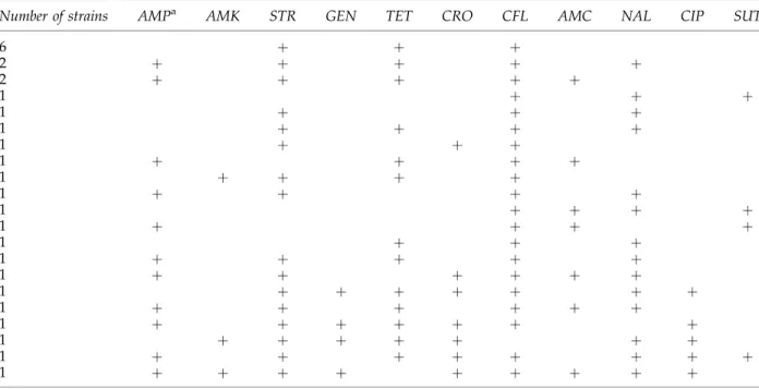
isolates, and the most common MDR pattern was to streptomycin, tetracycline, and cephalo-thin (Table 2).
Discussion
E. colistrains are part of the microbiota of the gastrointestinal tract of cattle raised for human meat consumption. Transfer of fecal material to the carcass at slaughtering leads to potential contamination of raw meat (Elderet al., 2000). In the present study, the collection dates of each material are not exactly comparable because of the size of samples and the fact that all three points were not collected on the same sam-pling days. Based on the first, second, and third sample collection, we verified that the isola-tion ofE. colifrom the carcasses examined was twice as high in the rainy season when com-pared to the dry season, confirming other reports (Barkocy-Gallagher et al., 2001; Varela-Hernandezet al., 2007). In the fourth collection at the dry season, a reduction in theE. coli
car-0 10 20 30 40 50 60 70 80 90 100
AMK GEN CIP CRO SUT AMC AMP TET NAL STR CFL
Antimicrobial drugs
Percentage
Percent of isolates
FIG. 1. Antimicrobial resistance patterns of 85 Escher-ichia colistrains of cattle from an abattoir in Brazil. AMC, amoxicillin=clavulanic acid; AMK, amikacin; AMP, am-picillin; CRO, ceftriaxone; CFL, cephalothin; CIP, cipro-floxacin; GEN, gentamicin; NAL, nalidixic acid; STR, streptomycin; TET, tetracycline; SUT, cotrimoxazole.
Table2. Resistance Patterns of28Multidrug-ResistantEscherichia coliStrains Isolated from Cattle
Carcasses During Slaughtering
Number of strains AMPa AMK STR GEN TET CRO CFL AMC NAL CIP SUT
6 þ þ þ
2 þ þ þ þ þ
2 þ þ þ þ þ
1 þ þ þ
1 þ þ þ
1 þ þ þ þ
1 þ þ þ
1 þ þ þ þ
1 þ þ þ þ
1 þ þ þ þ
1 þ þ þ þ
1 þ þ þ þ
1 þ þ þ
1 þ þ þ þ þ
1 þ þ þ þ þ þ
1 þ þ þ þ þ þ þ
1 þ þ þ þ þ þ
1 þ þ þ þ þ þ þ
1 þ þ þ þ þ þ þ
1 þ þ þ þ þ þ þ þ
1 þ þ þ þ þ þ þ þ þ
aAntimicrobial drugs.
casses contamination was detected at the post-evisceration stage in agreement with the data reported by Elderet al.(2000) for an U.S. abattoir. In the present study, a superficial contamina-tion of the carcass by STEC strains was estab-lished, but at a low level (1.4%) that agrees with reports by other authors. Rogerie et al. (2001) reported a low (1.9%) postprocessing non-O157 STEC prevalence in carcasses sampled during the summer in processing plants in France. Si-milarly, the non-O157 STEC prevalence in car-casses processed in Hong Kong has been reported as being of 1.7% (Leung et al., 2001). However, a different situation has been reported by carcasses processed in Mexico and the United States; Varela-Hernandez et al. (2007) and Ar-thur et al. (2002) reported a high level of con-tamination with non-O157 STEC, of 20.5% and 54.0%, respectively. Because a large number of variables (e.g., management practices, diets fed, animal factors, and methods of STEC detection) can influence STEC prevalence, comparisons among studies should be carefully evaluated.
Traditionally, Brazil is characterized as a beef cattle producer. Animals are fed mainly at pas-ture, considering they evolved as grazing her-bivores. However, cereal grains ferment at a faster rate than fiber, and grain can be a valuable supplement for cattle production. Several stud-ies have suggested continuous feeding of high grain diets that promotes the proliferation of
E. coli population, lowers the pH of gut con-tents, and selects for acid-resistant STEC (Diez-Gonzales and Russell, 1999; Vanselow et al., 2005), increasing the shedding of enterohemor-rhagic E. coli (EHEC O157:H7). These condi-tions, in addition to the high animal density in feedlot (traditional in the United States), make it reasonable to assume that a selection of acid-resistant E. coli serotypes in grain-fed cattle differ from those isolated from grazing-fed an-imals, in number as well as in serotypes.
The low level of STEC strains detected as contaminants in the carcass, in this study, contrasts with the high number of these strains detected in feces of healthy or diarrheic cattle in Brazil (Irino et al., 2005; Rigobelo et al., 2006; Aidar-Ugrinovichet al., 2007) what could suggest a efficient work during removal of the hide or the gastrointestinal tracts during slaughtering.
Absence or rarity of theeaegene observed in STEC isolates coincides with earlier reports in Brazil (Liraet al., 2004; Irinoet al., 2005). Absence of serotype O157:H7 in STEC isolates is not unexpected; it is extremely rare (0.6%) in Bra-zilian cattle (Irino et al., 2005), although a gold standard method as a immunomagnetic sepa-ration using beads coated with O157 antibodies was already used to select the STEC O157 strains in feces of cattle (Aidar-Ugrinovichet al., 2007). Magnetic beads labeled with antibodies to alternative non-O157 serotypes are now available commercially, but other aspects of their isolation (e.g., their optimum enrichment media and enrichment temperature) are still in development (Drysdaleet al., 2004).
It is not clear to what extent non-O157 STEC bacteria detected in cattle feces or on beef car-casses are able to cause disease in humans. Gyles
et al.(1998) proposed that all STEC bacteria could become pathogenic according to the presence or absence of favorable conditions; Bettelheim (2007) claims that non-O157 STEC’s ability to cause diseases is, in general, underestimated.
For over 4 decades, it has been a common practice in farms to use antimicrobial agents for animal disease prevention and growth promo-tion. Pathogenic organisms are clearly the anti-microbial drug’s target bacterial population on which selection pressure can be exerted. It is also important to consider that antimicrobial drugs may exert selection pressure on com-mensal bacteria (Catryet al., 2003).
Levels of antimicrobial resistance in fecal commensal bacteria can reflect the selection pressure exerted by the use of antimicrobial agents in a certain environment (Van den Bo-gaard and Stobberingh, 1999). In the present study, high levels of resistance as well as MDR were detected among the isolates agreeing with other reports from Brazil (Lira et al., 2004; Rigobelo et al., 2006), all of them showing re-sistance predominantly to cephalothin, tetracy-cline, streptomycin, and less frequently to nalidixic acid. These findings agree with data from previous studies showing that resistance is common among strains isolated from food, an-imals, and humans (Sa´enzet al., 2001; Schroeder
et al., 2002).
have resulted from the spread of mobile genetic elements. For example, the observation that nearly 62% of ampicillin-resistantE. coliisolates were also resistant to streptomycin and tetra-cycline suggests that resistance genes for these drugs are linked on plasmids, agreeing with data previously reported by Schroeder et al.
(2002) for genericE. coliand STEC strains. High levels of resistance to antimicrobial agents have also been reported for STEC strains isolated in India (Khanet al., 2002), in Europe (Moraet al., 2005), and in Palestine (Adwan and Adwan, 2004) with some strains also exhibiting MDR.
It is generally accepted that antimicrobial re-sistance in veterinary medicine could form a potential public health hazard. Indeed, the commensal gastrointestinal flora of healthy an-imals harbors a reservoir of resistance genes (Witte, 2000) that can colonize human flora through the food chain or by direct contact. Underlying resistance horizontal gene transfer to human pathogenic bacteria can result in treatment failures, which constitute a reason for concern (Van den Bogaard and Stobberingh, 2000; Catryet al., 2003).
In conclusion, we report here a small (1.4%) level of STEC strains on beef carcasses during processing at an abattoir in Brazil. Analyzed
E. coli isolates showed a high level of antimi-crobial resistance as well as MDR, again causes a reason for concern.
Acknowledgment
The authors thank FAPESP (Grant 04= 15600-4) for financial support.
Disclosure Statement
No competing financial interests exist.
References
Acheson DW. How doesEscherichia coliO157:H7 testing in meat compare with what we are seeing clinically? J. Food Prot. 2000;63:819–821.
Adwan GM, and Adwan KM. Isolation of Shiga toxigenic Escherichia colifrom raw beef in Palestine. Int. J. Food Prot. 2004;97:81–84.
Aidar-Ugrinovich L, Blanco J, Blanco M, Blanco JE, Leomil L, Dahbi G, Mora A, Onuma DL, Silveira WD, and Pestana de Castro AF. Serotypes, virulence genes, and intimin types of Shiga toxin-producingEscherichia coli (STEC) and enteropathogenic E. coli (EPEC) isolated
from calves in Sa˜o Paulo, Brazil. Int. J. Food Microbiol. 2007;115:297–306.
Arthur TM, Barkocy-Gallagher GA, Rivera-Betancourt M, and Koohmaraie M. Prevalence and characterization of Non O157 Shiga toxin-producing Escherichia coli on carcasses in commercial beef cattle processing plants. Appl. Environ. Microbiol. 2002;68:4847–4852.
Barkocy-Gallagher GA, Arthur GA, Siragusa GR, Keen JE, Elder RO, Laegreid WW, and Koohmaraie M. Genotype analyses ofEscherichia coliO157:H7 and O157 nonmotile isolates recovered from beef cattle and carcasses at processing plants in the midwestern states of the United States. Appl. Environ. Microbiol. 2001;67:3810–3818. Bell RG. Distribution and sources of microbial
contami-nation of beef carcasses. J. Appl. Microbiol. 1997;82:292– 300.
Bettelheim KA. Role of non-O157 VTEC. J. Appl. Micro-biol. 2000;88:385–505.
Bettelheim KA. The non-O157 Shiga toxigenic (Verotoxi-genic)Escherichia coli;under-rated pathogens. Clin. Mi-crobiol. Rev. 2007;33:67–87.
Beutin L, Geier D, Zimmermann S, Aleksic S, Gillespie HA, and Whittam TS. Epidemiological relatedness and clonal types of natural populations of Escherichia colistrains producing Shiga toxins in separate populations of cattle and sheep. Appl. Environ. Microbiol. 1989;63:2175–2180. Blanco M, Blanco JE, Blanco J, Mora A, Prado C, Alonso MP, Mourino M, Madrid C, Balsalobre C, and Juarez A. Distribution and characterization of faecal verotoxin producingEscherichia coli(VTEC) isolated from healthy cattle. Vet. Microbiol. 1997;54:309–319.
Caprioli A, Morabito S, Brugere H, and Oswald H. En-terohaemorrhagic Escherichia coli: emerging issues on virulence and modes of transmission. Vet. Res. 2005;36: 289–311.
Catry B, Laevens H, Devriese LA, Opsomer G, and De Kruif A. Antimicrobial resistance in livestock. J. Vet. Pharmacol. Ther. 2003;26:81–93.
China B, Pirson V, and Mainil J. Typing of bovine attaching and effacingEscherichia coliby multiplex amplification of virulence-associated genes. Appl. Environ. Microbiol. 1996;82:3462–3463.
Diez-Gonzalez F, and Russell JB. Factors affecting the ex-treme acid resistance ofEscherichia coliO157: H7. Food Microbiol. 1999;16:367–374.
Drysdale M, MacRae M, Strachan NJC, Reid TMS, and Ogden ID. The detection of non-O157E. coliin food by immunomagnectic separation. J. Appl. Microbiol. 2004; 97:220–224.
Elder RO, Keen JE, Siragusa GR, Barkocy-Gallagher GA, Koohmarale M, and Laegreid WW. Correlation of en-terohemorrhagic Escherichia coli O157 prevalence in feces, hides, and carcasses of beef cattle during process-ing. Proc. Natl. Acad. Sci. USA 2000;97:2999–3003. Farah SMSS, de Souza EM, Pedrosa FO, Irino K, da Silva
Gyles C, Johnson R, Gao A, Ziebell K, Pierard D, Aleksic S, and Boerlin P. Association of enterohemorrhagic Es-cherichia colihemolysin with serotypes of Shiga toxin producingE. coliof humans and bovine origins. Appl. Environ. Microbiol. 1998;64:4134–4141.
Hussein HS, and Bollinger LM. Prevalence of Shiga toxin-producing Escherichia coli in beef. Meat Sci. 2005;71: 676–689.
Irino K, Kato MAMF, Vaz TMI, Ramos II, Souza MAC, Cruz AS, Gomes TAT, Vieira MAM, and Guth BEC. Serotypes and virulence markers of Shiga toxin-producing Escherichia coli (STEC) isolated from dairy cattle in Sa˜o Paulo State, Brazil. Vet. Microbiol. 2005;105: 29–36.
Khan A, Das SC, Ramamurthy T, Sikdar A, Khanam J, Yamasaki S, and Nair GB. Antibiotic resistance, viru-lence gene, and molecular profiles of Shiga toxinpro-ducing Escherichia coli isolates from diverse source in Calcutta India. J. Clin. Microbiol. 2002;40:2009–2015. Koneman EW, Allen SD, Schrekenberger PC, Janda WM,
and Winn WC. Color atlas and textbook microbiology, fifth edition. Philadelphia, PA: Lippincott Company, 1997.
Leung PH, Yam WC, Ng WW, and Peiris JS. The prevalence and characterization of verotoxin-producingEscherichia coliisolated from cattle and pigs in an abattoir in Hong Kong. Epidemiol. Infect. 2001;126:173–179.
Lira WM, Macedo C, and Marin JM. The incidence of Shiga toxin-producingEscherichia coliin cattle with mastitis in Brazil. J. Appl. Microbiol. 2004;97:861–866.
Monroe S, and Polk R. Antimicrobial use and bacterial resistance. Curr. Opin. Microbiol. 2000;3:496–501. Mora A, Blanco JE, Blanco M, Alonso Pilar M, Dhabi G,
Echeita A, Gonzalez EA, Bernardez MI, and Blanco J. Antimicrobial resistance of Shiga toxin (verotoxin)-producingEscherichia coliO157:H7 and non-O157 strains isolated from humans, cattle, sheep and food in Spain. Res. Microbiol. 2005;156:793–806.
Nataro JP, and Kaper JB. Diarrheagenic Escherichia coli. Clin. Microbiol. Rev. 1998;11:142–201.
National Committee for Clinical Laboratory Standards. Performance Standards for Antimicrobial Disk Dilu-tion Susceptibility Test for Bacteria Isolated from Ani-mals Approved Standard M31A2. Wayne, MI: NCCLS, 2002.
Rigobelo EC, Gamez HJ, Marin JM, Macedo C, Ambrosin JA´ , and A´vila FA. Virulence factors of Escherichia coli isolated from diarrheic calves. Arq. Bras. Med. Vet. Zootec. 2006;58:305–310.
Riley LW, Remis RS, Helgerson SD, McGee HB, Wells GJ, Herbert RJ, Olcott ES, Johnson LM, Hargrett NT, Blake PA, and Cohen ML. Hemorrhagic colitis associated with a rareEscherichia coliserotypes. N. Engl. J. Med. 1983; 308:681–685.
Rodolpho D, and Marin JM. Isolation of Shiga toxigenic Escherichia coli from butcheries in Taquaritinga City,
State of Sa˜o Paulo, Brazil. Braz. J. Microbiol. 2007; 38:599–602.
Rogerie F, Marecat A, Gambade S, Dupond F, Beaubois P, and Lange M. Characterization of Shiga toxin producing Escherichia coli and O157 serotype E. coli isolated in France from healthy domestic cattle. Int. J. Food Micro-biol. 2001;63:217–223.
Sa´enz Y, Zarazaga M, Brinas I, Lantero M, Ruiz-Larrea F, and Torres C. Antibiotic resistance in Escherichia coli isolates obtained from animals, foods and humans in Spain. Int. J. Antimicrob. Agents 2001;18:353–358. Schmidt H, Beutin L, and Karch H. Molecular analysis
of the plasmid-encoded hemolysis of Escherichia coli O157:H7 strains, EDL 933. Infect. Immun. 1995;63:1055– 1061.
Schroeder CM, Meg J, Zhao S, DebRoy C, Torcolini J, Zhao C, McDermott PF, Wagner DD, Walker RD, and White DG. Antimicrobial resistance of Escherichia coli O26, O103, O111, O128, and O145 from animals and humans. Emerg. Infect. Dis. 2002;8:1409–1414.
Van den Bogaard AEJM, and Stobberingh EE. Antibiotic usage in animals: impact on bacterial resistance and public health. Drugs 1999;58:589–607.
Van den Bogaard AEJM, and Stobberingh EE. Epidemiol-ogy of resistance to antibiotic. Links between animals and humans. Int. J. Antimicrob. Agents 2000;14:327–335. Vanselow BA, Krause DO, and McSweeney CS. The Shiga toxin-producingEscherichia coli,their hosts, and poten-tial on-farm interventions: a review. Aust. J. Agric. Res. 2005;56:219–244.
Varela-Hernandez JJ, Cabrera-Diaz E, Cardona-Lopez MA, Ibarra-Vela´quez LM, Rangel-Villalobos H, Castillo A, Torres-Vitela MR, and Ramirez-Alvarez A. Isolation and characterization of Shiga toxin-producing Escher-ichia coliO157: H7 and non-O157 from beef carcasses at a slaughter plant in Me´xico. Int. J. Food Microbiol. 2007;113:237–241.
Wani SA, Bhat MA, Samanta I, Nishikawa Y, and Buchh AS. Isolation and characterization of Shiga toxin-producingEscherichia coli(STEC) and enteropathogenic Escherichia coli(EPEC) from calves and lambs with di-arrhea in India. Lett. Appl. Microbiol. 2003;37:121–126. Witte W. Ecological impact of antibiotic use in animals on
different complex microflora: environment. Int. J. Anti-microb. Agents 2000;14:321–325.
Address reprint requests to: