ISSN 0100-879X
CLINICAL INVESTIGATION
www.bjournal.com.br
www.bjournal.com.br
Volume 44 (1) 1-83 January 2011
Institutional Sponsors
The Brazilian Journal of Medical and Biological Research is partially financed by
Hotsite of proteomics metabolomics developped by:
Braz J Med Biol Res, January 2011, Volume 44(1) 46-52
doi: 10.1590/S0100-879X2010007500150
Three-year follow-up study of respiratory and systemic manifestations
of chronic obstructive pulmonary disease
Three-year follow-up study of respiratory
and systemic manifestations of chronic
obstructive pulmonary disease
R. Ferrari
1, S.E. Tanni
1, M.M. Faganello
2, L.M.O. Caram
1,
P.A. Lucheta
1and I. Godoy
11Disciplina de Pneumologia, Departamento de Clínica Médica, Faculdade de Medicina de Botucatu,
Universidade Estadual Paulista, Botucatu, SP, Brasil
2Departamento de Fisioterapia, UNISALESIANO, Lins, SP, Brasil
Abstract
Few studies show patient outcomes over time in chronic obstructive pulmonary disease (COPD).In the present study, we
moni-tored forced expiratory volume in the irst second (FEV1) and other manifestations of the disease over 3 years in 133 COPD
patients (69% males, age = 65 ± 9 years, FEV1 = 59 ± 25%) evaluated at baseline. During follow-up, 15 patients (11%) died and
23 (17%) dropped out. Measurements for 95 (72%) COPD patients alive after 3 years were analyzed. FEV1,body mass index
(BMI), 6-min walking distance (6MWD), Medical Research Council scale (MRC), Saint George’s Respiratory Questionnaire (SGRQ), Charlson Comorbidity index, and BODE index were obtained at baseline and after 3 years. At baseline, 17 patients
(18%) presented mild, 39% moderate, 19% severe, and 24% very severe COPD. Predicted FEV1 % and BMI did not change
over the period (P > 0.05). FEV1 in liters [1.25 (0.96-1.72) vs 1.26 (0.88-1.60) L; P < 0.001], 6MWD (438 ± 86 vs 412 ± 100 m;
P < 0.001), MRC [1 (1-2) vs 2 (1-3); P = 0.002], Charlson index [3 (3-4) vs 4 (3-5); P = 0.009], BODE index (2.2 ± 1.8 vs 2.6 ± 2.3; P = 0.008), and total SGRQ (42 ± 19 vs 44 ± 19%; P = 0.041) worsened after 3 years compared to baseline measure-ments. These data show that COPD patients deteriorated during the 3-year follow-up despite the fact that they had only minor
modiications in airway obstruction and body composition. They support the need for comprehensive patient assessment to
better identify disease progression.
Key words: Chronic obstructive pulmonary disease; Markers of disease severity; Quality of life; Exercise capacity; BODE index
Introduction
Correspondence: R. Ferrari, Departamento de Clínica Médica, Faculdade de Medicina de Botucatu, UNESP, Distrito de Rubião Junior,
s/n, 18618-970 Botucatu, SP, Brasil. Fax: +55-14-3882-2238. E-mail: [email protected]
Received August 26, 2010. Accepted December 8, 2010. Available online December 24, 2010. Published January 17, 2011.
Chronic obstructive pulmonary disease (COPD) is characterized by chronic airlow limitation and a range of pathological changes in the lungs, some signiicant extra-pulmonary effects, and important comorbidities, which may contribute to the severity of the disease (1). The recognition and quantitation of disease manifestations provide a more comprehensive assessment of disease severity and are important for the determination of prognosis (1). However, in comparison to physiological changes in pulmonary func-tion, the progression of other patient outcomes over time has been examined less frequently.
The traditional pulmonary function test assessed by the forced expiratory volume in the irst second (FEV1) is known
to correlate poorly with dyspnea (2), health status (3), and
exercise intolerance (4). Furthermore, longitudinal studies have shown that changes in the dyspnea sensation occur independently of alterations in airway obstruction assessed by FEV1 (5,6). Oga et al. (7) showed deterioration of health
status over 5 years, with changes being only weakly cor-related with changes in FEV1 (7). Another longitudinal study
showed that exercise tolerance declined over time without correlation with the rate of decline in FEV1 (8).
COPD markers after three years 47
previous investigators in international studies.
Patients and Methods
Patients
A total of 133 consecutive COPD patients with mild to very severe disease were recruited from the outpatient clinic of Hospital Universitário, Faculdade de Medicina de Botucatu. Disease severity was categorized according to the Brazilian Thoracic Society (BTS) and GOLD stages taking into consideration the values of FEV1 (% predicted)
and arterial blood gases (Table 1) (1,9).
Major inclusion criteria were clinical diagnosis of COPD according to the 2006 GOLD report and BTS (1,9): age ≥40 years, smoking history of ≥10 pack-years, and a post-bronchodilator FEV1/forced expiratory vital capacity
(FVC) ratio <70%. The following factors were grounds for exclusion: a history of asthma and/or FEV1 increase >12%
or a 200-mL post-bronchodilator test, associated restrictive disorders (tuberculosis sequelae, interstitial ibrosis), other clinically signiicant concomitant respiratory diseases (sleep apnea/hypopnea syndrome, lung cancer), noncompliance with treatment, myocardial infarction within the preceding 4 months, and unstable angina or congestive heart failure (New York Heart Association class III or IV). Patients not considered to be clinically stable (i.e.,with disease exac-erbation, hospital admissions in the preceding 6 weeks or changes in medication dose or frequency) were also excluded at baseline. All patients were optimized in terms of standard medical therapy according to GOLD and BTS guidelines (1,9). Patients with chronic hypoxemia were receiving a stable oxygen therapy dose over a period of 6 months before enrollment in the study.
All patients were evaluated at baseline and attended at the clinics every 6 months for 3 years or until death. In addition, patients or their families were contacted by telephone every 3 months to determine the occurrence of exacerbations, and to inquire about the cause of death when the patient died. Data were conirmed during visits at the clinics and by reviewing medical records. Basic causes of death were reviewed on death certiicates when available. Evaluation after 3 years was similar to the initial assessment, and was performed when patients were considered to be clinically stable. Smoking history was obtained by patient interview using standardized instruments at baseline and
smoking cessation was checked during patients’ contacts. Clinical assessment, collection of arterial blood gases (in patients with severe and very severe COPD) and spirom-etry (pre- and post-bronchodilator) were performed on the irst day, and nutritional status, the 6-min walking distance (6MWD), health status and dyspnea perception were as-sessed on an additional day. All procedures were approved by the Research Ethics Committee ofHospital Universitário, Faculdade de Medicina de Botucatu, and all patients gave written informed consent to participate in the study.
Pulmonary function, pulse oximetry, and arterial blood gas analysis
Spirometry was performed using a KOKO Spirometer before and 15 min after the inhalation of 400 µg salbutamol (Ferrari KOKO, USA), according to criteria of the American Thoracic Society (10). FEV1 valuesare reported in liters
as percentages of FVC and of the reference values (11). Pulse oximetry (SpO2) was assessed using anOnyx
oxi-meter (Model 9500 Oxioxi-meter; Nonin Medical Inc., USA) while patients were breathing room air. Blood gas mea-surements were performed only to characterize GOLD III and IV patients. Blood was drawn from the brachial artery when patients were at rest and breathing room air. Partial pressure of oxygen (PaO2) and partial pressure of carbon
dioxide (PaCO2) were assessed with a blood analyzer (Stat
Proile 5 Plus; Nova Biomedical, USA).
Nutritional assessment
Body weight and height were measured and body mass index [BMI = weight (kg)/height (m)2] was calculated. Body composition was evaluated by bioelectrical impedance (BIA 101A; RJL systems, USA) according to the guidelines of the European Society for Parenteral and Enteral Nutrition (ESPEN) (12). Fat-free mass (FFM, kg) was calculated us-ing a group-speciic regression equation developed by Kyle et al. (13). The FFM index (FFMI = FFM/height2) was also
calculated. Lean body mass depletion was deined as an FFMI <15 kg/m2 for women and <16 kg/m2 for men (14).
Health status and dyspnea
A translated version of the Saint George’s Respiratory Questionnaire (SGRQ), validated for use in Brazil, was used to evaluate patient health status (15).Clinically sig-niicant improvement was deined as a decrease of ≥4%
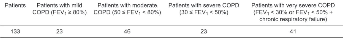
Table 1. Distribution of chronic obstructive pulmonary disease (COPD) patients according to disease severity (1,9).
Patients Patients with mild
COPD (FEV1 ≥ 80%)
Patients with moderate
COPD (50 ≤ FEV1 < 80%)
Patients with severe COPD
(30 ≤ FEV1 < 50%)
Patients with very severe COPD
(FEV1 < 30% or FEV1 < 50% +
chronic respiratory failure)
133 23 46 23 41
in total SGRQ score (16). Dyspnea was assessed using a validated Brazilian version of the Medical Research Council (MRC) scale (17).
Exercise tolerance
The 6MWD was performed according to the guidelines of the American Thoracic Society (18). Patients were instructed to walk, attempting to cover as much ground as possible within 6 min. A research assistant timed the walk, and standardized verbal encouragement was given. Following a rest of at least 30 min, each subject performed a second 6MWD in the same manner as the irst. Patient SpO2 was
monitored throughout the test. Patients who were hypoxic at baseline and patients whose SpO2 decreased to <85%
during the test were given oxygen by a physical therapist who wheeled an oxygen tank in a handcart alongside the patient. Before and after the test, data were obtained for SpO2, heart rate, respiratory rate, dyspnea sensation (Borg
scale dyspnea score), and blood pressure. The distance covered is reported in meters. There are two different refer-ences to deine the minimum clinically signiicant decline in 6MWD (19,20): a reduction ≥54 m (19) or ≥35 m (20) from baseline. In this study, we evaluated the 6MWD decline according to both deinitions.
BODE index and comorbidity evaluation
The BMI/airlow obstruction/dyspnea/exercise capacity (BODE) index was calculated using the model described by Celli et al. (21). BODE scores were categorized as class 1 (score: 0 to 2), class 2 (score: 3 to 4), class 3 (score: 5 to 6), or class 4 (score: 7 to 10) (21). Each patient’s BODE was classiied as being decreased (≥1 point), stable, or increased (≥1 point) based on its absolute change from baseline, as reported by Martinez et al. (22). Data on comorbidities were collected from the patients’ medical records and quantiied according to the Charlson index (23).
Statistical analysis
All data were analyzed using the SigmaStat 3.2 (Inc., USA) software. Means ± SD or median interquartile range (25-75%) was used depending on the data distribution. When comparing excluded patients to those who completed the study, the unpaired t-test was used for continuous variables and the Mann-Whitney test for ordinal variables. The paired t-test or Wilcoxon test was applied to compare the characteristics at baseline to those observed after 3 years. Chi-square tests were used to evaluate qualitative variables with a frequency higher than ive. The level of signiicance was set at P < 0.05.
Results
Of the 133 patients initially evaluated, 38 were excluded from the inal analyses; 15 patients died and 23 dropped out. The causes of deaths were pulmonary complications
resulting from COPD in 8 patients, cardiovascular disease in 5 patients, splenic abscess/septic shock in one patient, and colon cancer in one patient. Dropouts were due to one of the following reasons: unable to contact (N = 9), declined to participate (N = 12), inability to perform the 6MWD (N = 1), and clinical worseningof congestive heart failure (N = 1). Thus, 95 patients were monitored for 3 years. Comparisons of the dropouts and of the patients who died versus those completing the study did not show signiicant differences at baseline (Table 2).
At baseline, the mean age of the 95 patients studied (66% men) was 64 ± 9 years and smoking exposure was 54 ± 28 pack-years; 32 patients (33%) were smokers, and 8 patients (25%) stopped smoking during follow-up. As to severity, 17 patients (18%) presented mild, 39% moder-ate, 19% severe, and 24% a very severe COPD. There was no difference in the proportion of patients within each disease severity category at baseline and after 3 years (P = 0.865).
The comparison of patient characteristics at baseline and after 3 years is shown in Table 3. There was no sig-niicant change in FEV1 or FVC values after the use of a
bronchodilator, expressed as % of predicted values, or in body composition. The FEV1 value, in liters, worsened
signiicantly, with a mean rate of decline of 26 mL/year (95%CI = 15-38 mL/year; P = 0.001). The FVC value, in liters, also worsened signiicantly during the study period. There were signiicant deteriorations of SpO2, MRC, and
BODE index compared to baseline. Twenty-seven patients presented weight loss (>1 unit BMI), 52% of whom were in severe to very severe stages of the disease.
Table 2. Comparison of the characteristics of patients who died or dropped out versus those who completed the study.
Variables Patients who died or dropped out (N = 38)
Patients who completed the study (N = 95)
Gender F/M (N) 9/29 32/63
Age (years) 65 (58-74) 64 (59-71) FEV1 (%) 61 (36-77) 54 (41-73)
FVC (%) 96 (69-114) 86 (74-104)
FEV1/FVC 52 (38-62) 53 (43-63)
BMI (kg/m²) 24 (21-27) 25 (22-29)
6MWD (m) 426 ± 103 438 ± 85
MRC scale (score) 1 (1-2) 1 (1-2) SpO2 (%) 95 (93-96) 94 (92-96)
BODE index 2 (0-3) 2 (1-4)
Charlson index 3 (3-4) 3 (3-4)
Data are reported as means ± SD or as median (25-75%
inter-quartile range). F/M = female/male; FEV1 = forced expiratory
volume in the irst second (% of predicted); FVC = forced vital
capacity (% of predicted); BMI = body mass index; 6MWD: 6-min walking distance; MRC = Medical Research Council; SpO2 =
pulse oximetry; BODE index = BMI/airlow obstruction/dyspnea/ exercise capacity. There was no signiicant difference between
COPD markers after three years 49
According to the criteria set for the study, 41 patients (43%) showed stability of BODE index, 37 (39%) worsened (increase ≥1 point) and 17 (18%) presented improvement ≥1 point. Of the 37 patients who deteriorated, 25 (67%)
presented severe or very severe COPD. As to the vari-ables that compose the BODE index, in 35 patients the increase in the index was due to an increase in the MRC, the 6MWD decreased in 6 patients while 7 showed con-comitant changes.
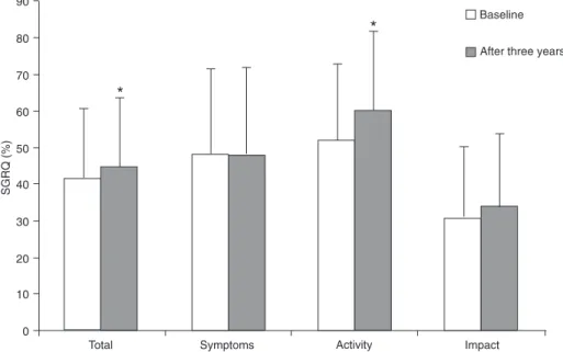
The 6MWD decreased signiicantly after 3 years. Ac-cording to the values proposed by different investigators (19,20), 31 patients (32%) presented clinically signiicant worsening (≥54 m) and in 36 patients (38%) the decrease in 6MWD was ≥35 m. The proportion of patients with a minimum clinically signiicant decline ranged from 50 to 58% among patients with severe or very severe disease. The Charlson index increased signiicantly during the study period [3 (3-4) vs 4 (3-5); P = 0.009]. In 20 patients, the in-crease was attributable only to the change in patient age. Health status presented a signiicant increase in the activity domain score (52 ± 21 vs 60 ± 22%; P < 0.001) and SGRQ total score (42 ± 19 vs 44 ± 19%; P = 0.041; Figure 1). Fifty-one percent of the patients presented clinically signiicant worsening (≥4%) in SGRQ total score while 58% showed deterioration in the activity domain.
Discussion
We have performed a 3-year follow-up of respiratory and systemic manifestations of COPD patients. Decreased exercise tolerance and increased dyspnea sensation may occur despite only minor modiications in airway obstruction and body composition. Furthermore, the multidimensional BODE index, which includes various aspects of the disease, showed signiicant worsening during the 3-year study
pe-Figure 1. Mean SGRQ (Saint George’s Respiratory Questionnaire) domains at baseline and after 3 years. *P < 0.05, baseline values compared to values observed after 3 years (paired t-test or Wilcoxon test).
Table 3. Characteristics of patients with chronic obstructive pul-monary disease followed up over a 3-year period.
Variables Initial assessment
(N = 95)
Final assessment (N = 95)
FEV1 (%) 54 (41-73) 54 (40-70)
FEV1 (L) 1.25 (0.96-1.72) 1.26 (0.88-1.60)*
FVC (%) 86 (74-104) 87 (71-106)
FVC (L) 2.6 (2.2-3.0) 2.4 (1.9-3.0)*
FEV1/FVC 52 ± 12 51 ± 10
BMI (kg/m²) 25 (22-29) 25 (22-29)
FFM (kg) 42 (38-47) 41 (37-47)
Depleted/not depleted (N) 38/57 41/54
SpO2 (%) 94 (92-96) 93 (90-95)*
MRC scale (score) 1 (1-2) 2 (1-3)*
BODE index (score) 2.2 ± 1.8 2.6 ± 2.3*
6MWD (m) 438 ± 86 412 ± 100*
Data are reported as means ± SD or as median (25-75%
inter-quartile range). FEV1 = forced expiratory volume in the irst sec
-ond (% of predicted); FVC = forced vital capacity (% of predicted);
BMI = body mass index; FFM = fat-free mass; Depleted/not de-pleted = patients with lean body mass depletion/patients without lean body mass depletion; SpO2 = pulse oximetry; MRC =
Medi-cal Research Council; BODE index = BMI/airlow obstruction/dys
-pnea/exercise capacity. *P < 0.05, inal assessment compared to
riod. Taken together, these data agree with international studies and support the concept that monitoring of patients with COPD should include other indicators in addition to those related to airway obstruction.
FEV1 is a simple and valid measurement of airlow
obstruction and is often used to stage disease severity in COPD (1). In our study, the value of FEV1, expressed
as percentage of predicted values, showed no signiicant change during the 3-year follow-up, while the value in liters decreased signiicantly. The mean rate of decline in FEV1
was 26 mL/year, and only 22 patients (23%) presented changes greater than 200 mL and 12% within a 3-year period. These results are consistent with those reported by Casanova et al. (8), who showed that FEV1 values, in
liters, declined signiicantly over 5 s, with a 23 mL/year mean rate of decline.
Despite minor modiications in airway obstruction and body composition, the exercise tolerance decreased and dyspnea sensation increased during the study period. The deterioration in exercise tolerance was clinically signiicant in 32% (≥54 m) to 38% (≥35 m) of the subjects and the proportion of patients with these declines was very similar among the different categories of disease severity. Another prospective study evaluated COPD patients with a wide range of airlow obstruction during a 5-year period. They also showed an annual decline of the 6MWD; however, in contrast to our indings, the 54-m decrease was really signiicant only in patients whose FEV1 was less than 50%
of predicted values (8).
Another symptom related to COPD severity that worsened signiicantly during follow-up was the dyspnea assessed by MRC. In 76 COPD patients monitored for 2 years, Mahler et al. (5) found that, despite improvement in lung function, dyspnea scores assessed by the Transition Dyspnea Index (TDI) increased signiicantly. Other studies have also shown uncoupling between dyspnea perception and airway obstruction over time (6,7). Lareau et al. (6) showed a signiicant decrease in mean FEV1 values (%
predicted) without change in dyspnea scores after 5 years of follow-up of 34 COPD patients. Our results agree with Oga et al. (7) who showed a signiicant worsening of dyspnea within 5 years of follow-up and a weak correlation between changes in dyspnea and the decline in pulmonary function. In summary, our data support the concept that changes in the dyspnea sensation occur independently of alterations in airway obstruction assessed by FEV1. Therefore,
as-sessment of dyspnea in the monitoring of COPD is crucial and should be undertaken independently of spirometry monitoring according to the recommendations of Brazilian and international guidelines (1,9). These data also sup-port the view of the imsup-portance of inclusion of pulmonary rehabilitation in the management of COPD patients since this treatment modality has a beneicial effect on dyspnea sensation and exercise tolerance (24).
Our results revealed no signiicant change in body
composition after the 3-year period. Prescott et al. (25) evaluated 1612 patients with COPD (736 women and 876 men) and reported that the mean weight loss was greater in patients with severe and very severe degrees compared to those with mild to moderate stages of the disease after 5 years. The proportion of patients who had a weight loss (>1 unit BMI) was 30% in patients with severe COPD. Our results showed that 27 patients (28%) presented weight loss (>1 unit BMI), and 52% of them presented severe to very severe COPD. We also showed a prevalence of malnutrition (BMI <21 kg/m2) at baseline (20%) similar to that previously reported in studies conducted in Brazil and reported in the international literature (26-28). The preva-lence of FFM depletion at baseline was on average 40%, with no difference between classes of disease severity. In the literature, the prevalence of FFM depletion varies from 18 (27,28) to 52% (29) depending on the patient group, illness severity and depletion criterion employed.
The BODE index increased signiicantly during the study period, supporting the importance of multidimensional as-sessment in monitoring COPD patients. The main variable determining the increase in BODE index was dyspnea, assessed by MRC. In our study, despite the signiicant decrease in mean 6MWD results, the proportion of patients who had an increased BODE index due to changes in the 6MWD was low, since the mean 6MWD values were greater than 365 m. These results are supported by the fact that the time spent walking in daily life in the Brazilian COPD group (56 ± 32 min/day) was markedly longer than that observed in the Austrian sample (40 ± 36 min/day) from the study of Pitta et al. (30).
In the present study, in addition to evaluating the compo-nents of the BODE index we investigated the health status of the patients. During follow-up we observed a signiicant worsening in the activity domain and SGRQ total score, in agreement with data reported by Oga et al. (7), who showed a deterioration of health status as indicated by an increased activity and impact domain, and SGRQ total score after a 5-year period. The increase in dyspnea sensation may partially explain these results since this symptom is an important determinant of health status (7,31). These results suggest that health status scores should be included as part of a comprehensive assessment to evaluate disease progression.
COPD markers after three years 51
greater than the proportion of those presenting deterioration in 6MWD and BODE index. In addition to the increase in dyspnea sensation, psychological factors may also have inluenced these results (32). However, these evaluations were not included in our design. We also recognize that comparisons with previous studies (7,8) may indicate some discrepancies due to differences in follow-up time; however, for most of the outcomes our indings were similar to those previously described. In addition, data about exacerbations during follow-up were evaluated for the entire group and have been presented in a previous publication (33). How-ever, their inluence on patient follow-up was not evaluated in the present study.
The present study has shown that, regardless of the development of airway obstruction and body composition, there is worsening of other systemic manifestations of COPD over time. These indings in Brazilian patients with COPD agree with other studies that evaluated COPD patients and support the need for a more comprehensive assessment of the patient to better identify disease progression.
Acknowledgments
Research supported by FAPESP (#04/00517-4). R. Ferrari was the recipient of a fellowship from FAPESP (#2008/52667-0).
References
1. Fabbri LM, Luppi F, Beghe B, Rabe KF. Update in chronic obstructive pulmonary disease 2005. Am J Respir Crit Care Med 2006; 173: 1056-1065.
2. Mahler DA, Weinberg DH, Wells CK, Feinstein AR. The mea-surement of dyspnea. Contents, interobserver agreement, and physiologic correlates of two new clinical indexes. Chest
1984; 85: 751-758.
3. Jones PW, Quirk FH, Baveystock CM, Littlejohns P. A
self-complete measure of health status for chronic airlow limita -tion. The St. George’s Respiratory Questionnaire. Am Rev Respir Dis 1992; 145: 1321-1327.
4. O’Donnell DE, Lam M, Webb KA. Measurement of
symp-toms, lung hyperinlation, and endurance during exercise
in chronic obstructive pulmonary disease. Am J Respir Crit Care Med 1998; 158: 1557-1565.
5. Mahler DA, Tomlinson D, Olmstead EM, Tosteson AN, O’Connor GT. Changes in dyspnea, health status, and lung function in chronic airway disease. Am J Respir Crit Care Med 1995; 151: 61-65.
6. Lareau SC, Meek PM, Press D, Anholm JD, Roos PJ. Dysp-nea in patients with chronic obstructive pulmonary disease: does dyspnea worsen longitudinally in the presence of declining lung function? Heart Lung 1999; 28: 65-73. 7. Oga T, Nishimura K, Tsukino M, Sato S, Hajiro T, Mishima
M. Longitudinal deteriorations in patient reported outcomes in patients with COPD. Respir Med 2007; 101: 146-153. 8. Casanova C, Cote CG, Marin JM, de Torres JP,
Aguirre-Jaime A, Mendez R, et al. The 6-min walking distance: long-term follow up in patients with COPD. Eur Respir J 2007; 29: 535-540.
9. Jardim JR, Oliveira JA, Nascimento O. II Consenso Brasil-eiro de DPOC. J Bras Pneumol 2004; 30: S1-S42. 10. Standardization of spirometry - 1987 update. Statement of
the American Thoracic Society. Am Rev Respir Dis 1987; 136: 1285-1298.
11. Knudson RJ, Lebowitz MD, Holberg CJ, Burrows B. Changes
in the normal maximal expiratory low-volume curve with
growth and aging. Am Rev Respir Dis 1983; 127: 725-734. 12. Kyle UG, Bosaeus I, De Lorenzo AD, Deurenberg P, Elia M,
Gomez JM, et al. Bioelectrical impedance analysis - part I: review of principles and methods. Clin Nutr 2004; 23: 1226-1243.
13. Kyle UG, Pichard C, Rochat T, Slosman DO, Fitting JW, Thiebaud D. New bioelectrical impedance formula for
patients with respiratory insuficiency: comparison to
dual-energy X-ray absorptiometry. Eur Respir J 1998; 12: 960-966.
14. Schols AM, Broekhuizen R, Weling-Scheepers CA, Wouters EF. Body composition and mortality in chronic obstructive pulmonary disease. Am J Clin Nutr 2005; 82: 53-59.
15. Sousa TC, Jardim JR, Jones P. Validation of the Saint
George Respiratory Questionnaire (SGRQ) in patients with chronic obstructive disease in Brazil. J Bras Pneumol 2000; 26: 119-125.
16. Schunemann HJ, Grifith L, Jaeschke R, Goldstein R, Stub -bing D, Guyatt GH. Evaluation of the minimal important difference for the feeling thermometer and the St. George’s
Respiratory Questionnaire in patients with chronic airlow
obstruction. J Clin Epidemiol 2003; 56: 1170-1176.
17. Kovelis D, Segretti NO, Probst VS, Lareau SC, Brunetto AF, Pitta F. Validation of the Modiied Pulmonary Functional Sta -tus and Dyspnea Questionnaire and the Medical Research Council scale for use in Brazilian patients with chronic obstructive pulmonary disease. J Bras Pneumol 2008; 34: 1008-1018.
18. ATS statement: guidelines for the six-minute walk test. Am J Respir Crit Care Med 2002; 166: 111-117.
19. Redelmeier DA, Bayoumi AM, Goldstein RS, Guyatt GH. Interpreting small differences in functional status: the Six Minute Walk test in chronic lung disease patients. Am J Respir Crit Care Med 1997; 155: 1278-1282.
20. Puhan MA, Mador MJ, Held U, Goldstein R, Guyatt GH, Schunemann HJ. Interpretation of treatment changes in 6-minute walk distance in patients with COPD. Eur Respir J
2008; 32: 637-643.
21. Celli BR, Cote CG, Marin JM, Casanova C, Montes de OM,
Mendez RA, et al. The body-mass index, airlow obstruction,
dyspnea, and exercise capacity index in chronic obstructive pulmonary disease. N Engl J Med 2004; 350: 1005-1012. 22. Martinez FJ, Han MK, Andrei AC, Wise R, Murray S, Curtis
JL, et al. Longitudinal change in the BODE index predicts mortality in severe emphysema. Am J Respir Crit Care Med
2008; 178: 491-499.
of a combined comorbidity index. J Clin Epidemiol 1994; 47: 1245-1251.
24. Ries AL, Bauldoff GS, Carlin BW, Casaburi R, Emery CF, Mahler DA, et al. Pulmonary Rehabilitation: Joint ACCP/
AACVPR Evidence-Based Clinical Practice Guidelines.
Chest 2007; 131: 4S-42S.
25. Prescott E, Almdal T, Mikkelsen KL, Tofteng CL, Vestbo J,
Lange P. Prognostic value of weight change in chronic ob-structive pulmonary disease: results from the Copenhagen City Heart Study. Eur Respir J 2002; 20: 539-544.
26. Dourado VZ, Antunes LC, Tanni SE, de Paiva SA, Padovani
CR, Godoy I. Relationship of upper-limb and thoracic muscle strength to 6-min walk distance in COPD patients. Chest
2006; 129: 551-557.
27. Schols AM, Soeters PB, Dingemans AM, Mostert R, Frantzen PJ, Wouters EF. Prevalence and characteristics of nutritional depletion in patients with stable COPD eligible for pulmonary rehabilitation. Am Rev Respir Dis 1993; 147: 1151-1156.
28. Engelen MP, Schols AM, Baken WC, Wesseling GJ, Wouters EF. Nutritional depletion in relation to respiratory and periph-eral skeletal muscle function in out-patients with COPD. Eur
Respir J 1994; 7: 1793-1797.
29. Steuten LM, Creutzberg EC, Vrijhoef HJ, Wouters EF. COPD
as a multicomponent disease: inventory of dyspnoea, under-weight, obesity and fat free mass depletion in primary care.
Prim Care Respir J 2006; 15: 84-91.
30. Pitta F, Breyer MK, Hernandes NA, Teixeira D, Sant’Anna TJ, Fontana AD, et al. Comparison of daily physical activity between COPD patients from Central Europe and South America. Respir Med 2009; 103: 421-426.
31. Schlecht NF, Schwartzman K, Bourbeau J. Dyspnea as clini-cal indicator in patients with chronic obstructive pulmonary disease. Chron Respir Dis 2005; 2: 183-191.
32. Hajiro T, Nishimura K, Tsukino M, Ikeda A, Koyama H, Izumi T. Comparison of discriminative properties among
disease-speciic questionnaires for measuring health-related quality
of life in patients with chronic obstructive pulmonary disease.
Am J Respir Crit Care Med 1998; 157: 785-790.