ULK1: A Promising Biomarker in Predicting
Poor Prognosis and Therapeutic Response in
Human Nasopharygeal Carcinoma
Miao Yun1,2‡, Hai-Yan Bai1‡, Jia-Xing Zhang1,2‡, Jian Rong3‡, Hui-Wen Weng2, Zhou-San Zheng2, Yi Xu2, Zhu-Ting Tong4, Xiao-Xia Huang1, Yi-Ji Liao1, Shi-Juan Mai1, Sheng Ye2*, Dan Xie1*
1The State Key Laboratory of Oncology in South China, Cancer Center, Sun Yat-sen University, Guangzhou, PR China,2Department of Oncology, the First Affiliated Hospital, Sun Yat-sen University, Guangzhou, Guangzhou, PR China,3Department of extracorporeal circulation, the First Affiliated Hospital, Sun Yat-sen University, Guangzhou, Guangzhou, PR China,4Department of Radiotherapy, the First Affiliated Hospital, Anhui Medical University, Hefei, PR China
‡These authors contributed equally to this work.
*[email protected](DX);[email protected](SY)
Abstract
Plenty of studies have established that dysregulation of autophagy plays an essential role in cancer progression. The autophagy-related proteins have been reported to be closely as-sociated with human cancer patients’prognosis. We explored the expression dynamics and prognostic value of autophagy-related protein ULK1 by immunochemistry (IHC) method in two independent cohorts of nasopharygeal carcinoma (NPC) cases. The X-tile program was applied to determine the optimal cut-off value in the training cohort. This derived cutoff value was then subjected to analysis the association of ULK1 expression with patients’ clini-cal characteristics and survival outcome in the validation cohort and overall cases. High ULK1 expression was closely associated with aggressive clinical feature of NPC patients. Furthermore, high expression of ULK1 was observed more frequently in therapeutic resis-tant group than that in therapeutic effective group. Our univariate and multivariate analysis also showed that higher ULK1 expression predicted inferior disease-specific survival (DSS) (P<0.05). Consequently, a new clinicopathologic prognostic model with 3 poor prognostic factors (ie, ULK1 expression, overall clinical stage and therapeutic response) could signifi-cantly stratify risk (low, intermediate and high) for DSS in NPC patients (P<0.001). These findings provide evidence that, the examination of ULK1 expression by IHC method, could serve as an effective additional tool for predicting therapeutic response and patients’ surviv-al outcome in NPC patients.
Introduction
Nasopharyngeal carcinoma (NPC), an Epstein-Barr virus (EBV)-related head and neck cancer, exhibits a high prevalence in Southeastern Asia and remains one of the leading lethal malignancies OPEN ACCESS
Citation:Yun M, Bai H-Y, Zhang J-X, Rong J, Weng H-W, Zheng Z-S, et al. (2015) ULK1: A Promising Biomarker in Predicting Poor Prognosis and Therapeutic Response in Human Nasopharygeal Carcinoma. PLoS ONE 10(2): e0117375. doi:10.1371/journal.pone.0117375
Academic Editor:Rossella Rota, Ospedale Pediatrico Bambino Gesu', ITALY
Received:July 14, 2013
Accepted:December 21, 2014
Published:February 25, 2015
Copyright:© 2015 Yun et al. This is an open access article distributed under the terms of theCreative Commons Attribution License, which permits unrestricted use, distribution, and reproduction in any medium, provided the original author and source are credited.
Funding:The project was supported by Research grants from the Nature Science Foundation of China (No. 81225018). The funders had no role in study design, data collection and analysis, decision to publish, or preparation of the manuscript.
in the Cantonese region of Southern China[1,2]. Compared with other head and neck cancers, the majority of NPC patients display adjacent region invasion as well as neck lymph nodes metastasis at the time of diagnosis[3]. Early-stage NPC is highly radiocurable. For locally advanced NPC, platinum-based induction chemotherapy (IC), followed by radiochemotherapy (RCT) or radio-therapy (RT) have become the backbone radio-therapy recently, however, the survival outcome of pa-tients with advanced stage remains poor[4,5,6]. The poor prognosis is in part related to the development of therapy resistance during conservative treatment[7,8,9]. Thus, plenty of study has focus on uncovering predictors of therapeutic response in NPC, which could identify patients who could benefit from a conservative treatment. To date, however, the promising biomarkers with great value in predicting patients’therapy efficiency still remains substantially limited.
Autophagy is an evolutionarily conserved cellular catabolic process that is characterized by the delivery of cytosolic material and organelles to lysosomes for bulk degradation[10,11]. Dys-regulation of autophagy is associated with diverse disease, including cancer, neuronal degener-ation, myopathies, and the adaptive immune response to various pathogens. Intriguingly, the function of autophagy in tumorigenesis is complicated and might have opposite consequences for tumor survival depending on certain circumstances[12]. Activation of autophagy may func-tion as a tumor suppressor by degrading defective organelles and other cellular components [13,14]. On the other hand, this pathway could also be exploited by cancer cells to generate nu-trients and energy during nutrient starvation, hypoxia, or other therapeutic stress reactions, and generally protects against cell death, facilitating adaptive survival[15,16]. Products of a se-ries of autophagy genes (ATGs) mediate and regulate various aspects of autophagy[17]. In mammals, five Atg1 homologues have been identified as uncoordinated (UNC) 51-like kinase 1 to 4 and STK36. ULK1, one of the core human autophagy-related genes, located on chromo-some 12q24.3, is a serine/threonine kinase, which promote autophagy signaling[18,19,20]. Under nutrient-rich conditions, the target of rapamycin (TOR) phosphorylates both ULK1 and ATG12, which represses ULK1 kinase activity and thus lead to autophary inhibition [21,22]. On the converse, upon nutrient deprivation, ULK1 is activated by the activated AMP activated protein kinase (AMPK) and subsequently lead to initiation of autophagy[23].
Previous studies have found that elevated ULK1 expression in human cancers is associated with poor clinical outcome, such as esophageal squamous cell carcinoma (ESCC) and breast cancer[24,25]. In light of its dual function as promising prognostic biomarker as well as novel molecular target for cancer therapy, ULK1 had attracted substantial attention over the last sev-eral years. However, till now, no study has reported the expression dynamics and prognostic value of ULK1 in NPC. Thus, we took upon the present study to explore the expression pattern of ULK1 in two independent cohorts of NPC.
Materials and Methods
Ethics statement
The study was approved by the Institute Research Medical Ethics Committee of Sun Yan-Sun University Cancer Center (Guangzhou, China) and the First Affiliated Hospital of Anhui Med-ical University (Hefei, China). No informed consent (written or verbal) was obtained for use of retrospective tissue samples from the patients in the study, since most of the patients were de-ceased and informed consent was not deemed necessary and waived by the Ethics Committee.
Patients and tissue specimens
Cancer Center (Guangzhou, China). In addition, 45 samples of normal nasopharyngeal muco-sa were used for controls. Ten fresh pairs of NPC tissue and the matched adjacent non-neoplas-tic nasopharyngeal mucosa tissue (ANT) were frozen and stored in liquid nitrogen until further use. We also collected 25 recurrent NPC and 23 liver/lung distant metastasis NPC sam-ples with the paired primary NPC tissues. In parallel, another 215 NPC patients were selected into the validation cohort. Samples from this patient cohort were obtained from the First Affili-ated Hospital of Anhui Medical University (Hefei, China) and Sun Yan-Sun University Cancer Center (Guangzhou, China) diagnosed between January 2002 and December 2006. All the cases selected were based on availability of biopsy specimens and follow-up data, no previous treatment, malignant disease or a second primary tumor; without radiotherapy, chemotherapy and surgery treatment history; Karnofsky70.
Treatment
In our RT group, the radiotherapy was administered for a total dose of 66–78Gy (2 Gy/fraction, 5 days a week). The neck received 50 Gy for lymph node-negative invaded cases and 60 to 70 Gy for lymph node-positive invaded cases. In the IC/RT group, patient received two cycles of PF regimen (floxuridine 750 mg/m2, d1–5; cisplatin 35–40 mg/m2 d1–3) chemotherapy and underwent radiotherapy thereafter at 1 week interval. In IC/RCT group, one week after com-pletion of two cycles of PF regimen, patient received radiotherapy and concurrent cisplatin (35 mg/m2 weekly) chemotherapy.
Evaluation and follow-up
The RT or RCT response was evaluated clinically for primary lesions based on fiber optic naso-pharyngeal and MRI one month after treatment according to the following criteria. Complete response (CR) was defined as the complete resolution of all assessable lesions. Partial response (PR) was defined as a reduction by 50% or more of the sum of the lesions and no progression of assessable lesions. No change (NC) was indicated by a reduction<50% or increase<25% in
tumor size. All these conditions had to last for at least 4 weeks and no appearance of new le-sions. Progressive disease (PD) was defined as an increase25% in tumor size or the appear-ance of new lesions.
The patients were followed up strictly in outpatient clinics: every 3 month for the first year and then every 6 months for the next 2 years, and finally annually. The disease-specific survival (DSS) was defined as the time from diagnosis to the date of cancer-related death or when cen-sured at the latest date if patients were still alive.
Immunohistochemistry (IHC)
Cell culture and RNA interference
The human NPC cell lines CNE1 and CNE2 were maintained in RPMI-1640 supplemented with 10% fetal bovine serum (Invitrogen, Carlsbad, CA) and 1% penicillin-streptomycin, and were cultured at 37°C in a humidified atmosphere of 5% CO2. siRNA against ULK1 (Santa Cruz Biotechnology) was transfected into NPC cells in six-well plates using Lipofectamine 2000 (Invitrogen) according to the manufacturer’s instructions.
MTT assay and apoptosis detection
Cell viability was measured by a 3-(4, 5-dimethylthiazol-2-yl)-2, 5-diphenyl tetrazolium bro-mide (MTT) assay (Sigma, St, Louis, Missouri, USA). The OD value was measured at 550 nm with a 655 nm reference filter by microplate reader (Bio Rad, Hercules, CA).
The apoptosis assay was conducted by flow cytometry using Annexin V–fluorescein isothio-cyanate (FITC) and propidium iodide (PI) stains according to manufacturers' instructions (BioVision Inc). Each sample was then subjected to analyses by flow cytometry (Beckman Coulter, cytomics FC 500, CA).
Western Blotting Analysis
Equal amounts of NPC tissue lysates were resolved by SDS-polyacrylamide gel electrophoresis (PAGE) and electrotransferred on a polyvinylidene difluoride (PVDF) membrane (Pall Corp., Port Washington, NY). The tissues were then incubated with primary rabbit monoclonal anti-bodies against ULK1 (Abcam, #ab65056, Cambridge, UK). The immunoreactive signals were detected with enhanced chemiluminescence kit (Amersham Biosciences, Uppsala, Sweden). The procedures followed were conducted in accordance with the manufacturer’s instructions.
Selection of Cutoff Score
X-tile plots were created for assessment of ULK1 expression and optimization of cutpoints based on outcome[28]. The X-tile program divided the cohorts randomly into a matched train-ing and validation set as a method for selecttrain-ing optimal cutpoints, respectively. Statistical sig-nificance was assessed by using the cutoff score derived from a training set to parse a separate validation set, using a standard log-rand method, with P values obtained from a lookup table. The X-tile plots allowed determination of an optimal cutoff value while correcting for the use of minimum P statistics by Miller-Siegmund P-value correction.
Statistical analysis
Statistical analysis was performed with SPSS software (SPSS Standard version 13.0, SPSS, Chi-cago, IL, USA). The chi-square test or Fisher’s exact test was employed to evaluate the relation-ship between ULK1 expression and clinicopathological variables. Pearson’s chi-squared test was used to analyze the relationship between ULK1 expression and therapeutic response. Sur-vival curves were plotted by the Kaplan–Meier method and compared by the log-rank test. Multivariate survival analysis was performed on all parameters that were found to be signifi-cant on univariate analysis using the Cox regression model. P-values of<0.05 were considered
Result
Patients’
Characteristics
The clinicopathologic characteristics of the NPC patients are presented inTable 1. In our train-ing cohort, a total of 141 patients were treated by RT only, where 101 patients were treated with IC plus RT. A total of 93 patients received IC prior to concurrent RCT. At the evaluation time, CR, PR, NC and PD were achieved in 258, 30, 29, and 8 patients, respectively. In the vali-dation cohort, 87 patients received radiotherapy only, where 66 patients were treated with IC plus RT. In addition, 62 patients received IC prior to concurrent RCT. At the evaluation time, CR, PR, NC and PD were achieved in 164, 20, 20 and 11 patients, respectively.
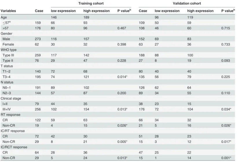
Table 1. The association of ULK1 expression with clinicopathological variables.
Training cohort Validation cohort
Variables Case low expression high expression P valueb Case low expression high expression P valueb
Age 146 189 96 119
57a
159 66 93 109 50 59
>57 176 80 96 0.467 106 46 60 0.715
Gender
Male 273 116 157 152 69 83
Female 62 30 32 0.398 63 27 36 0.733
WHO type
Type III 259 117 142 188 88 100
Type II 76 29 47 0.228 27 8 19 0.093
T status
T1–2 140 72 68 80 40 40
T3–4 195 74 121 0.014* 135 56 79 0.225
N status
N0–1 191 89 102 126 62 64
N2–3 144 57 87 0.200 89 34 55 0.110
Clinical stage
I+II 79 44 35 38 23 15
III+IV 256 102 154 0.013* 176 72 104 0.034*
RT response
CR 122 59 63 66 34 32
Non-CR 19 4 15 0.026* 21 5 16 0.026*
IC/RT response
CR 72 42 30 51 28 23
Non-CR 29 8 21 0.005* 15 3 12 0.017*
IC/RCT response
CR 64 28 36 47 25 22
Non-CR 29 5 24 0.013* 15 1 14 0.001*
a Mean age b Chi-square test
RT, radiotherapy; CR, complete response; Non-CR (including PR, partial response; NC, no change; PD, progressive disease); IC, induction chemotherapy; RCT, radiochemotherapy
*Statistically significant difference
Up-regulation of ULK1 in NPC tissues
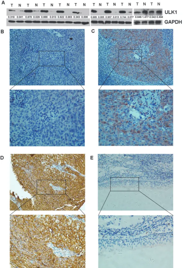
The expression of ULK1 was examined in 10 paired of NPC and ANTs. Our western blotting analysis demonstrated that the 8 out of 10 (80.0%) NPC cases also exhibited up-regulated ULK1 expression in protein level as compared to that in ANTs (Fig. 1A). For IHC staining, the positive ULK1 protein immunoreactivity was mainly detected in the cytoplasm pattern of tumor cells (Fig. 1B-D). Immunoreactivity score of ULK1 protein ranged from 0 to 12. On the other hand, among the 50 non-malignant tissues, absent or weak immunoreactivity staining of ULK1 protein was detected (Fig. 1E). In addition, we collected pairs of distant metastasis as well as pairs of recurrent NPC to compare their expression level in comparison with the prima-ry NPC respectively. As shown inS1 Fig., in both distant metastasis and recurrent NPC, the ex-pression level of ULK1 was much higher (P<0.05).
Selection of Cutoff Score for High Expression of ULK1
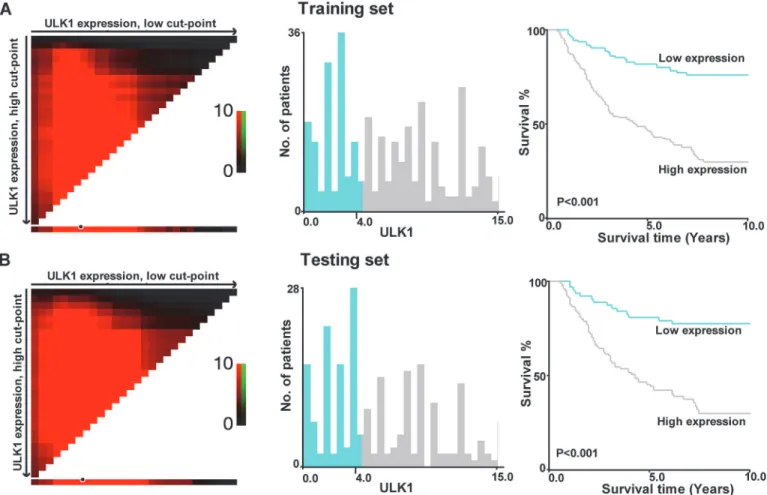
To develop a reasonable cutoff score and avoid the problems of multiple cutpoint selection, the X-tile program was employed to determine cutoff score for ULK1 expression. According to the X-tile plots, we dichotomized the training cohort into low (IHC score4) and high (IHC score>4) expression subgroups based on a cut-point of more than IHC score 4 for high ULK1
expression, which achieved high statistical significance (Fig. 2). In the training cohort, high expression of ULK1 was observed in 189/335 (56.4%) of NPC samples, and in 7/45 (15.6%) normal nasopharyngeal mucosal tissues (P<0.001); in the validation cohort, high expression
of ULK1 was observed in 119/215 (55.3%) of NPC samples according this generated cut-off point.
Correlation of ULK1 expression with clinicopathologic variables
Table 1summarized the detailed information about the rates of high ULK1 expression with re-spect to several standard clinicopathological features in these two cohorts. A statistically signifi-cant correlation between ULK1 expression with T status, and overall clinical stage was
observed in training cohort (P<0.05). In validation cohort, high ULK1 expression was
ob-served to be associated with overall clinical stage (P<0.05). However, we failed to detect a
rela-tionship between ULK1 expression and other patient characteristics, including patient's age, gender, and WHO pathological type (P>0.05).
Correlation between ULK1 expression and therapeutic response
In the training and validation cohort, primary CR was achieved in 259/335 (77.3%) and 161/ 215 (74.9%) of the NPC patients, respectively. And the remaining cases were included in the Non-CR group (PR/NC/CD). Moreover, using the optimal cut-off value of more than 4 score, ULK1 expression was also the factor that showed a negative correlation with treatment re-sponse in the RT, IC/RT as well as IC/RCT groups both in the training and validation cohort, in which high expression of ULK1 was observed more frequently in Non-CR subset than in CR subset (P<0.05,Table 1).
Association between Clinicopathologic Features, ULK1 Expression, and
Patient Survival: Univariate Survival Analysis
As shown inFig. 4, Kaplan-Meier analysis showed that the DSS difference between subsets with high and low ULK1 expression was marginal in the validation and overall patients (P<0.001). To confirm the representativeness of the NPCs, we first tested well-established
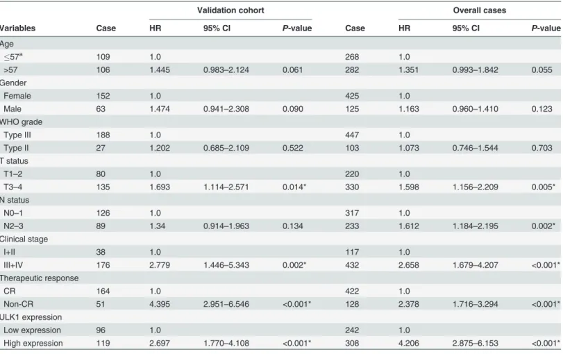
prognostic factors of patient survival. In the validation cohort, univariate analysis evaluated a significant impact of well-known clinical pathological prognostic parameters on patients’ sur-vival, such as T status, overall clinical stage, as well as therapeutic response (P<0.05,Table 2).
Assessment of patient survival also revealed that high expression of ULK1 was significantly correlated with poor DSS in the validation cohort (HR = 2.697, 95%CI (1.770–4.018); P<0.001,
Table 2).
Results in the overall cases were similar to those in the validation cohort. Patients with high ULK1 expression also displayed a significant trend toward worse survival compared with
cytoplasm pattern of the NPC cancer cells (upper panel ×200). (E) Normal nasopharyngeal mucosa tissue showed negative expression of ULK1 protein (upper panel ×200). The lower panels indicated the higher magnification (× 400) from the area of the box in B., C., D. and E., respectively.
doi:10.1371/journal.pone.0117375.g001
Fig 2. X-title plots of ULK1 expression for optimal cut-point selection in the training cohort.Patients in the training cohort were randomly divided into equal training (A) and pretesting subsets (B). The cut-point for the training set (IHC score>4, P<0.001) is demarcated by the circle (black with white border),
which was then used as the point separating low ULK1 expression (blue) from high ULK1 expression (gray) in the expression frequency of the whole set (middle panel). The right panel presents the Kaplan-Meier curve for examining the survival of cohort subsets defined by ULK1 expression4(blue line) and>4 (gray line).
patients with low ULK1 expression (HR = 4.206, 95%CI (2.875–6.153) P<0.001,Table 2). Of
the other prognostic factors, univariate analysis demonstrated that T stage, N stage, overall clinical stage, well as therapy response were also significant predictors for patients’DSS (P<0.05,Table 2).
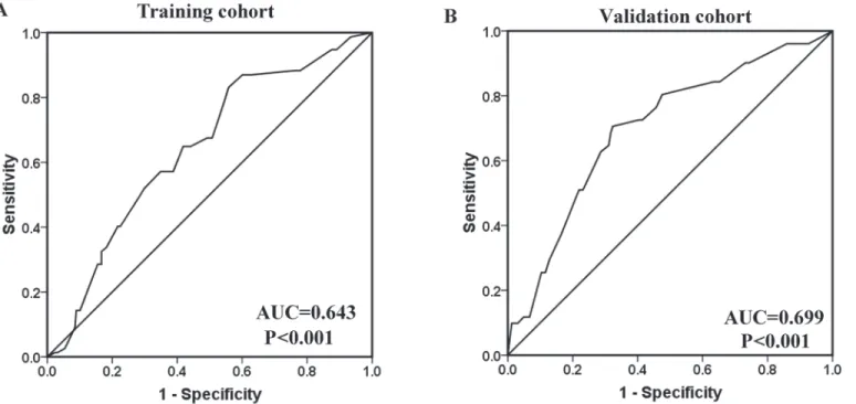
Fig 3. The predictive value of ULK1 expression regarding NPC patients’treatment response.Receiver operating characteristic curve analysis for ULK1 expression was performed to assess NPC treatment response in the training cohort (AUC = 0.643, P<0.001) (A) and in the validation cohort (AUC =
0.699, P<0.001) (B).
doi:10.1371/journal.pone.0117375.g003
Fig 4. The association between ULK1 expression and NPC patients’survival.Kaplan-Meier survival analysis of ULK1 expression for disease-specific survival in the validation cohort (A) and in the overall cases (B) (log-rank test).
Independent Prognostic Factors of NPC: Multivariate Survival Analysis
Since variables observed to have prognostic influence by univariate analysis might covariate, the expression of ULK1 as well as other clinicopathologic features that were significant in univariate analysis was examined in multivariate analysis. We found that high expression of was evaluated as an independent risk factor for adverse DSS in validation cohort (hazard ratio, 3.653; 95% con-fidence interval [CI], 1.996; 95% CI, 1.286–3.097, P = 0.002;Table 3). The same results were ob-tained in our overall cases (hazard ratio, 3.609; 95% CI, 2.442–5.332, P<0.002;Table 3). Of the
other variables, CRT response and overall clinical stage were found as independent prognostic factors for patient survival in both validation cohort and overall patients (P<0.05).
Silencing of ULK1 inhibits cellular growth and promotes apoptosis in
NPC cells
To exam biological role of ULK1 in NPC, we used ULK1 genes-specific siRNA in CNE1 and CNE2 cell lines, which effectively knocked down the expression of endogenous ULK1 protein Table 2. Univariate analysis of ULK1 expression and various clinicopathological parameters in validation and overall cases of NPC patients.
Validation cohort Overall cases
Variables Case HR 95% CI P-value Case HR 95% CI P-value
Age
57a 109 1.0 268 1.0
>57 106 1.445 0.983–2.124 0.061 282 1.351 0.993–1.842 0.055
Gender
Female 152 1.0 425 1.0
Male 63 1.474 0.941–2.308 0.090 125 1.163 0.960–1.410 0.123
WHO grade
Type III 188 1.0 447 1.0
Type II 27 1.202 0.685–2.109 0.522 103 1.073 0.746–1.544 0.703
T status
T1–2 80 1.0 220 1.0
T3–4 135 1.693 1.114–2.571 0.014* 330 1.598 1.156–2.209 0.005*
N status
N0–1 126 1.0 317 1.0
N2–3 89 1.34 0.914–1.963 0.134 233 1.612 1.184–2.195 0.002*
Clinical stage
I+II 38 1.0 117 1.0
III+IV 176 2.779 1.446–5.343 0.002* 432 2.658 1.679–4.207 <0.001*
Therapeutic response
CR 164 1.0 422 1.0
Non-CR 51 4.395 2.951–6.546 <0.001* 128 2.378 1.716–3.294 <0.001*
ULK1 expression
Low expression 96 1.0 242 1.0
High expression 119 2.697 1.770–4.108 <0.001* 308 4.206 2.875–6.153 <0.001*
a Mean age.
Cox propotional hazard regression model, enter; HR, Hazard ratio; CI, confidence interval; CR, complete response, Non-CR (including PR, partial response; NC, no change; PD, progressive disease)
*Statistically significant difference
(S2A Fig.). A MTT assay revealed that knock down of ULK1 could cause a significant reduction of cellular growth in cell lines (S2B Fig.). Furthermore, we used flow cytometry analysis to detect that the percentage of apoptosis cells were significantly increased in both the cell lines. These re-sults suggested that suppression of pro-oncogenic role of ULK1 in NPC cell lines (S2C Fig.).
New prognostic model
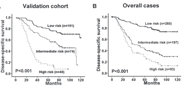
Based on the results of multivariate survival analysis, we proposed a new clinico-pathological prognostic model composed of three variables: high ULK1 expression, advanced clinical stage and poor CRT response. Patients were categorized into three risk group: low-risk (0 risk fac-tor), intermediate-risk (1 risk facfac-tor), and high risk (2–3 risk factors) groups. On Kaplan-Meier analysis, the 3 risk groups showed clear separation into 3 survival groups in the validation co-hort (P<0.001, the specificity and sensitivity were 65.1% and 71.7%,Fig. 5A). Patients in the
low- (n = 101), intermediate- (n = 74), and high- (n = 40) risk groups had a 5-year DSS of 80%, 61% and 15% respectively. For the overall cases, the new prognostic model also could differen-tiate different patients with different outcomes (5-year DSS of low- (n = 260), intermediate-(n = 197), and high- intermediate-(n = 93) risk group was 83%, 45%, and 30% respectively) (P<0.001, the
specificity and sensitivity were 70.1% and 77.2%,Fig. 5B).
Discussion
Previous studies have shown autophagy-related proteins are associated with patients prognosis in several human cancers. ULK1, a key protein in autophagy, has been suggested the pro-onco-geneic role in human ESCC and breast cancer[24,25]. To best of our knowledge, the prognostic value of ULK1 in NPC has never been studied. Thus, in the present study, we selected the NPC specimens from two independent cohorts to detect their ULK1 protein expression pattern and survival probability. Of note, to avoid predetermined arbitrary cutpoint, we applied X-tile plots to obtain the optimal IHC score for high UKL1 expression. For validation, this X-tile-derived cutoff point was subjected to analysis of the association of ULK1 expression with patient out-come and clinical characteristics in validation cohort and overall cases.
Table 3. Results of multivariate Cox proportional-hazards analysis for disease-specific survival for NPC patients.
Variable HR 95% CI P-value
Validation cohort
Tumor status (T1+2 vs. T3+4) 1.088 0.675–1.7563 0.729
Clinical stage (I+II vs. III+IV) 2.198 1.036–4.664 0.040
Therapeutic response (CR vs. Non-CR) 3.493 2.307–5.287 <0.001*
ULK1 expression (Low vs. High) 1.996 1.286–3.097 0.002*
Overall cases
T status (T1+2 vs. T3+4) 1.008 0.669–1.521 0.968
Nodel status (N0+1 vs. N2+3) 1.153 0.791–1.682 0.459
Clinical stage (I+II vs. III+IV) 1.904 1.021–3.551 0.043*
Therapeutic response (CR vs. Non-CR) 1.509 1.068–2.133 0.020*
ULK1 expression (Low vs. High) 3.609 2.442–5.332 <0.001*
Cox propotional hazard regression model, enter; HR, Hazard ratio; CI, confidence interval; CR, complete response, Non-CR (including PR, partial response; NC, no change; PD, progressive disease)
*Statistically significant difference
The role of autophagy with the context of cancer remains somewhat controversial and ap-pears to be quite divergent in the pre- and postmalignant states[29,30]. Authophagy has been well established to play an important role in the development and progression of cancer. How-ever, whether antuophagy in cancer cells results in death or cell-protection remains controver-sial[12,31]. For example, high expression of Beclin 1, one of the first identified mammalian autophagy proteins, has been observed to be associated with either favourable or inferior prog-nosis in different types of human cancers[32,33,34]. As a key autophagosomal modulating pro-tein, ULK1 was generally identified as an oncogene and has been detected to be up-regulated in human ESCC and breast cancer[24,25]. Consistent with the previous result, our study also re-vealed elevated ULK1 expression in NPC tissues. In addition, further correlation analysis dem-onstrated that high expression of ULK1 in NPC was positively correlated with tumor
aggressive and advanced clinical stage. These data, taken together, suggested that up-regulation of a major autophagy component, ULK1 might facilitate the progressive process of human maligancies, including NPC.
We know that the maliganent cells always have a high demand for nutrient and oxygen to facilitate their high proliferation rate during tumor development and progression. However, during the long term of therapy courses, the NPC tumor cells often encounter unfavourable condition, such as metabolic stress and severe hypoxia due to the decreased microvascular den-sity (MVD) [35,36]. The function of autophagy to provide energy, maintain cellular hemeosta-sis and degrade toxic cytoplasmic constituents helps to keep cancer cells alive during nutrient and oxygen deprivation and other stressfull condition. Therefore, induction of autophagy has emerged as a drug resistance mechanism that promotes cancer cell survival and evades hypoxic cell death via self-digestion. Accordingly, NPC cells with elevated ULK1 expression, interpreted as presenting an autophagy phenomenon, possess the potential to activate initiation of autop-hagy and protect them form both apoptosis and necrosis. This might provide a better under-standing of why NPC patents have a high expression of ULK1 was closely linked with poor therapeutic response. In this respect, ULK1 had been suggested as a promising therapeutic Fig 5. Comparison of disease-specific survival based on a novel clinico-pathological prognostic model (including ULK1 expression, therapy response and overall clinical stage) in the validation cohort (A) and overall cases (B) (log-rank test).
molecular target for NPC diseasing control. Furthermore, it is noteworthy that acquisition of chemo/radio-resistance under therapy may affect the biology of tumor cell, leading toward a more malignant phenotype in cancer progression. This is could explain the phenomenon that a higher frequency of elevated ULK1 protein was observed in recurrent and distant metastasis NPCs which were acquired with a more aggressive phenotype and worse survival outcome. Our finding, combined together, suggested that high ULK1 expression may favor the NPC cells a more aggressive phenotype, surviving form chemo/radiotherapy and metastastsizing to dis-tant organs, suggesting a potential molecular target for NPC therapy. Of note, more detailed investigation are required to the development of a novel strategy for the adjuvant therapy of NPC.
As for the prognostic impact of ULK1 on NPC, our study demonstrated that for the first time high ULK1 expression was closely correlated with adverse survival for NPC patients. This result is closely consistent with the previous studies, which have reported that elevated ULK1 expression is associated with poor survival in ESCC and breast cancer patients[24,25]. IHC is perhaps the most readily adaptable method to clinical practice, as it is already widely used to guide treatment of patients[24,25]. Recently, IHC staining of ULK1 was validated for the detec-tion of antophagy in several different human paraffin-embedded tumors. Thus, ULK1 is an at-tractive biomarker for autophagy that could be used in many diseases, especially cancer. Our reports suggested that the examination of ULK1 expression, as detected by IHC method, could be used as an effective additional tool to predict the therapeutic response and prognostic out-come of NPC and make the optimal clinical decisions. For example, those high-risk patients with high ULK1 expression could be offered for higher-dose radiation and adjuvant chemo-therapy earlier at the course of chemo-therapy modality. By contrast, low-risk NPC patients with lower ULK1 expression, may benefit from mild treatment without unnecessary radical therapy.
Our study also suffered from some limitations. As we know, ULK1 kinase activity affects its autophary functions, however we only analyzed the total ULK1 expression level in NPC tissues in the present study. If phosphorylated levels of ULK1 in therapeutic response or resistant NPC tissues could be examined, the result could be more convincing.
Our study demonstrated that ULK1 was an independent prognostic biomarker for thera-peutic response in NPC, and examination of ULK1 expression by IHC method might be help-ful in determining the prognosis of this tumor in clinical practice. Furthermore, inhibitor of ULK1 could be emerged as a novel promising cancer therapeutic strategy by compromising autophagic cell survival.
Supporting Information
S1 Fig. ULK1 expression is upregulated in recurrent (A) and distant metastasis (B) NPC samples.Left panel: IHC staining of representative of ULK1 expression in recurrent/distant metastasis NPC with the paired primary NPC sample. Right panel: statistical analysis revealed that a significant increase of ULK1 expression in recurrent/distant metastasis NPC relative to expression in primary NPC.
(JPG)
S2 Fig. Knockdown of endogenous ULK1 inhibited NPC cellular growth and promoted apoptosis.(A) Western blotting analysis for ULK1 expression in NPC cells transfected with ULK1 or control siRNA, respectively. (B) Silencing of ULK1 inhibited cellular growth as deter-mined by MTT assay. Each bar represented the average + SD of three independent experi-ments. (C) Silencing of ULK1 promoted more apoptosis in NPC cells as determined by flow cytometry analysis.
Author Contributions
Conceived and designed the experiments: DX SY. Performed the experiments: MY JXZ HYB ZTT. Analyzed the data: JXZ HYB JR ZTT. Contributed reagents/materials/analysis tools: JXZ HWW ZSZ YX XXH YJL SJM. Wrote the paper: MY JXZ. Supervised all the work: DX SY.
References
1. Chang ET, Adami HO (2006) The enigmatic epidemiology of nasopharyngeal carcinoma. Cancer Epi-demiol Biomarkers Prev 15: 1765–1777. PMID:17035381
2. Wei WI, Sham JS (2005) Nasopharyngeal carcinoma. Lancet 365: 2041–2054. PMID:15950718 3. Ahmad A, Stefani S (1986) Distant metastases of nasopharyngeal carcinoma: a study of 256 male
pa-tients. J Surg Oncol 33: 194–197. PMID:3773537
4. Petrovich Z, Cox JD, Roswit B, MacKintosh R, Middleton R, et al. (1982) Advanced carcinoma of the nasopharynx. A clinical study of 274 patients. Radiology 144: 905–908. PMID:6810406
5. Baujat B, Audry H, Bourhis J, Chan AT, Onat H, et al. (2006) Chemotherapy in locally advanced naso-pharyngeal carcinoma: an individual patient data meta-analysis of eight randomized trials and 1753 pa-tients. Int J Radiat Oncol Biol Phys 64: 47–56. PMID:16377415
6. Langendijk JA, Leemans CR, Buter J, Berkhof J, Slotman BJ (2004) The additional value of chemother-apy to radiotherchemother-apy in locally advanced nasopharyngeal carcinoma: a meta-analysis of the published literature. J Clin Oncol 22: 4604–4612. PMID:15542811
7. Farias TP, Dias FL, Lima RA, Kligerman J, de Sa GM, et al. (2003) Prognostic factors and outcome for nasopharyngeal carcinoma. Arch Otolaryngol Head Neck Surg 129: 794–799. PMID:12874084 8. Yamashita S, Kondo M, Hashimoto S (1985) Squamous cell carcinoma of the nasopharynx. An
analy-sis of failure patterns after radiation therapy. Acta Radiol Oncol 24: 315–320. PMID:2994387 9. Chua DT, Ma J, Sham JS, Mai HQ, Choy DT, et al. (2005) Long-term survival after cisplatbased
in-duction chemotherapy and radiotherapy for nasopharyngeal carcinoma: a pooled data analysis of two phase III trials. J Clin Oncol 23: 1118–1124. PMID:15657403
10. Kroemer G, Jaattela M (2005) Lysosomes and autophagy in cell death control. Nat Rev Cancer 5: 886–897. PMID:16239905
11. Klionsky DJ (2007) Autophagy: from phenomenology to molecular understanding in less than a de-cade. Nat Rev Mol Cell Biol 8: 931–937. PMID:17712358
12. Baehrecke EH (2005) Autophagy: dual roles in life and death? Nat Rev Mol Cell Biol 6: 505–510. PMID:15928714
13. Liu Y, Schiff M, Czymmek K, Talloczy Z, Levine B, et al. (2005) Autophagy regulates programmed cell death during the plant innate immune response. Cell 121: 567–577. PMID:15907470
14. Jin S, White E (2008) Tumor suppression by autophagy through the management of metabolic stress. Autophagy 4: 563–566. PMID:18326941
15. Morselli E, Galluzzi L, Kepp O, Vicencio JM, Criollo A, et al. (2009) Anti- and pro-tumor functions of autophagy. Biochim Biophys Acta 1793: 1524–1532. doi:10.1016/j.bbamcr.2009.01.006PMID: 19371598
16. Lum JJ, Bauer DE, Kong M, Harris MH, Li C, et al. (2005) Growth factor regulation of autophagy and cell survival in the absence of apoptosis. Cell 120: 237–248. PMID:15680329
17. Suzuki K, Ohsumi Y (2007) Molecular machinery of autophagosome formation in yeast, Saccharomy-ces cerevisiae. FEBS Lett 581: 2156–2161. PMID:17382324
18. Alers S, Loffler AS, Wesselborg S, Stork B (2012) Role of AMPK-mTOR-Ulk1/2 in the regulation of autophagy: cross talk, shortcuts, and feedbacks. Mol Cell Biol 32: 2–11. doi:10.1128/MCB.06159-11 PMID:22025673
19. Lin SY, Li TY, Liu Q, Zhang C, Li X, et al. (2012) GSK3-TIP60-ULK1 signaling pathway links growth fac-tor deprivation to autophagy. Science 336: 477–481. doi:10.1126/science.1217032PMID:22539723 20. Kraft C, Kijanska M, Kalie E, Siergiejuk E, Lee SS, et al. (2012) Binding of the Atg1/ULK1 kinase to the
ubiquitin-like protein Atg8 regulates autophagy. EMBO J 31: 3691–3703. doi:10.1038/emboj.2012. 225PMID:22885598
21. Kim J, Kundu M, Viollet B, Guan KL (2011) AMPK and mTOR regulate autophagy through direct phos-phorylation of Ulk1. Nat Cell Biol 13: 132–141. doi:10.1038/ncb2152PMID:21258367
23. Egan D, Kim J, Shaw RJ, Guan KL (2011) The autophagy initiating kinase ULK1 is regulated via oppos-ing phosphorylation by AMPK and mTOR. Autophagy 7: 643–644. PMID:21460621
24. Jiang S, Li Y, Zhu YH, Wu XQ, Tang J, et al. (2011) Intensive expression of UNC-51-like kinase 1 is a novel biomarker of poor prognosis in patients with esophageal squamous cell carcinoma. Cancer Sci 102: 1568–1575. doi:10.1111/j.1349-7006.2011.01964.xPMID:21518141
25. Pike LR, Singleton DC, Buffa F, Abramczyk O, Phadwal K, et al. (2013) Transcriptional up-regulation of ULK1 by ATF4 contributes to cancer cell survival. Biochem J 449: 389–400. doi:10.1042/BJ20120972 PMID:23078367
26. Zhang JX, Tong ZT, Yang L, Wang F, Chai HP, et al. (2013) PITX2: a promising predictive biomarker of patients' prognosis and chemoradioresistance in esophageal squamous cell carcinoma. Int J Cancer 132: 2567–2577. doi:10.1002/ijc.27930PMID:23132660
27. Zhu ZH, Sun BY, Ma Y, Shao JY, Long H, et al. (2009) Three immunomarker support vector machines-based prognostic classifiers for stage IB non-small-cell lung cancer. J Clin Oncol 27: 1091–1099. doi: 10.1200/JCO.2008.16.6991PMID:19188679
28. Camp RL, Dolled-Filhart M, Rimm DL (2004) X-tile: a new bio-informatics tool for biomarker assess-ment and outcome-based cut-point optimization. Clin Cancer Res 10: 7252–7259. PMID:15534099 29. Kondo Y, Kanzawa T, Sawaya R, Kondo S (2005) The role of autophagy in cancer development and
re-sponse to therapy. Nat Rev Cancer 5: 726–734. PMID:16148885
30. Shintani T, Klionsky DJ (2004) Autophagy in health and disease: a double-edged sword. Science 306: 990–995. PMID:15528435
31. Levine B (2007) Cell biology: autophagy and cancer. Nature 446: 745–747. PMID:17429391
32. Huang JJ, Li HR, Huang Y, Jiang WQ, Xu RH, et al. (2010) Beclin 1 expression: a predictor of prognosis in patients with extranodal natural killer T-cell lymphoma, nasal type. Autophagy 6: 777–783. PMID: 20639699
33. Zhou WH, Tang F, Xu J, Wu X, Yang SB, et al. (2012) Low expression of Beclin 1, associated with high Bcl-xL, predicts a malignant phenotype and poor prognosis of gastric cancer. Autophagy 8: 389–400. doi:10.4161/auto.18641PMID:22240664
34. Wan XB, Fan XJ, Chen MY, Xiang J, Huang PY, et al. (2010) Elevated Beclin 1 expression is correlated with HIF-1alpha in predicting poor prognosis of nasopharyngeal carcinoma. Autophagy 6: 395–404. PMID:20150769
35. Chen FH, Chiang CS, Wang CC, Tsai CS, Jung SM, et al. (2009) Radiotherapy decreases vascular density and causes hypoxia with macrophage aggregation in TRAMP-C1 prostate tumors. Clin Cancer Res 15: 1721–1729. doi:10.1158/1078-0432.CCR-08-1471PMID:19240176