Outcome of Asian Age-Related Macular Degeneration
and Polypoidal Choroidal Vasculopathy
Chui Ming Gemmy Cheung1,2,3,4*, Xiang Li2,5, Ranjana Mathur1, Shu Yen Lee1, Choi Mun Chan1, Ian Yeo1, Boon Kwang Loh1, Rachel Williams6, Edmund Yick-Mun Wong1, Doric Wong1, Tien Yin Wong1,2,3,4 1Vitreoretinal Service, Singapore National Eye Centre, Singapore, Singapore,2Singapore Eye Research Institute, Singapore, Singapore,3Department of Ophthalmology, Yong Loo Lin School of Medicine, National University of Singapore, Singapore, Singapore,4Ophthalmology Academic Clinical Program, Duke-NUS Graduate Medical School, Singapore, Singapore,5Department of Statistics and Applied Probability, National University of Singapore, Singapore, Singapore,6Worldwide Epidemiology, R&D Projects, Clinical Platforms & Sciences, GlaxoSmithKline, Pennsylvania, United States of America
Abstract
Objective:To study the treatment patterns and visual outcome over one year in Asian patients with choroidal neovascular membrane secondary to age-related macular degeneration (AMD-CNV) and polypoidal choroidal vasculopathy (PCV).
Design:Prospective cohort, non-interventional study.
Methods: 132 treatment-naı¨ve patients who received treatment for AMD-CNV and PCV were included. All patients underwent standardized examination procedures including retinal imaging at baseline and follow-up. AMD-CNV and PCV were defined on fundus fluorescein angiography and indocyanine green angiography at baseline. Patients were treated according to standard of care.We report the visual acuity (VA) and optical coherence tomography (OCT) measurements at baseline, month 3 and month 12 The factors influencing month 12 outcomes were analyzed.
Main Outcome Measure:Type of treatment, number of Anti-vascular endothelial growth factor (VEGF) treatments, visual outcome over one year.
Results:Anti-VEGF monotherapy was the initial treatment in 89.1% of AMD-CNV, but only 15.1% of PCV. The mean number of anti-VEGF injections up to month 12 was 3.97 (4.51 AMD-CNV, 3.43 PCV, p = 0.021). Baseline OCT, month 3 OCT and month 3 VA were significant in determining continuation of treatment after month 3. At month12, mean VA improved from 0.82 (,20/132) at baseline to 0.68 (,20/96) at month 12 (mean gain 6.5 ETDRS letters, p = 0.002). 34.2% of eyes (38/113
eyes) gained$15 ETDRS letters and 14.4% (16/113 eyes) lost$15 ETDRS letters. There were no significant differences in visual outcome between AMD-CNV and PCV (p = 0.51). Factors predictive of month 12 visual outcome were baseline VA, baseline OCT central macular thickness, month 3 VA and age.
Conclusions: There is significant variation in treatment patterns in Asian eyes with exudative maculopathy. There is significant visual improvement in all treatment groups at one year. These data highlight the need for high quality clinical trial data to provide evidence-based management of Asian AMD.
Citation:Cheung CMG, Li X, Mathur R, Lee SY, Chan CM, et al. (2014) A Prospective Study of Treatment Patterns and 1-Year Outcome of Asian Age-Related Macular Degeneration and Polypoidal Choroidal Vasculopathy. PLoS ONE 9(6): e101057. doi:10.1371/journal.pone.0101057
Editor:Pedro Gonzalez, Duke University, United States of America
ReceivedDecember 29, 2013;AcceptedJune 2, 2014;PublishedJune 30, 2014
Copyright:ß2014 Cheung et al. This is an open-access article distributed under the terms of the Creative Commons Attribution License, which permits unrestricted use, distribution, and reproduction in any medium, provided the original author and source are credited.
Funding:This study was supported by National Medical Research Council grant: NMRC/NIG/1003/2009; GlaxoSimthKline Research and Development Protocol WEUSKOP5855. The funders had no role in study design, data collection and analysis, decision to publish, or preparation of the manuscript.
Competing Interests:REW is employed by and holds stock in GlaxoSmithKline. The remaining authors have no competing interest. The authors confirm that this does not alter their adherence to PLOS ONE policies on sharing data and materials.
* Email: [email protected]
Introduction
Age-related macular degeneration (AMD) is one of the major causes of blindness worldwide [1–4]. The efficacy of ranibizumab, bevacizumab and aflibercept, have been confirmed by landmark clinical trials and these agents are now the mainstay of treatment for exudative AMD [5–11]. However, frequent follow-up and retreatment remain challenging in clinical setting. Data from the US Medicare and several European registries have highlighted
these in the form of high treatment discontinuation rate within the first year and low mean number of injections [11–18].
government funded reimbursement in many Asian countries, potential differences in patient understanding and expectation, and uncertainties of the role of anti-VEGF mono-therapy in PCV, may all affect the pattern of therapy in an Asian setting [20–24]. Furthermore, because photodynamic therapy (PDT) has been suggested to have superior angiographic, and possibly visual outcome in the PCV subtype [25–28], PDT is recommended as preferred treatment in PCV [29], in contrast to declining usage in Western populations. Therefore, significant heterogeneity remains in the management of Asian eyes with exudative maculopathy, in terms of diagnosis, optimal treatment and outcome.
There are few prospective studies which have examined treatment patterns and outcomes in a ‘‘real life’’ setting in Asians. To address this gap, we performed a prospective observational clinical study to document the current treatment pattern and outcomes of Asian exudative maculopathy, comparing in partic-ular AMD-CNV and PCV.
Methods
Study Design and Population
The Asian AMD Phenotyping Study is a prospectively planned, cohort study approved by the SingHealth Centralized Institutional Review Board (protocol number R697/47/2009). All patients provided written informed consent to participate in this research. Specifically, the Asian AMD Phenotyping Study aimed to investigate prospectively a consecutive series of treatment-naı¨ve Asian patients with exudative maculopathy secondary to AMD-CNV or PCV [30]. Consecutive patients were recruited from the retinal clinic of the Singapore National Eye Centre. Recruitment started on March 01 2010 and is still ongoing.
Clinical Examination and Investigations
Participants received treatment according to standard of care by individual physicians, and treatment was not altered by entering into the study. Patients were followed up prospectively according to clinical need, but minimum reviews at month 1, month 3 and month 12 were mandated by the protocol to ensure minimal data collection. Additional visits were allowed if indicated by clinical needs. All patients underwent a complete standardized ophthalmic examination at baseline and follow-up. This included visual acuity, dilated fundus examination, color fundus photography and optical coherence tomography (OCT), fundus fluorescein angiography (FFA), and indocyanine angiography (ICGA), according to a standardized protocol at baseline.
Fundus photography was performed using a digital mydriatic retinal camera (TRC-50X/IMAGEnet 2000, Topcon, Tokyo, Japan). Spectral domain optical Coherence Tomography (OCT) was performed with the Cirrus OCT (Carl Zeiss Meditec, Dublin, CA) using the 5126128-volume cube setting. Central macular thickness (CMT) was recorded. Fundus angiography with fluores-cein and indocyanine green is performed using a fundus camera (TRC-50X/IMAGEnet 2000, Topcon, Tokyo, Japan) or confocal SLO (Heidelberg Retina Angiograph Spectralis; Heidelberg Engineering, Heidelberg, Germany).
Best corrected visual acuity (BCVA) was tested with Snellen chart and converted to LogMAR. Repeat fundus photography and OCT were performed at each follow-up visit and repeat fluorescein and ICGA was performed as deemed appropriate by the treating retinal specialist.
Angiographic Grading
AMD-CNV and PCV were diagnosed clinically and by FFA and ICGA. Both the primary diagnosis and treatment decision
were made by the treating clinician, all of whom are fellowship-trained retinal specialist. Presence or absence of CNV was graded using criteria from the Macular Photocoagulation Study [31]. The diagnosis of definitive PCV lesions was based on the angiographic criteria from the Japanese Study Group guidelines, which defined PCV as characteristic polypoidal lesions on ICGA [32] To reflect the ‘real-world’ scenario, no secondary level grading was used in this analysis.
Treatment Strategies
Treatment was recommended by one of the retinal specialists in the department, all of whom were fellowship-trained. However, patients decided their final treatment after considering their visual need and financial situation. Typically, anti-VEGF therapy was recommended in the presence of subretinal or intraretinal fluid or hemorrhage associated with active AMD-CNV. In cases of PCV, anti-VEGF was recommended where there was significant fluid or hemorrhage subfoveally. In addition, focal laser was recom-mended if polyps were localized extrafoveally. Where PCV lesions were extensive and not amenable to focal laser, PDT was recommended. Anti-VEGF monotherapy was used in patients who could not afford PDT for subfoveal or juxtafoveal PCV. In selected cases, individual retinal specialist might recommend anti-VEGF monotherapy if polyps were small and vision was good.
Patients were followed-up at intervals determined by their treating physician. A variety of regimens were used, including monthly with prn retreatment, treat-and –extend, and sometimes less frequent follow-up. Retreatment was based on visual acuity, clinical examination and OCT morphology, usually if there is persistent subretinal or intraretinal fluid or hemorrhage. Loading with 3 initial monthly injections was not compulsory.
Anti-VEGF treatment was given intravitreally either as ranibizumab (Lucentis, Novartis) 0.5 mg/0.05 ml or bevacizumab (Avastin, Roche) 1.25 mg/0.05 ml in pre-aliquoted syringes using local compounding pharmacy.
All focal laser treatment was performed by retinal specialists. Generally, treatment was performed with a green laser with a focal contact lens, using the following parameters: 100 to 200mm spot size, 0.15 to 0.3 millisecond duration to achieve a greyish-white burn to the active polypoidal lesions identified on ICGA. Laser may be repeated at more than one session if closure is felt to be incomplete at follow-up. Laser was not performed on the associated branching network.
PDT was performed with verteporfin (Visudyne, Novartis, Basel, Switzerland) according to the protocol of the TAP study [33]. The greatest linear dimension (GLD) was measured based on FA for CNV lesions, according to TAP protocol. For eyes with PCV, the GLD included the entire area of abnormal choroidal vasculature on the ICGA. The diameter of the laser spot size selected to be 1000mm more than the GLD. Patients were
followed-up 3-monthly after the initial PDT.
Statistical analysis
month 3 were analyzed by applying recursive partitioning tree model using R package ‘‘rpart’’ (Recursive Partitioning), to the factors of age, gender, initial treatment, OCT CMT (baseline, month 3 and change at month 3) and VA (baseline, month 3 and change at month 3). Decision tree was pruned after built to avoid overfitting.
Linear mixed-effect model was used to analyze longitudinal data of visual acuity over the first year to account for the repeated measurements made on the same eye. Both random and fixed effects of number of injections were added to control the heterogeneous effect among eyes. Stepwise model selection (forward and backward selection) was performed to investigate the effect on visual acuity at month 12, from a set of variables, including age, gender, VA at baseline, VA at month 3, change of VA at month 3, OCT CMT at baseline, OCT CMT at month 3, change of OCT CMT at month 3. Akaike information criterion (AIC) and recursive partitioning and regression tree model were used to select the important predictors for visual acuity at month 12.
Results
From March 1st2010 to July 31st2011, a total of 167 study eyes from 167 patients were included (participation rate of 77.7%). The mean age of the subjects was 69.5 years, with a slight male dominance (58.1%). The presenting visual acuity was 0.85 (,20/
132). The proportion presenting with 0.3 (,6/12) or better was
13.2%, and the proportion presenting with vision 1.0 (,6/60) or
worse was 35.3%. Sixty five eyes (38.9%) had typical AMD-CNV and 102 eyes (61.1%) had PCV. Baseline characteristics are summarized inTable 1.
Initial Treatment Pattern for All Eyes
Treatment was initiated between baseline visit and 3-month visit in one hundred and thirty-two eyes (79%). At least 1 anti-VEGF injection was received by 97 of these 132 eyes (73.5%) during the first 3 months (Table 2). Of these 97 eyes treated with anti-VEGF, 54 eyes were as monotherapy, and the remainder was in combination with laser (12 eyes) or PDT (29 eyes) or both PDT and laser (2 eyes). After the initial 3 months, only 46.2% (61 eyes) received further treatment, most with continuation of anti-VEGF monotherapy (70.5%, 43 eyes). Data at 1 year was available from 87/97 eyes that were commenced on anti-VEGF therapy. The mean number of injections was 2.46 (3 months), 3.15 (6 months) and 3.97 (12 months) (Table 3). The mean cumulative numbers of out-patient visits were 3.08, and 8.03 at month 3 and month 12 (Table 3). Among eyes treated with any anti-VEGF, 83.2% received bevacizumab, and the remaining received ranibizumab. Thirty-four out of 41 patients initially treated with PDT remained on follow-up at one year. The mean cumulative number of PDT sessions was 1.29 (range 1–3) during the first year of treatment. Of the 35 eyes that initially did not receive treatment before their month-3 visit, 10 were eventually treated due to increase in activity or patient’s change of mind, 8 patients defaulted and the remaining patients continued to be observed.
One-Year Treatment Outcomes for All Eyes
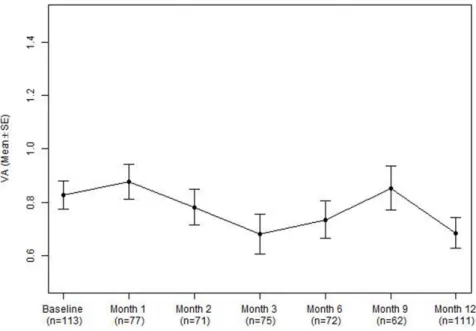
VA at month 12 was available in 113 (of 132) treated patients. The mean VA improved from 0.82 (,20/132) at baseline to 0.68
(,20/96) at month 12 (mean gain 6.5 ETDRS letters, p = 0.002, Table 4, Fig 1). 34.2% gained $15 ETDRS letters and 14.4% lost$15 ETDRS letters. VA at month 3 was available in 75 eyes (Table 4) and generally are similar to VA at month 12. At month 12, 48% of 98 eyes (including 58.5% of AMd-CNV eyes and 41.3% of PCV eyes) had activity when graded into categories of active and inactive. Central macular thickness measured using
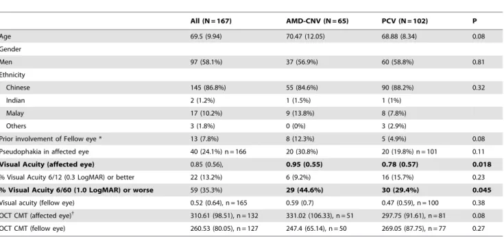
Table 1.Baseline Characteristics, comparing eyes with Age-related Macular Degeneration (AMD-CNV) and Polypoidal Choroidal Vasculopathy (PCV).
All (N = 167) AMD-CNV (N = 65) PCV (N = 102) P
Age 69.5 (9.94) 70.47 (12.05) 68.88 (8.34) 0.08
Gender
Men 97 (58.1%) 37 (56.9%) 60 (58.8%) 0.81
Ethnicity
Chinese 145 (86.8%) 55 (84.6%) 90 (88.2%) 0.32
Indian 2 (1.2%) 1 (1.5%) 1 (1%)
Malay 17 (10.2%) 9 (13.8%) 8 (7.8%)
Others 3 (1.8%) 0 (0%) 3 (2.9%)
Prior involvement of Fellow eye * 13 (7.8%) 8 (12.3%) 5 (4.9%) 0.08
Pseudophakia in affected eye 40 (24.1%) n = 166 20 (30.8%) 20 (19.8%) n = 101 0.11
Visual Acuity (affected eye) 0.85 (0.56), 0.95 (0.55) 0.78 (0.57) 0.018
% Visual Acuity 6/12 (0.3 LogMAR) or better 22 (13.2%) 6 (9.2%) 16 (15.7%) 0.23
% Visual Acuity 6/60 (1.0 LogMAR) or worse 59 (35.3%) 29 (44.6%) 30 (29.4%) 0.045
Visual acuity (fellow eye) 0.52 (0.64), n = 165 0.59 (0.7) 0.47 (0.59), n = 100 0.38
OCT CMT (affected eye){
310.61 (98.51), n = 132 331.02 (106.33), n = 51 297.75 (91.61), n = 81 0.08 OCT CMT (fellow eye) 260.53 (80.05), n = 127 247.4 (65.14), n = 50 269.05 (87.75), n = 77 0.27
*Indicating whether a scar was present in the fellow eye.
{
Exclude statistically outlier values of 18, 23, 785.
Cirrus OCT (available in 74 eyes) was significantly reduced from baseline to month 12 (300.86mm vs 264.68mm, p,0.001). The proportion of eyes with vision 0.3 (,6/12) or better increased
from 15.3% to 31.5%, and the proportion of eyes with vision 1.0 (,6/60) or worse decreased from 33.3% at baseline to 22.5%. In
the subgroup of 85 eyes treated with anti-VEGF, visual outcome was similar compared to the whole group, with mean gain of 7.0 letters at month 12. 32.9% (28/85) gained$15 letters and 12.9% (11/85) lost$15 letters.
Comparison of Treatment Patterns for AMD-CNV and PCV
Presenting vision was significantly worse in AMD-CNV than PCV (0.95 vs 0.78, p = 0.018) (Table 1) and more eyes with AMD-CNV had vision worse than 6/60 (44.6%) than PCV (29.4%, p = 0.045). However, within the group of treated eyes with at least one year follow-up (n = 113), the baseline vision was similar. There was no significant difference in age, gender, OCT thickness, proportion of previous involvement in fellow eyes and lens status. However after excluding 35 patients who did not receive treatment (29 AMD-CNV, 16 PCV), the presenting vision was similar in the remaining eyes (0.79 AMD-CNV, 0.83 PCV, p = 0.89) (Table 4).
Initial treatment choice was markedly different between AMD-CNV and PCV groups (Table 2, Figure 2). Anti-VEGF monotherapy was used in the majority of AMD-CNV (89.1%, 41/46 eyes), but only 15.1% (13/86) of PCV. Within the PCV group, the single most commonly used treatment was anti-VEGF and PDT combination (33.7%, 29/86), followed by laser monotherapy (29.1%, 25/86). Overall, 59.3% of eyes with PCV received anti-VEGF therapy alone or in combination. The mean cumulative number of anti-VEGF injections was significantly lower in PCV group (4.51 for AMD-CNV, 3.43 for PCV, p = 0.021).
Mean vision at month 12 was similar between AMD-CNV and PCV group (0.70 AMD-CNV, 0.67 PCV, p = 0.94) (Table 3).
The mean change in vision and proportion gaining and losing$
15 ETDRS letters were also similar. (Table 3)
Factors Influencing Treatment Patterns
Factors that influenced the choice of initial therapy were diagnosis (see above, AMD-CNV compared to PCV) and OCT CMT at baseline. Eyes treated with anti-VEGF monotherapy had thicker OCT CMT at baseline compared to eyes treated with combination. (367.62mm vs 291.68mm, p = 0.001). These two
factors remained significant (diagnosis, p,0.001; OCT CMT p = 0.004) after adjusting for age, gender, baseline OCT and diagnosis Baseline VA and previous fellow eye involvement were not significantly related to treatment choice.
Factors that influenced the cumulative number of injections included diagnosis, choice of initial treatment and OCT CMT measurement at month 3. PCV eyes had significantly less injections than AMD eyes (3.43 vs 4.51 at 1 year, p = 0.021) This difference remained after controlling age, gender and the OCT CMT at month 3. Eyes treated with anti-VEGF combinations had significantly lower number of injections than eyes treated with anti-VEGF monotherapy only in the PCV group (3.03 vs 4.70 at 1 year, p = 0.008). Each 100mm increase in OCT CMT at month 3
was associated with additional 0.925 injections after month 3 (p = 0.029).This remains significant after controlling for diagnosis and the baseline OCT CMT (p = 0.025).
Factors Influencing One-Year Visual Outcome
Using recursive partitioning and regression tree method, baseline VA, baseline OCT thickness and month 3 VA were significant predictors of VA at month 12. Eyes with baseline VA of
$1.0 had worse vision at month 12 compared to eyes with better baseline VA (1.13 vs 0.46, p,0.001). However they experienced larger improvement in VA (18.5 letters vs 1 letter, p = 0.002) and had a larger proportion experiencing at least 15 letters gain (54.1% vs 24.3%, p = 0.002).
In addition, we performed a longitudinal analysis, utilizing all available data at different time points over the first year. Visual Table 2.Initial Treatment Pattern from Baseline to Month 3, comparing eyes with Age-related Macular Degeneration (AMD-CNV) and Polypoidal Choroidal Vasculopathy (PCV).
All (N = 167) AMD-CNV (N = 65) PCV (N = 102) P value{
Initial treatment* 0.036
No 35 (21%) 19 (29.2%) 16 (15.7%)
Yes 132 (79%) 46 (70.8%) 86 (84.3%)
Eyes with initial treatment n = 132 n = 46 n = 86 ,0.001
Any anti-VEGF 97 (73.5%) 44 (95.7%) 51 (59.3%)
Anti-VEGF monotherapy 54 (40.9%) 41 (89.1%) 13 (15.1%)
PDT monotherapy 9 (6.8%) 2 (4.3%) 7 (8.1%)
Laser monotherapy 25 (18.9%) 0 (0%) 25 (29.1%)
Anti-VEGF & PDT 29 (22.0%) 0 (0%) 29 (33.7%)
Anti-VEGF & laser 12 (9.1%) 3 (6.5%) 9 (10.5%)
PDT & laser 1 (0.8%) 0 (0%) 1 (1.2%)
Anti-VEGF & PDT & laser 2 (1.5%) 0 (0%) 2 (2.3%)
* Treatment given before month 3 (inclusive).
{
P value to test the distribution of initial treatment between AMD and PCV, Fisher’s exact test.
acuity was weakly correlated with number of injections and age. For each additional injection received, there was on average an additional 1.7 letters improvement in VA at month 12 (p = 0.05). Every year increase in age was associated with a 0.6 letter worsening of VA (p = 0.05). OCT CMT measurement was significantly correlated with visual acuity (p,0.001). Each 100mm increase in OCT CMT was correlated to a drop of 3.62
ETDRS letters in visual acuity. There was no significant difference in Month 12 VA or change in VA whether anti-VEGF therapy was alone or in combination.
Discussion
This prospective study reports the current treatment pattern and outcome for exudative maculopathy in an Asian population over a one year period. While it has been documented that anti-VEGF monotherapy is now the most common treatment for exudative AMD in the USA and Europe [11], similar data from an Asian population are limited. In addition, it is unclear how anti-VEGF usage may be affected by issues including the high prevalence of PCV in Asian populations, the apparent superior
efficacy of PDT in causing polyp regression, and the uncertainty of the role of anti-VEGF monotherapy [20–29]. Recently, health authorities from some Asian countries have excluded PCV from the treatment label of anti-VEGF agents. Therefore, a clearer understanding of the management patterns and outcome of Asian eyes with exudative maculopathy is important in accurately planning eye care provision, research strategies, labelling of medications and re-imbursement of therapies.
Our study population can be compared to similar registry and insurance-based data, mostly from Western countries with largely white populations (Table 5). Our study population was generally younger (mean age 69.6 years), had higher male subjects (58.1%) and had lower presenting vision (44.0 ETDRS letters). Overall, 72.0% of treated eyes received at least one anti-VEGF treatment. While the widespread use of anti-VEGF is comparable to data from US [11], only 40.9% of cases received anti-VEGF as monotherapy. This pattern is remarkably different compared to reports in white people [11]. However, when stratified by diagnosis, the vast majority of AMD-CNV cases were treated with anti-VEGF monotherapy (89.1%). Therefore the lower monotherapy rate can be attributed largely to the higher Table 3.Cumulative number of Anti-vascular endothelial growth factor (anti-VEGF) injections and follow-up visits to month 12, comparing eyes with Age-related Macular Degeneration (AMD-CNV) and Polypoidal Choroidal Vasculopathy (PCV).
Month 3 Month 6 Month 12
All Anti-VEGF (N = 87)
Number of injections 2.46 (1.13) 3.15 (1.47) 3.97 (2.07)
Number of visits 3.08 (1.31) 4.11 (2.00) 8.03 (1.90)
Stratified by Diagnosis & Treatment mode AMD-CNV (N = 43)
Number of injections 2.70 (1.19) 3.63 (1.63) 4.51 (2.25)
Number of visits 2.794 (1.41) 3.86 (2.17) 5.51 (2.76)
AMD-CNV treated with Anti-VEGF monotherapy (n = 33)
Number of injections 2.73 (1.13) 3.55 (1.6) 4.45 (2.31)
Number of visits 2.52 (1.42) 3.33 (2.16) 4.91 (2.82)
AMD-CNV treated with combination therapy (n = 10)
Number of injections 2.6 (1.43) 3.9 (1.79) 4.7 (2.21)
Number of visits 3.7 (0.95) 5.6 (0.97) 7.5 (1.27)
P value* for number of injections comparing monotherapy vs combination in AMD-CNV
1.000 0.536 0.511
PCV (N = 44)
Number of injections 2.23 (1.03) 2.68 (1.14) 3.43 (1.73)
Number of visits 3.36 (1.16) 4.36 (1.81) 6.20 (2.36)
PCV treated with Anti-VEGF monotherapy (n = 10)
Number of injections 3.1 (0.57) 3.8 (1.03) 4.8 (1.99)
Number of visits 3.7 (0.95) 5.2 (1.81) 7.3 (2.67)
PCV treated with combination therapy (n = 34)
Number of injections 1.97 (1) 2.35 (0.95) 3.03 (1.45)
Number of visits 3.26 (1.21) 4.12 (1.75) 5.88 (2.2)
P value* for number of injections comparing monotherapy vs combination in PCV
0.002 0.001 0.008
P value* for number of injections between AMD-CNV and PCV
0.003 0.002 0.017
*P value based on Wilcoxon rank sum test.
Figure 1. Mean visual acuity of all treated eyes from baseline to month 12.The number of eyes with OCT available at each timepoint is indicated by n.VA Visual acuity; SE standard error.
doi:10.1371/journal.pone.0101057.g001
Table 4.Visual Acuity and Optical Coherence Tomography Central Macular Thickness at Month 3 and Month 12, stratified by Diagnosis.
All AMD-CNV PCV P
Visual acuity N = 113 N = 43 N = 70
Baseline 0.82 (0.57) 0.79 (0.53) 0.83 (0.59) 0.89
Month 3 0.68 (0.65) (n = 75) 0.76 (0.79) (n = 25) 0.64 (0.57) (n = 50) 0.94
Month 12 0.68 (0.6) 0.7 (0.64) 0.67 (0.57) 0.94
Change at month 3 20.16 (0.47) 0.03 (0.52) 20.26 (0.42) 0.15
Change at month 12 20.13 (0.51) 20.09 (0.52) 20.16 (0.51) 0.51
Gain$15 letters at Month 3 25 (33.3%) 9(36.0%) 16 (32.0%) 0.73
Gain$15 letters at Month 12 38 (34.2%) 14 (34.1%) 24 (34.3%) 0.99
Loss$15 letters at month 3 7 (9.3%) 6 (24.0%) 1 (2.0%) 0.005 Loss$15 letters at Month 12 16 (14.4%) 6 (14.6%) 10 (14.3%) 0.96 Proportion with VA 0.3 or better
Baseline 17 (15.3%) 7 (17.1%) 10 (14.3%) 0.69
Month 3 29 (38.7% 11 (44.0%) 18 (36.0%) 0.50
Month 12 35 (31.5%) 14 (34.1%) 21 (30%) 0.65
Proportion with VA 1.0 or worse
Baseline 37 (33.3%) 13 (31.7%) 24 (34.3%) 0.781
Month 3 17 (22.7%) 6 (24.0%) 11 (22.0%) 0.85
Month 12 25 (22.5%) 11 (26.8%) 14 (20%) 0.406
OCT CMT N = 76 N = 26 N = 50
Baseline 300.86 (98.29) 317.62 (94.79) 292.14 (99.88) 0.26
Month 12 264.68 (89.82) 272.81 (71.18) 260.46 (98.54) 0.16
Change at month 12 236.17 (93.99) 244.81 (79.52) 231.68 (101.17) 0.68
P value based Wilcoxon rank sum test or Chi-square test.
proportion of patients needing specific PCV treatment, of which only 15.1% received anti-VEGF monotherapy. Despite the lower proportion of anti-VEGF monotherapy, a further 44.2% of PCV eyes received anti-VEGF in combination with PDT (33.7%) or laser (10.5%). Thus, an key observation of this study is that anti-VEGF therapy constitutes a significant part of treatment for PCV eyes (59.3%), although more commonly in combination with other treatments.
The overall number of injections was low at 3.97 (range 1–9) over 12 months. Even after separating PCV eyes from AMD-CNV eyes, the mean number of injections was only 4.51 injections over 12 months in AMD-CNV eyes. However, these figures are not far off published data from USA and European registries (Table 5) [11–18]. These data highlight the challenges in translating clinical trial results into ‘‘real life’’ clinical practice. As in most countries, the utilization of anti-VEGF is affected by reimbursement structure and retreatment criteria. In addition to low retreatment number, the number of follow-up visits during the first year was also low (mean 8 visits). Although only three mandatory visits were included in the study protocol (at months 1, 3 and 12), most patients attended much more frequently due to clinical need (mean 8.03 visits). However,it is likely this lack of follow-up and under-treatment contributed significantly towards the low injec-tion numbers, as 48% of eyes still had activity on OCT at month 12, However, similar low follow-up visits (8.06 visits) had been reported from France [17], highlighting the adherence to follow-up is also a challenge in the delivery of AMD care.
We report the mean VA improved significantly by 6.5 letters, with 34.2% gaining 15 ETDRS letters and 14.4% losing $15 ETDRS letters. When outcome was analyzed by diagnosis of AMD-CNV versus PCV, there was no significant difference in mean VA change, or of the proportion gaining and losing 15 letters. Visual outcome from several European registries have been summarized in Table 5 for comparison. In addition to mean change in VA, it is also important to note that patients from the current study had generally worse presenting visual acuity than those from European registries [12–18]. Despite significant mean visual gain, the vision at 1 year remained significantly lower than those from European registries [12–18]. Lower public awareness of AMD and generally later presentation in Asian setting is likely to be a significant explanation for these findings. It is also interesting to note that 35 patients did not receive treatment initially (before their month-3 visit). The most common reasons were poor prognosis (10/19 eyes in the AMD-CNV group and 6/ 15 eyes in the PCV group), and limited activity at presentation (3/ 19 in the AMD-CNV group and 8/15 in the PCV group) reason
in the PCV group (8/15 eyes). The remaining patients defaulted their follow up. Of these 35 eyes, 10 eyes eventually received treatment from month 4 onwards.
In terms of predictive factors for visual outcome, we identified OCT measurement at baseline and VA at month 3 as significant predictors for both the need to continue treatment beyond month 3 and also vision at month 12. Each 100mm increase in OCT
CMT was correlated to a drop of 3.6 ETDRS letters in visual acuity (p,0.001). OCT CMT measurement at month 3 was also predictive of whether treatment was continued after 3 months. Each 100mm increase in OCT CMT at month 3 was associated with 0.925 additional injections after month 3 (p = 0.025). The number of injection was only weakly correlated with visual outcome at month 12. Each additional injection was associated with a further 1.7 letters gain in vision at month 12.
In our study, 61.1% of eyes had PCV. Apart from features on ICGA and worse baseline VA in AMD-CNV (0.95 in AMD-CNV vs 0.78 in PCV, p = 0.018), there were no significant differences in baseline characteristics in terms of age, gender, fellow eye involvement or lens status between AMD-CNV and PCV eyes. These results highlight the importance to screen for PCV with ICGA in an Asian population. Our results showed several major findings regarding PCV treatment in our centre. First, a variety of treatment modalities are currently employed in treating PCV. Second, anti-VEGF use was common, but appears to require less injection number when used as combination therapy. Third, the visual outcome was similar in PCV and AMD-CNV eyes in the anti-VEGF era. Therefore, the role of anti-VEGF therapy in PCV, as monotherapy or as adjunct to occlusive therapies, should be further studied.
There are limitations to this study. The observational design aims to capture the current treatment pattern of exudative AMD in an Asian setting in Singapore. Variations in treatment preference between individual clinicians, between countries and regions are to be expected. Variations in reimbursement systems between countries will also influence the treatment pattern. Practice pattern, such as injection number, is likely to evolve with time and injection numbers may increase with increasing acceptance of the need for long-term therapy. We did not perform secondary grading of PCV but decided to follow the initial clinical diagnosis by the treating clinician. Therefore PCV diagnosis may be less stringent, and some of the PCV cases in this series may represent possible/probable PCV (mixed cases) in addition to definite PCV cases. Due to logistic constraints, OCT examination was performed using SD-OCT other than the Cirrus OCT in some cases. Therefore the OCT thickness measurements in these Figure 2. Distribution of Initial Treatment Modalities by diagnosis of Age-related Macular Degeneration (AMD-CNV) (left) and Polypoidal Choroidal Vasculopathy (PCV) (right).
Study origin
USA (2008) [11]
Germany (2008) [12,13]
UK
(2007) [14] UK (2007) [14]
Sweden (2007) [12,15]
Belgium (2008) [12,16]
Netherlands (2008) [12,16]
France (2007) [17]
Beirut (2005) [18]
Korea (2007) [19]
Singapore, Current study (2010)
Singapore, Current study (2010)
Sample size 91,628 3,470 897 (pretreated
n = 125)
897 (no pretreatment n = 772)
471 253 243 122 60 41 132 (AMD-CNV,
n = 43)
132 (PCV, n = 87)
Mean age, years (SD)
81.1 (7.0) 77.6 (7.8) 75.0 (8.4) 78.1 (8.0) 78.7 (6.8) 77.9 (8.0) 78.3 (7.0) 72.2 70.3 (7.9) 69.6 (10.5)
Female, % 63.8% 64.6% - 66.0% 62.1% 59.3% 70.0% 43.1% 51.2% 41.9%
Mean baseline VA ETDRS letters (SD)
NA 48.8 (18.7) 50.4 54.1 58.3 (12.2) 56.3 (14.2) 45.1 (21.5) 56.2 (14.0) 45.7 42.1 45.5 43.5
Mean VA at 1 year ETDRS letters (SD)
NA 48.0 (11.7) 53.1 57.9 59.3 (16.2) 58.8 (17.9) 50.7 (24.0) 56.9 (17.0) 53.1 46.0 50.0 51.5
Mean number of injections at 1 year (SD)
6.2 (2.6) 5.2 (2.7) 4.51 (2.25) 3.43 (1.73)
Formula of conversion from LogMAR to ETDRS letters: 85-50LogMAR.
AMD - age-related macular degeneration; PCV- polypoidal choroidal vasculopathy; SD standard deviation. doi:10.1371/journal.pone.0101057.t005
Asian
AMD
Treatmen
t
and
Outcome
ONE
|
www.ploson
e.org
8
June
2014
|
Volume
9
|
Issue
6
|
eyes were not incorporated into the current analysis. Post treatment ICGA would have been informative in all cases. However ICGA was only repeated post treatment if there is clinical suspicion (based on fundoscopy, visual aculty and OCT findings) of residual activity.
In conclusion, our prospective study showed significant varia-tion in treatment patterns in Asian eyes with exudative maculop-athy. AMD-CNV was treated predominantly with anti-VEGF monotherapy, whereas a variety of treatment modalities were used to treat PCV. Low overall injection numbers over 1 year suggest that many patients may be under-treated in the ‘‘real life’’ setting, a situation not dissimilar to US and European countries. Despite these variations, relatively good visual outcomes at 1 year were achieved in both AMD-CNV and PCV. Nevertheless, these data highlight the need for high quality clinical trial data to provide
evidence-based guidelines to optimize the management of Asian AMD.
Acknowledgments
The authors would like to acknowledge Mr. Joseph Ho, principal ophthalmic imaging specialist, Singapore National Eye Centre, for performing the angiography for the Singapore patients.
Author Contributions
Conceived and designed the experiments: CMGC TYW. Performed the experiments: CMGC RM SYL CMC IY BKL EW DW TYW. Analyzed the data: CMGC XL RW TYW. Contributed reagents/materials/analysis tools: CMGC RM SYL CMC IY BKL RW EW DW TYW. Wrote the paper: CMGC.
References
1. Lim LS, Mitchell P, Seddon JM, Holz FG, Wong TY (2012) Age-related macular degeneration. Lancet379: 1728–38
2. Klein R, Klein BE, Knudtson MD, Wong TY, Cotch MF, et al. (2006)Prevalence of age related macular degeneration in 4 racial/ethnic groups in the Multi-ethnic Study of Atherosclerosis. Ophthalmology 113: 373–80. 3. Mitchell P, Smith W, Attebo K, Wang JJ (1995) Prevalence of age-related
maculopathy in Australia. The Blue Mountains Eye Study. Ophthalmology 102: 1450–60.
4. Kawasaki R, Yasuda M, Song SJ, Cheng SJ, Jonas JB, et al (2010) The Prevalence of Age-Related Macular Degeneration in Asians. Ophthalmology 117: 921–927.
5. Rosenfeld PJ, Brown DM, Heier JS, Boyer DS, Kaiser PK, et al (2006) Ranibizumab for neovascular age-related macular degeneration. N Eng J Med 355: 1419–31.
6. Brown DM, Kaiser PK, Michels M, Soubrane G, Heier JS, et al (2006) Ranibizumab versus verteportin for neovascular age-related macular degener-ation. N Eng J Med 355: 1432–44.
7. The CATT Research Group (2011) Ranibizumab and bevacizumab for neovascular age-related macular degeneration. N Eng J Med 364: 1897–908. 8. The CATT Research Group (2012) Ranibizumab and bevacizumab for
neovascular age-related macular degeneration: Two-year results. Ophthalmol-ogy 119: 1388–98.
9. Chakravarthy U, Harding SP, Rogers CA, Downes SM, Lotery AJ, et al (2013) Alternative treatments to inhibit VEGF in age-related choroidal neovascular-isation: tow-year findings of the IVAN randomised controlled trial. The Lancet. 382: 1258–67.
10. Heier JS, Brown DM, Chong V, Korobelnik JF, Kaiser PK, et al (2012) Intravitreal aflibercept (VEGF trap-eye) in wet age-related macular degenera-tion. Ophthalmology 119: 2537–48.
11. Curtis LH, Hammill BG, Qualls LG, DiMartino LD, Wang F, et al (2012) Treatment patterns for neovascular age-related macular degeneration: Analysis of 284380 Medicare Beneficiaries. Am J Ophthalmol 153: 1116–1124. 12. Holz FG, Bandello F, Gillies M, Mitchell P, Osborne A, et al (2013) Safety of
ranibizumab in routine clinical practice: 1-year retrospective pooled analysis of four European neovascular AMD registries within the LUMINOUS pro-gramme. Br J Ophthalmol 97: 1161–7.
13. Finger RP, Wiedemann P, Blumhage F, Pohl K, Holz FG (2013) Treatment patterns, visual acuity and quality-of-life outcomes of the WAVE study- a noninterventional study of ranibizumab treatment for neovascular age-related macular degeneration in Germany. Acta Ophthalmologica 91: 540–6. 14. Pushpoth S, Sykakis E, Merchant K, Browning AC, Gupta R, et al (2012)
Measuring the benefit of 4 years of Intravitreal Ranibizumab treatment for neovascular age-related macular degeneration. Br J Ophthalmol 96: 1469–73. 15. Hjelmqvist L, Lindberg C, Kanulf P, Dahlgren H, Johansson I, et al (2011)
One-year outcomes using ranibizumab for neovascular age-related macular degeneration. J Ophthalmol 2011: 405724.
16. Rakic JM, Leys A, Brie H, Denhaerynck K, Pacheco C, et al (2013). Real-world variability in ranibizumab treatment and associated clinica, quality of life, and safety outcomes over 24 months in patients with neovascular age-related macular degeneration: the HELIOS study. Clin Ophthalmol 7: 1849–58. 17. Cohen SY, Dubois L, Tadayoni R, Fajnkuchen F, Nghiem-Buffet S, et al (2009)
Results of one-year’s treatment with ranibizumab for exudative age-related macular degeneration in a clinical setting. Am J Ophthalmol 148: 409–413.
18. Bashshur ZF, Haddad ZA, Schakal A, Jaafar RF, Saab M, et al (2008) Intravitreal bevacizumab for treatment of neovascular age-related macular degeneration: A one-year prospective study. Am J Ophthalmol 145: 249–256. 19. Kang S, Roh YJ (2011) Ranibizumab treatment administered as needed for
occult and minimally classic neovascular membranes in age-related macular degeneration. Jpn J Ophthalmol 55: 123–7.
20. Cheung CM, Wong TY (2011) Anti-VEGF Debate: Limitation in Asia New Eng J Med 365: 2237.
21. Lamoureux EL, Mitchell P, Rees G, Cheung G, Yeo I, et al (2011) Impact of early and late age-related macular degeneration on vision-specific function. Br J Ophthalmol 95: 666–70.
22. Laude A, Cackett PD, Vithana EN, Yeo IY, Wong D, et al (2010) Polypoidal choroidal vasculopathy and neovascular age-related macular degeneration: Same or deifferent disease? Prof Ret Eye Res 29: 19–29.
23. Gomi F, Sawa M, Sakaguchi H, Tsujikawa M, Oshima Y, et al (2008) Efficacy of intravitreal bevacizumab for polypoidal choroidal vasculopathy. Br J Ophthalmol 92: 70–73.
24. Lai TY, Chan WM, Liu DT, Luk FO, Lam DS (2008) Intravitreal bevacizumab (Avastin) with or without photodynamic therapy for the treatment of polypoidal choroidal vasclopathy. Br J Ophthalmol 92: 661–666.
25. Koh A, Lee WK, Chen LJ, Chen SJ, Hashad Y, et al (2012) EVEREST STUDY: Efficacy and Safety of Verteporfin Photodynamic Therapy in Combination with Ranibizumab or Alone Versus Ranibizumab Monotherapy in Patients with Symptomatic Macular Polypoidal Choroidal Vasculopathy. Retina. 32: 1453–64
26. Chan WM, Lam DS, Lai TY, Liu DT, Li KK, et al (2004) Photodynamic therapy with verteporfin for symptomatic polypoidal choroidal vasculopathy one-year results of a prospective case series. Ophthalmology 111: 1576–1584. 27. Gomi F, Ohji M, Sayanagi K, Sawa M, Sakaguchi H, et al (2008) One-year
outcomes of photodynamic therapy in age-related macular degeneration and polypoidal choroidal vasculopathy in Japanese patients. Ophthalmology 115: 141–146.
28. Kurashige Y, Otani A, Sasahara M, Yodoi Y, Tamura H, et al (2008) Two-year results of photodynamic therapy for polypoidal choroidal vasculopathy. Am J Ophthalmol 146: 513–519.
29. Koh AH, Chen LJ, Chen SJ, Chen Y, Giridhar A, et al (2013) Polypoidal choroidal vasculopathy: Evidence-based guidelines for clinical diagnosis and treatment. Retina 33: 686–716.
30. Cheung CM, Bhargava M, Li X, Mathur R, Mun CC, et al (2013) Six Month Visual Prognosis in Eyes with Submacular Hemorrhage secondary to Age-Related Macular Degeneration or Polypoidal Choroidal Vasculopathy. Graefes Arch Clin Exp Ophthalmol 251: 19–25.
31. Macular Photocoagulation Study Group (1991) Laser photocoagulation of subfoveal neovascular lesions in age-related macular degeneration: Results of a randomized clinical trial. Arch Ophthalmol 109: 1219–1230.
32. Japanese Study Group of Polypoidal Choroidal Vasculopathy (2005) Criteria for diagnosis of polypoidal choroidal vasculopathy. Nippon Ganka Gakkai Zasshi [Japanese] 109: 417–427.