1 Department of Internal Medicine, Ministry of Health Yıldırım Beyazıt Education and Research Hospital, Ankara, Turkey. 2 Gazi University Faculty of Medicine, Ophtalmology Department, Ankara, Turkey.
Yazışma Adresi /Correspondence: Ezgi Coşkun Yenigün,
Balıkesir Atatürk Devlet Hastanesi Nefroloji Klinigi Balıkesir-Türkiye Email: drezgi_76@hotmail.com ORIGINAL ARTICLE / ÖZGÜN ARAŞTIRMA
Increased mean platelet volume in type 2 diabetes mellitus
Tip 2 diyabetes mellitusda artmış ortalama trombosit hacmi
Ezgi Coşkun Yenigün1, Gülay Ulusal Okyay1, Atakan Pirpir1, Ahmet Hondur2, İ.Safa Yıldırım1
ÖZET
Amaç: Diyabetik hastalarda vasküler komplikasyonların gelişiminde trombositler önemli rol oynamaktadır. Büyük trombositler küçüklere oranla daha aktif olup, ortalama trombosit hacmi (MPV) trombosit aktivitesini göstermede kullanılan bir belirteçdir. Biz bu çalışmamızda MPV’nin tip 2 diyabetes mellituslu hastalarda ve diyabetin mikrovas-küler ve makrovasmikrovas-küler komplikasyonlardaki ilişkisi araş-tırmayı amaçladık.
Yöntemler: Çalışmaya Dışkapı Eğitim ve Araştırma Has-tanesi Dahiliye polikliniğinde takipli olan 48 tip 2 diyabetli ve 30 sağlıklı hasta dahil edilmiştir. Tüm hastalarda tam kan sayımı, açlık kan şekeri ve lipid parametreleri çalı-şıldı. Diyabetik hastalarda diyabet süresi, HbA1c düze-yi, mikrovasküler ve makrovasküler komplikasyon varlığı araştırıldı. Ortalama trombosit hacmi diyabetik ve sağlıklı kontrol grubu arasında ve diyabetik kolda komplikasyon olan olmayan grup arasında karşılaştırıldı.
Bulgular: Diyabetik hastalarda MPV, non-diyabetik sağ-lıklılarla karşılaştırıldığında anlamlı yüksek saptandı. En az bir mikrovasküler komplikasyonu olan hastalarda ol-mayanlara göre MPV belirgin yüksek saptandı. Makro-vasküler komplikasyonu olan hastalarda MPV olmayan hastalara göre yüksek bulundu.
Sonuç: Hem diyabetiklerde hem de mikro-makrovasküler komplikasyonları olan diyabetik hastalarda MPV yüksek bulunmuştur.
Anahtar kelimeler: Diyabetes mellitus; diyabetik kompli-kasyon; ortalama trombosit hacmi
ABSTRACT
Objective: Platelet functions have important roles in the development of vascular complications in diabetic pa-tients. Platelets with increased volume have increased activity compared to smaller ones; therefore, mean plate-let volume (MPV) is used as a marker for plateplate-let activity. In the present study, we evaluated MPV in patients with type II diabetes mellitus (DM) and its associations with diabetic microvascular and macrovascular complications. Methods: Consecutive type II diabetic patients were screened from outpatient clinic of Internal Medicine De-partment of Diskapı Yıldırım Beyazıt Education and Re-searsch Hospital, Ankara, Turkey. A total of 48 patients with type II DM and 30 age and gender matched healthy subjects constituted the study population. For all subjects a complete blood count including MPV, fasting blood glu-cose level and lipid parameters were studied. In diabetic patients, duration of diabetes and HbA1C level, presence of microvascular and macrovascular complications were noted additively. Mean platelet volume was compared between diabetic patients and healthy counterparents. Then, among diabetic patients, MPV was compared be-tween the ones with and without microvascular and mac-rovascular complications.
Results: Mean platelet volume was found significantly higher in diabetic patients compared to non-diabetic healthy subjects. Diabetic patients with at least one of the microvascular complications had significantly higher MPV than those without microvascular damage.Higher MPV levels have also been shown in diabetics with mac-rovascular complications compared to the ones without macrovascular disease.
Conclusion: Mean platelet volume was found to be high-er in type II diabetics and those having any of microvas-cular or macrovasmicrovas-cular diabetic complications.
INTRODUCTION
Diabetes mellitus (DM) impairs glucose tolerance. As such it is a genetically and clinically heteroge -neous disease requiring continuous follow up. Pa -tients with DM and vascular complications face an increased risk of mortality. Many studies are being conducted on the pathogenetic factors that play a role in complication development in DM. It is thought that platelets have an effective role in the development of vascular complications. It has been shown that diabetic patients have increased throm -botic adhesion and aggregation, thromboxane syn -thesis and platelet factor 4 plasma levels [1,2].
Platelets express procoagulant proteins such as P-selectin and glycoprotein IIIa on their surfaces [3]. Large platelets that contain denser granules are metabolically and enzymatically more active than smaller ones and have higher thrombotic poten -tial; hence, increased MPV might be linked with increased thrombotic potential [4]. Several studies focusing on MPV and DM have suggested a relation between the presence of vascular complications and MPV.
The aim of the present study was to evaluate MPV in patients with type II DM in comparison with a healthy control group, the determination of the association between MPV and vascular compli -cations, the estimation of the correlation between MPV and HbA1c, fasting blood glucose and dura -tion of diabetes.
METHODS Patients
Consecutive forty-eight patients (59.35 ± 9.04 years of age, 15 male and 33 female) with the diagnosis of type II DM were enrolled from outpatient clinic of Internal Medicine Department of Dıskapı Yıldırım Beyazıt Education and Researsch Hospital, Ankara, Turkey during 4 months period. In the same time frame, 30 age and gender matched healthy subjects (13 male and 17 female) were recruited as the con -trol group. Subjects with anemia (Hb <11g/dl for females and Hb <12 g/dl for males) and thrombocy -topenia (platelet count <150.000/μL) were excluded from the study.
Hypertension was defined as current use of anti-hypertensive drugs or systolic blood pressure
> 140 mmHg, diastolic blood pressure > 90 mmHg. Weight and height measurements of the patients were performed without heavy outer garments and shoes. Body mass index was calculated with the weight (kg)/length²(m) formula.
Coronary artery disease was defined as the presence of angiographically proven coronary ar -tery stenosis, history of myocardial infarction or coronary artery bypass grafting operation and pres -ence of current ischemic changes indicated by elec -trocardiography. Presence of cladicatio was used as a sign of peripheral arterial disease and lower limb doppler ultrasonography was performed for these patients. Presence of coronary artery disease and/or peripheral arterial disease were accepted as macro -vascular complications.
Retinopathy, nephropathy and neuropathy were assessed as microvascular complications of DM. Fundoscopic examinations were performed for all diabetic patients. At least two microaneurysms and/ or retinal hemorrhage and/or other signs of retinal damage were recognised as diabetic retinopathy. Twenty four hour urinary albumin excretion rate (after exclusion of infection with urine culture) was classified as normoalbuminuria (<30 mg/day), mi -croalbuminuria (30-300 mg/day) and macroalbu -minuria (≥300 mg/day) according to the criteria of ADA (5). Patients with micro- and macroalbumin -uria were accepted as having diabetic nephropathy. Symmetrical sensorineural neuropathy on neurolog -ical examination that was also confirmed on electro -myogram was accepted as diabetic neuropathy. The control group was constituted from subjects without laboratory abnormalities and known chronic-meta -bolic diseases.
The study was approved by the institutional ethics committee and all contributors gave their in -formed consent.
Laboratory examinations
was measured using high-performance liquid chro -matography.
Statistical Analysis
Data were analyzed using the Statistical Package for Social Sciences (SPSS) software version 11.5 for Windows (SPSS Inc., Chicago, IL). For continuous variables, the suitability of parametric test condi -tions was checked with Kolmogorov Smirnov test. Student’s t test was used for parametric data and Mann-Whitney U test was used for non-parametric data. The Chi-square and Fisher’s Exact tests were used to establish the differences between categori -cal variables of the study population and the control group. Parametric data were presented as mean ± standard deviations (SD) and non-parametric data were presented as median and interquartile range (IQR; the range of values lying between the 25th and 75th centiles). Categorical variables were shown as frequency and percentages. The Pearson correlation
test was used for the correlations of MPV with BMI, fasting blood glucose level and HbA1C. A p value <0.05 was considered statistically significant.
RESULTS
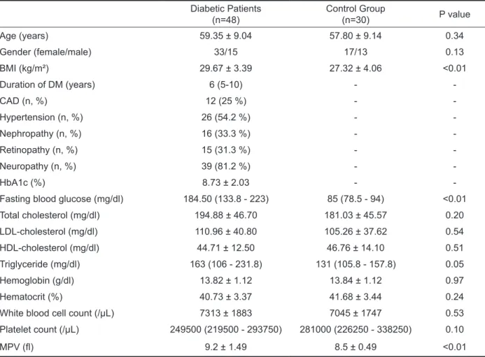
The baseline characteristics of the patients and con -trol group were given in table-1. Duration of DM was 8.33 ± 5.4 years. Twelve of the diabetics (25 %) had macrovascular complications, 26 patients (54.2 %) had HT, 15 patients (31.3 %) had retinopathy, 16 patients (33.3 %) had nephropathy and 39 patients (81.2 %) had neuropathy. Mean HbA1c was 8.73 ± 2.03 %. Mean platelet volume was significantly higher in patients with type II DM than the healthy controls (9.25 ± 1.49 and 8.47 ± 0.49, respectively) (p<0.01) (table-1). Platelet count was somewhat lower in the diabetic group; however, this differ -ence was not significant (249.729 ± 73.479 /μL and 279.466 ± 73.294 /μL, respectively) (p= 0.10).
Table 1 . Baseline characteristics and laboratory results of type II DM patients and healthy control group. Diabetic Patients
(n=48) Control Group(n=30) P value
Age (years) 59.35 ± 9.04 57.80 ± 9.14 0.34
Gender (female/male) 33/15 17/13 0.13
BMI (kg/m²) 29.67 ± 3.39 27.32 ± 4.06 <0.01
Duration of DM (years) 6 (5-10) -
-CAD (n, %) 12 (25 %) -
-Hypertension (n, %) 26 (54.2 %) -
-Nephropathy (n, %) 16 (33.3 %) -
-Retinopathy (n, %) 15 (31.3 %) -
-Neuropathy (n, %) 39 (81.2 %) -
-HbA1c (%) 8.73 ± 2.03 -
-Fasting blood glucose (mg/dl) 184.50 (133.8 - 223) 85 (78.5 - 94) <0.01
Total cholesterol (mg/dl) 194.88 ± 46.70 181.03 ± 45.57 0.20
LDL-cholesterol (mg/dl) 110.96 ± 40.80 105.26 ± 37.62 0.54
HDL-cholesterol (mg/dl) 44.71 ± 12.50 46.76 ± 14.10 0.51
Triglyceride (mg/dl) 163 (106 - 231.8) 131 (105.8 - 157.8) 0.05
Hemoglobin (g/dl) 13.82 ± 1.12 13.84 ± 1.12 0.97
Hematocrit (%) 40.73 ± 3.37 41.68 ± 3.44 0.24
White blood cell count (/μL) 7313 ± 1883 7045 ± 1747 0.53
Platelet count (/μL) 249500 (219500 - 293750) 281000 (226250 - 338250) 0.10
MPV (fl) 9.2 ± 1.49 8.5 ± 0.49 <0.01
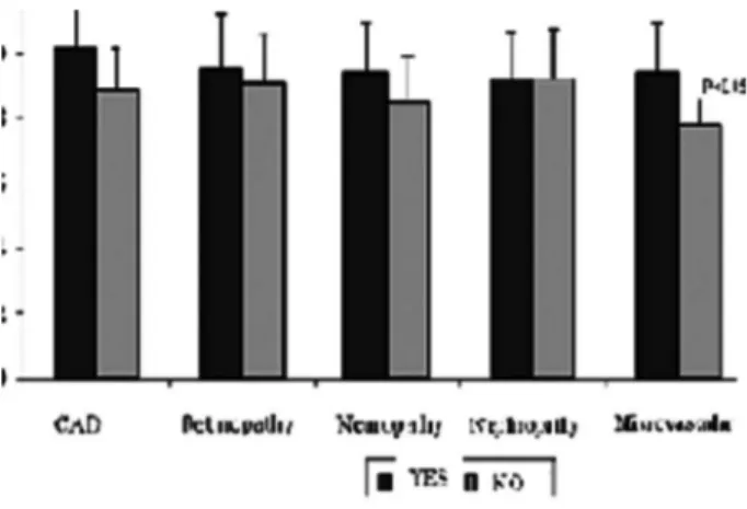
The diabetic patients were divided into sub -groups depending on the presence of microvascular complications. Patients with at least one of the mi -crovascular complications had slightly higher MPV compared to the ones without any of the complica -tions (9.38 ± 1.47 fl and 7.85 ± 0.88 fl, respectively) (p= 0.048). When the groups were analysed indi -vidually as patients with and without retinopathy (9.48 ± 1.60 and 9.15 ± 1.45, p= 0.48), nephropathy (9.25 ± 1.45 and 9.25 ± 1.53, p= 0.99) and neuropa -thy (9.43 ± 1.47 and 8.49 ± 1.39, p= 0.09), there were no significance between the groups regarding MPV (Figure 1). The diabetic patients were classi -fied into subgroups depending on the presence of macrovascular complications. Twelve patients with macrovascular complications showed higher MPV compared to the ones without macrovascular com -plications (10.23 ± 1.66 fl and 8.93 ± 1.29 fl, respec -tively) (p<0.01).
Figure 1. MPV levels between groups
In type II diabetic patients there was no asso -ciation between MPV and age (p= 0.62, r: 0.07), duration of diabetes (p= 0.75, r: -0.05), total cho -lesterol level (p= 0.23, r: -0.18), LDL-cho-lesterol level (p= 0.34, r: -0.14), HDL-cholesterol level (p= 0.22, r: -0.18), triglyceride level (p= 0.96, r: -0.01), HbA1C (p= 0.18, r: 0.20) and fasting blood glucose level (p= 0.37, r: 0.13). Mean platelet volume was found as similar between smoking (n= 12) and non-smoking II diabetic patients (n= 36) (9.68 ± 1.72 and 9.11 ± 1.40, respectively) (p= 0.26). Diabetic males have similar MPV values with females (9.43 ± 1.90 and 9.2 ± 1.30, respectively) (p= 0.62).
DISCUSSION
Diabetes mellitus is a chronic disease that causes in -creased morbidity and mortality due to its vascular complications. There is a need to develop risk factor modification to reduce the impact of complications. Diabetic patients are at risk of increased thrombosis and atherogenesis. Changes in hemostatic balance constitute a pathogenetic factor with a role in com -plication development in DM. Owing to the role of blood platelets in hemostatic balance, changes in platelets in diabetic patients have been studied ex -tensively and an increase in thrombotic adhesion, aggregation and secretion has been shown in many of these [6-8].
-ference was not found between mean MPV values. This finding was similar to the results of previous studies by Keskin et al. and Hekimsoy et al. [2,21]. It is still debated whether platelett activation plays a primary pathogenetic role in the development of diabetic vascular complications or whether the in -creased activity is secondary to vascular complica -tions. Based on our findings, we are of the opinion that higher MPV cannot be attributed solely to the existence of diabetes and platelets play a primary role in complication development.
In our study we found no association between MPV and HbA1c, fasting blood glucose, patient age, HT, hyperlipidemia and duration of diabetes. These findings were in agreement with the previous reports [21,24]. They suggested vascular damage to be due to more reactive platelets and claimed the rate of damage to be constant for the duration of the disease and independent of diabetic control.
Additionally, we found an association between higher MPV and macrovascular complications. This association has, to our knowledge, not been report -ed previously.
Increases in platelet volume are often associ -ated with decreases in platelet count [18,25], per -haps as a result of small platelets being consumed in order to maintain a constant platelet functional mass [26]. Although the relationships is not com -pletely understood. Association of increased plate -let volume and reduced plate-let survival in diabetic patients has been reported by Jones et al [27]. We found the number of platelets were smaller in dia -betic patients similar to the results of Tschöpe et al [28], this difference was not statistically meaning -ful. Our examination of the diabetic group showed a negative linear relationship between MPV and the number of platelets (p=0.006).
An important point in MPV measurement is the ability of platelets to change their volume follow -ing blood collection. The anticoagulant used and the time between sample collection and the study affect MPV measurement. The platelets in blood samples collected in EDTA tubes swell after a while. Even though volume change with sodium citrate is much less than EDTA, this agent is not appropriate for the counters used in practice. If samples collected in EDTA tubes can be studied within 2 hours, the results are acceptable [26]. In light of the literature,
we have performed and recommend the analysis of samples collected in EDTA tubes within 2 hours.
In conclusion, MPV is a marker of platelet function and activity. Diabetes is a complex disease which affects many vascular systems, and increased platelet volume is likely to be associated with the pathological processes and increased risk of vascu -lar disease.
Acknowledgement: We would like to acknowledge for valuable contributions of Dr. Yıldız Arslan due to their kindly efforts of neurological examination and assesment of EMG reports.
REFERENCES
1. Alessandrini P, McRae J, Feman S, FitzGerald GA. Throm
-boxane biosynthesis and platelet function in type 1 diabetes mellitus. N Engl J Med 1988;319:208-212.
2. Keskin A, Özgen AG, Sermez Y, et al. Tip II diabetes mellitu
-sta trombosit fonksiyonları ve glisemi kontrolü ile ilişkisi. Ulusal Endokrinoloji Dergisi 1995;5:179-185.
3. Mathur A, Robinson MS, Cotton J, et al. Platelet reactivity in acute coronary syndromes: evidence for differences in platelet behaviour between unstable angina and myocardial infarction. Thromb Haemost 2001;85:989-994.
4. Endler G, Klimesch A, Sunder-Plassmann H, et al. Mean platelet volume is an independent risk factor for myocardial infarction but not for coronary artery disease. Br J Haema
-tol 2002;117:399-404.
5. American Diabetes Association. Diabetes Care 2005;28. 6. Colwell JA, Winocour PD, Halushka PV. Do platelets have
anything to do with diabetic microvascular disease? Diabe
-tes 1983;32:14-19.
7. Jindal S, Gupta S, Gupta R, et al. Platelet indices in diabetes mellitus: indicators of diabetic microvascular complica
-tions. Hematology 2011;16:86-89.
8. Unubol M, Ayhan M, Güney E. The relationship between mean platelet volume with microalbuminuria and glycemic control in patients with type II diabetes mellitus. Platelets 23:475-480.
9. Gawaz M, Langer H, May AE. Platelets in inflammation and atherogenesis. J Clin Invest 2005;115:3378-384.
10. Coppinger JA, Cagney G, Toomey S, et al. Characteriza
-tion of the proteins released from activated platelets leads to localization of novel platelet proteins in human athero
-sclerotic lesions. Blood 2004;103:2096-2104.
11. Senaran H, Ileri M, Altinbas A, et al. Thrombopoietin and mean platelet volume in coronary artery disease. Clin Car
-diol 2001;24:405-408.
12. Trowbridge EA, Martin JF. The platelet volume distribu
-tion: a signature of the prethrombotic state in coronary heart disease? Thromb Haemost 1987;58:714-717. 13. Erne P, Wardle J, Sanders K, et al. Mean platelet volume
-tients with coronary artery disease and congestive heart failure. Thromb Haemostas 1988;59:259-263.
14. Valkila EH, Salenius JP, Koivula TA. Platelet indices in patients with occlusive carotid artery disease. Angiology 1994;45:361-365.
15. Coban E, Ozdogan M, Yazicioglu G, Akcit F. The mean platelet volume in patients with obesity. Int J Clin Pract 2005;59:981-982.
16. Nadar SK, Blann AD, Kamath S, et al. Platelet indexes in relation to target organ damage in high-risk hypertensive patients: a substudy of the Anglo-Scandinavian Cardiac Outcomes Trial (ASCOT). J Am Coll Cardiol 2004;44:415-422.
17. Nadar S, Blann AD, Lip GY. Platelet morphology and plas
-ma indices of platelet activation in essential hypertension: effects of amlodipinebased antihypertensive therapy. Ann Med 2004;36:552-557.
18. Yang A, Pizzulli L, Luderitz B. Mean platelet volume as marker of restenosis after percutaneous transluminal coro
-nary angioplasty in patients with stable and unstable angina pectoris. Thromb Res 2006;117:371-377.
19. Henning BF, Zidek W, Linder B, Tepel M. Mean Platelet Volume and Coronary Heart Disease in Hemodialysis Pa
-tients. Kidney & Blood Pressure Research 2002;25:103-108.
20. Sharpe PC, Trinic T. Mean platelet volume in diabetes mel
-litus. Quarterly Journal of Medicine 1993;86: 739-742. 21. Hekimsoy Z, Payzin B, Örnek T, Kandogan G. Mean plate
-let volume in Type 2 diabetic patients. Journal of Diabetes and Complications 2004;18:173-176.
22. Saigo K, Yasunaga M, Ryo R, Yamaguchi N. Mean platelet volume in diabetics. Rinsho Byori 1992;40:215-217. 23. Tschoepe D, Roesen P, Schwipper B, Gries FA. Platelets in
diabetes: the role in the hemostatic regulation in atheroscle
-rosis. Semin Thromb haemost 1993;19:122-128.
24. Papanas N, Symeonidis G, Maltezos E, et al. Mean platelet volume in patients with type 2 diabetes mellitus. Platelets 2004;15:475-478.
25. Huczek Z, Kochman J, Filipiak KJ, et al. Mean platelet vol
-ume on admission predicts impaired reperfusion and long
-term mortality in acute myocardial infarction treated with primary percutaneous coronary intervention. J Am Coll Cardiol 2005;46: 284-90.
26. Chu SG, Becker RC, Berger PB, et al. Mean platelet volume as a predictor of cardiovascular risk: a systematic review and meta-analysis. J Thromb Haemost 2010;8:148-156. 27. Jones R L, Paradise C, Peterson C M. Platelet survival in
patients with diabetes mellitus. Diabetes 1981;30:486-489. 28. Tschöpe D, Langer E, Schauseil S. Increased platelet vol