EGL-13/SoxD Specifies Distinct O
2
and CO
2
Sensory
Neuron Fates in
Caenorhabditis elegans
Jakob Gramstrup Petersen1, Teresa Rojo Romanos1, Vaida Juozaityte1, Alba Redo Riveiro1, Ingrid Hums2, Lisa Traunmu¨ller2, Manuel Zimmer2, Roger Pocock1*
1Biotech Research and Innovation Centre, University of Copenhagen, Copenhagen, Denmark,2Research Institute of Molecular Pathology, Vienna, Austria
Abstract
Animals harbor specialized neuronal systems that are used for sensing and coordinating responses to changes in oxygen (O2) and carbon dioxide (CO2). In Caenorhabditis elegans, the O2/CO2 sensory system comprises functionally and
morphologically distinct sensory neurons that mediate rapid behavioral responses to exquisite changes in O2or CO2levels
via different sensory receptors. How the diversification of the O2- and CO2-sensing neurons is established is poorly
understood. We show here that the molecular identity of both the BAG (O2/CO2-sensing) and the URX (O2-sensing) neurons
is controlled by the phylogenetically conserved SoxD transcription factor homolog EGL-13.egl-13mutant animals fail to fully express the distinct terminal gene batteries of the BAG and URX neurons and, as such, are unable to mount behavioral responses to changes in O2and CO2. We found that the expression ofegl-13is regulated in the BAG and URX neurons by
two conserved transcription factors—ETS-5(Ets factor) in the BAG neurons and AHR-1(bHLH factor) in the URX neurons. In addition, we found that EGL-13 acts in partially parallel pathways with both ETS-5 and AHR-1 to direct BAG and URX neuronal fate respectively. Finally, we found that EGL-13 is sufficient to induce O2- and CO2-sensing cell fates in some
cellular contexts. Thus, the same core regulatory factor,egl-13, is required and sufficient to specify the distinct fates of O2
-and CO2-sensing neurons inC. elegans. These findings extend our understanding of mechanisms of neuronal diversification
and the regulation of molecular factors that may be conserved in higher organisms.
Citation:Gramstrup Petersen J, Rojo Romanos T, Juozaityte V, Redo Riveiro A, Hums I, et al. (2013) EGL-13/SoxD Specifies Distinct O2and CO2Sensory Neuron
Fates inCaenorhabditis elegans. PLoS Genet 9(5): e1003511. doi:10.1371/journal.pgen.1003511
Editor:Susan E. Mango, Harvard University, United States of America
ReceivedNovember 2, 2012;AcceptedApril 3, 2013;PublishedMay 9, 2013
Copyright:ß2013 Gramstrup Petersen et al. This is an open-access article distributed under the terms of the Creative Commons Attribution License, which permits unrestricted use, distribution, and reproduction in any medium, provided the original author and source are credited.
Funding:This work was supported by a grant from the Lundbeck Foundation (www.lundbeckfonden.dk) (grant number R93-A8391) and by an ERC Starting Grant (281869—elegansNeurocircuits). The funders had no role in study design, data collection and analysis, decision to publish, or preparation of the manuscript.
Competing Interests:The authors have declared that no competing interests exist.
* E-mail: [email protected]
Introduction
The capacity of the nervous system to sense and respond to fluctuations in the external and internal environment is essential for homeostasis and survival. Neuronally controlled homeostatic buffering is delivered through cellular and systemic physiological adjustments and by seeking optimal environmental conditions through behavioral strategies [1–4]. A crucial homeostatic capacity of animals is the ability to sense and respond to changes in concentration of the respiratory gases oxygen (O2) and carbon
dioxide (CO2) [5,6]. O2is essential for the generation of energy in
the form of adenosine triphosphate (ATP); however, O2also exerts
toxicity through the production of reactive oxygen species (ROS) [1–4,7]. CO2 is a by-product of oxidative metabolism and
prolonged exposure leads to acidosis [5,6,8]. CO2 is also an
environmental cue used in host- and mate- finding and can initiate both aversive or attractive behaviors [9–11]. The evolution of mechanisms required to sense and respond to O2 and CO2 is
therefore paramount for survival.
InDrosophila, specific sensory systems respond to external O2
levels [1,7,12]. In addition,Drosophilauses specialized olfactory and gustatory neurons to detect CO2changes via specialized
chemo-sensory receptors called Gr21a/Gr63a [9,13]. In humans, O2,
CO2 and pH levels are monitored by specific regions of the
brainstem and by specialized neurosecretory glomus cells of the
carotid body [14], whereas in non-human mammals CO2is also
sensed by specific olfactory neurons that target the necklace glomeruli in the olfactory bulb via the guanylyl cyclase GC-D [15]. It is poorly understood how the specification of such specialized sensory neurons is regulated. However, recent work inDrosophila has shown that epigenetic mechanisms play an important role [16].
Respiratory gas sensing is a crucial modality for Caenorhabditis elegans whose natural environment, such as rotting fruit and compost, can have wide ranges of O2 and CO2 levels [17].
Previous work has shown that in the laboratory, worms have a behavioral preference for 5%–10% O2 and are exquisitely
sensitive to minor changes in O2 concentration [18,19]. In
addition, worms mount avoidance responses to CO2levels above
0.5% [4,11]. Of the 302 neurons in the C.elegansnervous system, at least six neurons are specifically dedicated to the detection and response to changes in O2 and CO2 levels. These include the
BAGL/R, URXL/R, AQR and PQR neurons. The BAG neurons are the primary CO2sensors and they also respond to
decrease in O2concentration [11,20–22]. The URX, AQR and
PQR neurons are specialized for responding to increasing O2
concentrations [20]. InC. elegans, members of the guanylyl cyclase family of proteins are crucial factors required for O2 and CO2
behavior via the URX, AQR and PQR neurons and that GCY-35 directly binds to molecular O2[18]. In contrast, the sGCs,
GCY-31 and GCY-33 function in the BAG neurons to sense decreases in O2[20]. Recent work found that the membrane-bound
receptor-type guanylyl cyclase GCY-9 acts specifically in the BAG neurons to mediate CO2avoidance behavior [21]. Other molecules such as
the Phe-Met-Arg-Phe-NH2(FMRF-amide)-related peptides
(FLP-8, FLP-13, FLP-17 and FLP-19) are expressed in either a subset or all of the O2- and CO2-sensing neurons; however their precise
molecular functions in O2 and CO2 sensing are not known
[23,24].
Neuronal specialization within the O2/CO2-sensing system in
C. elegans is an excellent model to study the control of neuron diversity. The O2-sensing (URX, AQR and PQR) and O2/
CO2-sensing (BAG) neurons have overlapping and
non-overlapping patterns of guanylyl cyclase and neuropeptide expression, which are reflected in their related, albeit distinct functionalities [20]. At present, it is unclear how the expression of these molecules is restricted to certain parts of the O2/CO2
-sensing nervous system, and how such restrictions coordinate neuronal fate and function.
Here, we have identified the Sox transcription factor EGL-13 as an important regulator of the O2and CO2-sensing neuron cell fate
decision. EGL-13 is required for the expression of distinct proteins required for sensing both O2and CO2and as such,egl-13mutant
animals are unable to mount behavioral responses to changes in O2 and CO2. We found that the expression of EGL-13 is
controlled by ETS-5 in the BAG neurons and by AHR-1 in the URX neurons, and acts in partially parallel pathways with these factors to drive neuronal fate. Finally, we found that EGL-13 is sufficient to drive O2- and CO2-sensing cell fates in certain cellular
contexts. Therefore, EGL-13 is a core regulatory factor that is both required and sufficient to drive O2- and CO2-sensing neuron
specification inC. elegans. As EGL-13 is a member of the SoxD family of transcription factors, we anticipate that the regulatory relationships described here will provide a paradigm for the control of neuronal fate specification by Sox proteins in other cellular contexts.
Results/Discussion
The SoxD transcription factoregl-13specifies distinct O2
-and CO2-sensing neurons
In order to identify molecules and pathways important for O2
-and CO2-sensing neuron specification, we have taken a forward
genetics approach in C. elegans. We isolated four independent allelic mutations (rp14, rp22, rp23 and rp26) that affect the expression of terminal differentiation markers in the O2and/or
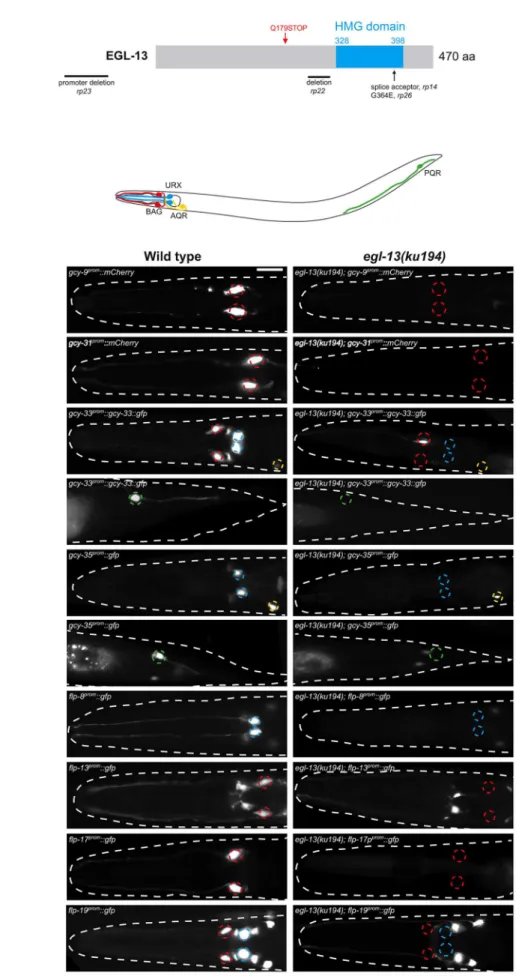
CO2-sensing neurons (Figure 1 and Table S1) Mutant
hermaph-rodites of each of these alleles are severely egg-laying defective (Egl) and form a bag-of-worms where embryos hatch inside the mother (Figure S1). We investigated their vulval phenotype and found that the anchor cell fails to fuse with the uterine seam cell, causing a blockage of the uterus and the resultant Egl phenotype (Figure S1). This anchor cell fusion defect is reminiscent of that observed inegl-13(ku194) mutant animals [25] which we found to also exhibit defects in O2and CO2reporter expression (Figure 1
and Figure S1). Subsequent Sanger sequencing ofrp14,rp22,rp23 andrp26revealed genetic lesions in theegl-13locus (Figure 1A). egl-13encodes theC. elegansortholog of the HMG-domain-containing SoxD family of transcription factors that has no previously reported role in the worm nervous system.
Loss ofegl-13affects terminal fate of O2- and CO2
-sensing neurons
The BAG, URX, AQR and PQR neurons in C. elegans are required for sensing and responding to fluctuations of O2and
CO2levels in the environment [11,18,19]. Distinct batteries of
genes are expressed in these neurons that are predicted to provide the optimal functionality required for O2 and CO2
sensing, however the role of only a subset of these genes has been analyzed in detail [20,21,26]. We used fluorescent reporter constructs to monitor expression of these gene batteries to understand howegl-13controls O2and CO2-sensing neuron cell
fate (Figure 1). We analyzed the expression of guanylyl cyclases (gcy-9, gcy-31, gcy-33, gcy-35and gcy-36) and Phe-Met-Arg-Phe-NH2(FMRF-amide)-related peptides (flp-8,flp-13,flp-17and
flp-19) that are all terminal differentiation genes expressed in all or a subset of O2 and CO2-sensing neurons [18,19,21,24]. We
crossed these reporter transgenes into egl-13 mutant animals (ku194allele) and found that none of the reporters were properly expressed in egl-13mutants (Figure 1 and Table S1). We also found similar effects in the fouregl-13mutant alleles we isolated (rp14,rp22,rp23andrp26) (Figure S1C). We noticed that some of the reporters were exquisitely sensitive toegl-13loss whereas others exhibited partially penetrant defects (Table S1). This suggests that the expression of some terminal differentiation factors are under the collaborative control of additional factors that are able to compensate for the loss ofegl-13.
The BAG and URX neurons are derived from the AB lineage and are posterior sisters of other neurons that have distinct fates [27] (Figure S2). We therefore asked whetheregl-13is also required for the specification of the sister cells of BAG or URX. We crossed egl-13(ku194)mutant animals into fluorescent reporter strains for the SMDV, zfIs2 (lgc-55::mCherry) and CEPD, vtIs1 (dat-1::gfp), sister cells for BAG and URX neurons respectively. We found that the expression of these reporters were unaffected by loss ofegl-13 suggesting a specific role foregl-13in the posterior branch of these lineages (Table S1 and Figure S2). Taken together, we conclude thategl-13 controls the expression of the distinct O2- and CO2
-sensing neuron terminal gene batteries that distinguish them from lineage-related neurons.
Author Summary
During the development of an organism, certain neurons are programmed to perform specific tasks. For example, motor neurons coordinate locomotion and sensory neu-rons recognize specific environmental cues. The molecular mechanisms that generate specific neuronal classes are not fully understood. We investigated mechanisms that control the development of two distinct classes of neurons that are required for the nematodeCaenorhabditis elegans to sense the respiratory gases O2or CO2. In this study, we
identified and characterized a conserved transcription factor, egl-13, that is required for the development of both of these classes of neurons.egl-13 is related to the SoxD family of transcription factor proteins in vertebrates. We found thategl-13 controls the production of specific proteins that provide these cells with the ability to sense both O2and CO2. Further, we found thategl-13works in
Figure 1.egl-13is required for O2- and CO2-sensing neuron specification.(A) Molecular identity of mutant alleles obtained from the forward
egl-13acts cell-autonomously in O2- and CO2-sensing
neurons
To monitor egl-13 expression, we generated two promoter-driven fluorescent reporters (egl-13prom1::mCherryandegl-13prom1::gfp) that contain 3.5 kb ofegl-13 upstream sequence (Figure 2A and Figure S3). Expression is first detected in 4 neuronal cells at around 350 min post-fertilization, which is the time at which the BAG and URX neurons are born (Figure S3). Expression is restricted to these 4 neurons during embryogenesis (Figure S3). At the first larval stage,egl-13expression is observed in the BAG and URX neurons plus occasionally in a small number of unidentified cells in the head and tail (including the AQR and PQR neurons) (Figure S3). Later during larval development,egl-13expression is observed in body wall muscle and vulval cells (data not shown). Neuronal expression is restricted to the O2 and CO2-sensing
neurons in the adult (Figure 2A). Using the 3.5 kbegl-13promoter (egl-13prom1) we transgenically expressed egl-13isoformAcDNA in egl-13(ku194)mutant animals and were able to rescue both the defect in O2and CO2-sensing neuron fate marker expression and the Egl
phenotype (Figure 2B–2C, Figure S4 and data not shown). To confirm thategl-13acts cell autonomously to control O2and CO2
-sensing neuron fate, we used neuron-specific promoters to drive egl-13isoformAcDNA expression in the BAG or URX neurons (Figure 2D). We found that indeed neuron-specific expression of egl-13rescued the O2and CO2-sensing neuron fate defect of
egl-13(ku194) mutant animals (Figure 2D). Therefore, we conclude thategl-13acts autonomously in the BAG and URX neurons to direct their fate.
Theegl-13gene has 4 predicted isoforms, all of which contain the same HMG DNA/protein binding domain, however they each have varying lengths of amino terminal tail. Such tails in SoxD proteins can cooperate with other factors to control gene expression [28]. We therefore tested whether the long N-terminal region of EGL-13isoformA is required for its rescuing ability. We usedegl-13prom1to drive EGL-13isoformD (lacking 157 amino acids of the N-terminal tail of isoformA) in egl-13(ku194)animals and found that it fully rescued the defect in O2 and CO2-sensing
neuron fate marker expression and the Egl phenotype (Figure S4 and data not shown). Thus, the EGL-13 N-terminal region is not required for its roles in vulval cell nor O2and CO2-sensing neuron
specification. We next asked whether SoxD proteins play specific roles in these decisions by attempting to rescue theegl-13mutant defects with the SoxB family member,sox-2. We expressedsox-2 cDNA under the control of egl-13prom1 in egl-13(ku194) mutant animals. We found that sox-2is unable to rescue O2 and CO2
-sensing neuron fate marker expression (Figure 2E). These data indicate that the SoxD HMG domain plays a specific role in the specification of O2and CO2-sensing neuron fate inC. elegans.
egl-13is required and sufficient to induce O2-sensing
neuron fate
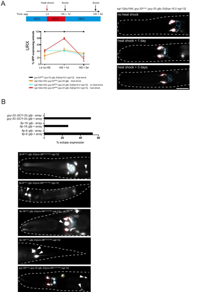
We have shown thategl-13is expressed throughout the life of the worm in the O2and CO2-sensing neurons; and is required to
induce terminal differentiation features. To ask whetheregl-13is required continuously to maintain the expression of the terminal gene battery of these neurons, we sought to postdevelopmentally remove egl-13 gene activity. egl-13 gene activity could not be removed by RNA-mediated interference in an RNAi sensitized background (data not shown) and there are no temperature-sensitive alleles ofegl-13available. Instead, we generated animals that lack endogenous EGL-13 protein but express heat-shock inducibleegl-13cDNA from an extrachromosomal array under the control of the hsp-16.2 promoter (Figure 3). We focused our analysis on the URX neurons and found that the loss of gcy-33prom::gcy-33::gfpreporter expression inegl-13(ku194)worms could be rescued through heat-shock induction of egl-13 during mid-larval stages (Figure 3A). This indicates that O2-sensing neurons
generated during embryogenesis persist in an egl-13-responsive state. These neurons are, therefore, not converted into another fate whenegl-13is lost; however, they do not acquire the terminal O2-sensing neuron differentiation program. Whenegl-13activity
was supplied transiently, through removal of heat-shock stimulus, we observed a gradual loss of reporter expression during adulthood in the URX neurons (Figure 3A). Therefore, egl-13 gene activity is continuously required to maintain URX cell fate. To ask whether misexpression of egl-13 in other neurons is sufficient to induce O2and CO2terminal fate we expressedegl-13
under the control of an early neuronal promoter (Figure 3B). We found thategl-13 is indeed sufficient to induce expression of O2
and CO2 terminal fate markers in some cellular contexts
(Figure 3B). This suggests thategl-13is not only required but also sufficient to induce O2 and CO2-sensing neuron fate in specific
contexts, which is similar to previous studies of terminal selector genes [29–31]. The restricted induction we observed may be dependent on the embryonic time-point of induction or the expression of other unknown co-factors that are required for induction of O2and CO2-sensing neuron fate.
egl-13mutants are defective in O2and CO2sensing
The crucial role foregl-13in O2and CO2-sensing neuron fate
determination suggested that egl-13 mutant animals would be defective in O2and CO2sensing. We applied three behavioral
paradigms that have been previously reported to be specific to either one of these neuron classes: BAG neurons modulate the animals’ locomotion speed in response to an oxygen downshift from 21% O2towards 10% O2(Figure 4A, 4E) [20]. In addition,
BAG neurons detect increases in CO2 concentrations, which
trigger reorientation movements (omega turns) (Figure 4G) [11,21]. URX neurons modulate the animals’ locomotion speed in response to O2upshifts towards 21% O2(Figure 4A, 4F) [20].
We applied these behavioral assays to test how BAG and URX neurons are functionally affected inegl-13 mutants. We tracked animals in a chamber without food, in an air-flow that switched between 21% O2and 10% O2, or between 0% CO2and 1% CO2.
In contrast to wild-type animals,egl-13(ku194)mutant animals do not slow their locomotion in response to O2upshift or downshift
(Figure 4A, 4C, 4E, 4F). We found thategl-13(ku194)mutants are red. The nature of the molecular lesions is as follows:rp14is a splice acceptor mutation between exons 8 and 9, causing a premature termination codon within the HMG domain.rp22is an out-of-frame 317 bp deletion between exons 6 and 7 that leads to a predicted premature STOP codon in the EGL-13 protein that lacks the HMG domain. Therp23allele is a promoter deletion, removing 1128 bp between21700 and2572 upstream of the ATG codon.rp26is a G-to-A transition that converts a highly conserved glycine to a glutamic acid in the HMG domain. The isolatedegl-13mutants are therefore predicted to either abrogate DNA binding (rp14,rp22andrp26) or reduce/eliminate expression ofegl-13transcript (rp23). (B) Schematic of the O2- and CO2-sensing system. Anterior to the left. (C) Fluorescence reporter expression of the O2- and CO2-sensing neuron terminal gene batteries in wild-type (left) andegl-13(ku194) mutant (right) at the young adult stage. Quantification of data and information on reporter strains is shown in Table S1. Neuron positions are marked with dashed circles: BAG(red), URX(blue), AQR(yellow) and PQR(green). Fluorescent cells not marked with circles are non-O2/CO2-sensing neurons in the respective strains and their expression is unaffected by loss ofegl-13. Scale bar, 20mm. Anterior to the left.
Figure 2. egl-13 functions cell autonomously to drive O2- and CO2-sensing neuron cell fate. (A) Dorsal view of a young adult
also defective in CO2sensing since they fail to slow or perform
omega turns in response to CO2 (Figure 4G). O2 and CO2
behavioral defects ofegl-13(ku194)mutants are fully rescued when egl-13 cDNA is resupplied under the control of egl-13prom1 (Figure 4D–4G). These data confirm thategl-13is crucial for the specification and function of O2 and CO2-sensing system in C.
elegans.
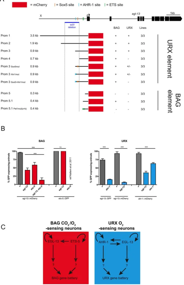
Theegl-13promoter contains neuron-specific regulatory modules
One of theegl-13mutant alleles retrieved from our screen was a promoter deletion mutant (rp23). The rp23 deletion removes 1128 bp ofegl-13promoter from21700 to2572 upstream of the translational start site (Figure 1A and Figure 5A). Intriguingly, the rp23mutation affects terminal marker expression in the URX but not BAG sensory neurons and is mostly defective in URX and less affected in BAG regulated behaviors (Figure 4B, 4E–4G; Figure S4C; and Table S1). This suggests that therp23promoter deletion removes element(s) required to driveegl-13in the URX neurons while leaving the BAG-specific element(s) intact. To identify which upstream factors drive expression ofegl-13in the molecularly and functionally distinct BAG and URX neurons we performed promoter deletion analysis, using the 3.5 kb upstream element (egl-13prom1) as a template. We generated transgenic worms expressing truncated versions ofegl-13prom1drivingmCherryorgfpprotein and focused our expression analysis on BAG and URX regulation (Figure 5A). A 900 bp fragment (egl-13prom3), which includes 360 bp corresponding to the 39 end of therp23 deletion, drove expression in BAG and URX. However, a 691 bp fragment (egl-13prom4), which lacks the missing region in therp23deletion, only drove expression in the BAG neurons. Therefore, an important element required for egl-13 expression specifically in the URX neurons lies within the 200 bp region included in egl-13prom3. Bioinformatic analysis of this region revealed that there are two conserved motifs that are potential binding sites for EGL-13/ SOX5 itself and AHR-1, an aryl hydrocarbon receptor bHLH protein. Interestingly,ahr-1was previously shown to be required for the expression of some URX terminal fate markers [32]. Site-directed mutagenesis of the predicted AHR-1 binding site significantly reduced egl-13prom1::mCherry expression and a subse-quent mutation in the putative EGL-13 binding site further reduced expression (Figure 5A). This suggests that both AHR-1 and EGL-13 regulate egl-13 expression. To test this, we crossed egl-13prom1mCherry/gfp-expressing animals into egl-13(ku194) and ahr-1(ia3)mutants and found that URX expression was reduced in both cases (Figure 5B). Therefore, AHR-1 and EGL-13 both contribute to the control ofegl-13expression in the URX neurons. To identify the regulatory module(s) that control egl-13 expression in the BAG neurons, we continued to dissect the
egl-13 promoter. We identified a 432 bp region (egl-13prom5.1), immediately upstream of the ATG codon, which is sufficient to drive expression in the BAG neurons. Intriguingly, we found two conserved ETS-5/Pet1 binding sites in this region (Figure 5). Previous work identified ETS-5 as a crucial factor required for the specification of the BAG neurons, suggesting that ETS-5 may regulateegl-13expression in these neurons [26,33]. We used site-directed mutagenesis to eliminate the ETS-5/Pet1 binding sites individually and in combination, and found that when both ETS-5/Pet1 binding sites are mutated the expression of egl-13 is abrogated in the BAG neurons (Figure 5A). This suggests that ETS-5 directly regulates the expression of egl-13 in the BAG neurons via conserved binding sites. We crossed theets-5(tm1734) mutant into theegl-13prom1::mCherry strain and indeed found that BAG expression was affected (Figure 5B). In addition, we found thategl-13 can regulate its own expression in the BAG neurons independently of ets-5 via an, as yet, unidentified mechanism (Figure 5B).
Taken together, these data indicate that control of egl-13 expression is coordinated by two independent regulatory mech-anisms. In the O2-sensing URX neurons, egl-13 expression is
predominantly regulated by AHR-1 (Figure 5C). In contrast, an independent promoter module controlled by ETS-5 regulates egl-13expression in the O2/CO2-sensing BAG neurons (Figure 5C).
In addition,egl-13is able to autoregulate in both the URX and BAG neurons (Figure 5C).
egl-13acts in partially parallel pathways withets-5and ahr-1
Previous work has identifiedets-5andahr-1as regulators of BAG and URX specification respectively [26,32,33] and we have shown that these factors are predominantly required to drive egl-13 expression in these cells. To understand how these factors function together to coordinate BAG and URX specification, we analyzed the expression of terminal fate markers in single and double mutant combinations, where appropriate. We analyzed three URX markers (flp-8::gfp, flp-19::gfp and gcy-33prom::gcy-33::gfp) and six BAG markers (flp-13::gfp, flp-17::gfp, flp-19::gfp, gcy-9::gfp, gcy-31::gfpand gcy-33prom::gcy-33::gfp) and compared the effect of the individual loss ofegl-13,ets-5andahr-1(Figure S5).
The first observation from this analysis was that the expression of a subset of terminal differentiation markers is completely dependent onegl-13and one of the other factors acting in a linear pathway. For example, we find that BAG expression offlp-13::gfp andflp-19::gfpis almost 100% affected in both theegl-13andets-5 single mutants (Figure S5). This suggests that for these markers egl-13andets-5act in the same pathway to drive marker expression. In contrast, expression ofgcy-9::mCherryis completely dependent on ets-5withegl-13playing a minor role in its regulation (Figure S5). marking BAGL/R, URXL/R, AQR and PQR (center panels) and a merge of the two pictures showing co-localization in the O2- and CO2-sensing neurons (bottom panels). Left hand panels show the head region and right hand panels the tail region. We also observed expression ofegl-13prom1::mCherryin
muscle and vulval cells (not shown). A: anterior, L: left. The scale bar in lower panel is 20mm. Theegl-13prom1::mCherrytransgene isrpEx272and the
gcy-33prom::GCY-33::gfptransgene isrpIs7. (B) Fluorescence micrographs ofgcy-33prom::gcy-33::gfpexpression in a wild-type animal (top panels), an egl-13(ku194)mutant animal (center panels) and anegl-13(ku194)mutant animal rescued by transgenic expression ofegl-13isoformAcDNAdriven by the endogenous 3.5 kb egl-13prom1(bottom panels). Left hand panels show the head region and right hand panels the tail region. (C) Transgenic
expression ofegl-13isoformAcDNAunder the control ofegl-13prom1rescuesegl-13mutant neuronal phenotypes, in thegcy-33prom::gcy-33::gfpstrain. n = 52–55. ***P,0.001. Lines 1–3 = independent transgenic rescue lines. See materials and methods for neuronal scoring criteria used. (D) Transgenic expression ofegl-13isoformAcDNAunder the control of thegcy-331kb(BAG-specific) orunc-86(700bp)(URX-specific) promoters rescuesegl-13mutant neuronal phenotypes in theflp-19prom::gfp(BAG) andgcy-33prom::gcy-33::gfp(URX) strains. n = 39–55. ***P,0.001. Lines 1–2 = independent transgenic
At the other end of the spectrum,ets-5andegl-13are minimally required to drivegcy-31::mCherry expression in the BAG neurons suggesting other factor(s) control the expression of this terminal fate marker (Figure S5). Taken together, these data indicate that egl-13andets-5act in partially parallel pathways to drive BAG cell fate and that other unknown factors possibly act in a combinatorial manner to drive specific aspects of BAG fate.
We also observed differential effects ofegl-13 loss with URX terminal fate markers. Expression of the flp-8::gfp reporter is partially affected by single loss ofegl-13andahr-1, whereas loss of both genes totally abrogates expression, suggesting thategl-13and ahr-1 act in parallel pathways to regulate flp-8::gfp expression (Figure S5). However, in the case offlp-19::gfp, loss ofegl-13causes complete loss of expression and ahr-1 plays a minor role in its regulation (Figure S5).
To further investigate the regulatory relationship between egl-13,ets-5andahr-1we analyzed how they affect the expression of each other. We have already shown thatets-5positively regulates the expression of egl-13 in the BAG neurons (Figure 5B). In a reciprocal experiment, we found that ets-5::gfp expression is unaffected in egl-13(ku194) mutant animals (Figure 5B). These data and other work [26,33] suggest thatets-5acts upstream and in parallel toegl-13to direct BAG cell fate (Figure 5C). In addition, we found thategl-13is able to regulate its own expression in the BAG neurons, in parallel toets-5; however, the mechanistic basis of this regulation is unclear (Figure 5B). In the URX neurons, we found thategl-13andahr-1regulate the expression of each other in addition to having autoregulatory capabilities (Figure 5B,C and Figure S5).
Our studies have elucidated a novel function for egl-13, the SoxD homolog, in the specification of distinct classes of O2and
CO2sensory neurons inC. elegans. We show thategl-13is expressed
in the O2- and CO2-sensing neurons and acts cell-autonomously to
regulate their distinct cell fates. We further show that egl-13 is continuously expressed in the O2- and CO2-sensing system to
maintain the expression of terminal features of these neurons. In certain cellular contexts,egl-13is also sufficient to induce O2- and
CO2-sensing neuron cell fate. We found that the regulatory inputs
controlling the expression ofegl-13 in the O2- and CO2-sensing
system are mechanistically distinct. Independent regulatory modules controlegl-13expression in the BAG neurons (CO2and
O2 downshift sensors) versus the URX neurons (O2 upshift
sensors). Interestingly, we found thategl-13expression in the BAG neurons is controlled by the ETS-5 transcription factor via conserved ETS binding sites. In contrast, in the URX neurons, egl-13 expression is controlled by the bHLH transcription factor AHR-1 via a conserved AHR1 binding site.
The influence EGL-13 exerts on the expression of the terminal gene batteries of the distinct O2- and CO2-sensing neurons is
diverse. Particular factors are exquisitely sensitive to loss ofegl-13, whereas others are only partially affected. These findings suggest that alternative unknown modes of regulation are in place to ensure that particular molecules are faithfully expressed in the O2
and CO2sensory neurons, which work in conjunction with and/or
in parallel toegl-13.
Sox transcription factors have diverse functions during devel-opment and play crucial roles in regulating neuronal fate [34–37]. In addition, Sox proteins act at different levels to preselect neuronal genes in embryonic stem cells and to direct the activation of these genes in neuronal precursors and fully differentiated neurons [38]. Here we describe a novel role for EGL-13, the SoxD transcription factor in C. elegans, in driving the specification of different but related sensory neuron identities. Closely related orthologs of EGL-13 are found in vertebrates, some of which are expressed in sensory neurons [39], therefore; SoxD proteins may have a previously unrecognized conserved function in the specification of gas-sensing neurons in higher organisms.
Materials and Methods
Strains used in this study
Strains were grown using standard growth conditions on NGM agar at 20uC onEscherichia coliOP50 [8,40]. Transgenic animals were created according to [41]. Strain information is detailed in Table S2.
Forward genetic screening approaches
In all screens, animals were mutagenized with EMS (ethyl methanesulfonate) according to standard protocols [42]. Worms were incubated at 25uC at all times. In the manual screens, 5 parental (P0) mutagenized animals were placed in each of 10 founder plates. Three days later, 400 F1 progeny of the mutagenized P0 animals were singled. Their ensuing F2 progeny were screened under a fluorescence stereomicroscope.
In the automated worm sorter screen, around 100,000 synchronized larval stage L4 animals were mutagenized with EMS, the following day the P0 young adult animals were bleached and their F1 progeny synchronized at larval stage L1 by starvation (approximately 1,000,000 animals). F1 animals were grown to the young adult stage, bleached and their F2 progeny synchronized at larval stage L1 by starvation (approximately 10,000,000 animals). The F2 progeny were grown until larval stage L4 and 10% of the population (approximately 1,000,000) was passed through a COPAS biosorter (Karolinska Institute, Stockholm, Sweden).
egl-13(ku194)mutant animals. A schematic describing the heat-shock protocol is shown at the top left. The number of positive neurons was assessed one day after heat-shock (L2/3+1 day). No increase in the number of positive neurons was observed inegl-13(ku194); gcy-33prom::gcy-33::gfp; Ex
[hsp-16.2::egl-13]animals in the absence of heat shock. A significant decrease in the number of positive neurons was observed inegl-13(ku194); gcy-33prom::gcy-33::gfp; Ex[hsp-16.2::egl-13]animals 3 days after heat shock with growth at 15
uC (L4+3 days) when compared to one day after heat shock
(L4+1 day). Quantification of the number ofgcy-33prom::gcy-33::gfppositive neurons is shown at the indicated time points. Error bars represent the standard error of the mean (SEM). Statistical model applied is a one-way ANOVA with Newman-Keuls multiple comparison test **P,0.05, ***P,0.005. Representative fluorescent micrographs on the right indicate the continual requirement foregl-13to maintain O2/CO2-sensing neuron fate. Neuron positions are marked with dashed circles: BAG(red), URX(blue). Two different extrachromosomal lines were analyzed for this experiment and showed similar effects (only one is shown). 53–113 animals were used per time point per genotype. See materials and methods section for heat shock protocol and for neuronal scoring criteria used. Scale bar, 20mm. Anterior to the left. (B)egl-13is sufficient to induce O2/CO2-sensing cell fate markers. Anunc-86promoter was used to drive broad neuronal expression ofegl-13cDNA in a wild-type background. Graph indicates the percentage of animals that exhibit ectopic expression O2/CO2-sensing cell fate marker (+array = transgenic animals expressingunc-86prom::egl-13cDNAarray;2 array = transgenic siblings not expressing the array). Representative fluorescent micrographs of ectopic expression of three O2/CO2-sensing cell fate markers:flp-8prom::gfp,flp-19prom::gfpandgcy-33prom::gcy-33::gfp. Neuron positions are marked with dashed circles: BAG(red), URX(blue) and ectopic expression of each reporter is marked with arrowheads. Scale bar, 20mm. Anterior to the left.
C. elegansexpression constructs and generation of transgenic worms
Reporter gene constructs were generated by PCR amplifying promoter elements and cloning into the pPD95.75-mCherry and gfp vectors (Fire Vector Kit). Mutagenesis was performed using the QuikChange II XL Site-Directed Mutagenesis Kit (Stratagene). Rescue constructs were generated by cloning promoter and cDNA sequences into the pPD49.26 expression vector (Fire Vector Kit). Constructs were injected into young adult hermaphrodites as either simple arrays (gcy-33prom::gcy-33::gfp (50 ng ul21) and pRF4 (50 ng ul21) as injection markers) or as complex arrays using 1– 10 ng ul21 of linearized plasmid, 150 ng ul21 of PvuII-digested bacterial genomic DNA and myo-2prom::dsRed (3–5 ng ul21), elt-2prom::gfp(3–15 ng ul21) as injection markers.
Behavioral assays
Animals were transferred without food to 14 cm NGM assay plates containing a cut out arena of Whatman filter paper soaked in 20 mM CuCl2to prevent them from leaving a 56 mm656 mm
center area. Sixty to seventy animals were used in a single experiment and starved for one hour prior to examination. Each experiment was carried out three times, except for wild-type, which was performed six times. A custom-made transparent plexiglass device with a flow arena of 60 mm660 mm60.7 mm
was placed onto the assay arena and animals were accustomed to a gas flow of 100 ml/min containing 21% (v/v) oxygen for 5 minutes. During the assays animals were exposed for 6 minutes to 21% O2 before and after a 6 minute stimulus
interval of either 10% O2 or 1% CO2 (+21% O2). All gas
mixtures were balanced with N2. Gases were mixed with a static
mixing element connected to mass flow controllers (Vo¨gtlin Instruments) that were operated by LabView software. Record-ings of freely behaving animals illuminated with flat red LED lights were made at 3 fps on a 4 megapixel CCD camera (Jai) using Streampix software (Norpix). Movies were analyzed by MatLab-based image processing and tracking scripts as previ-ously described [43,44]. The resulting trajectories were used to calculate instantaneous speed during continuous forward movements (1 second binning). Omega turns were detected based on characteristic changes in object eccentricity and their frequency was calculated in 15 second bins. For quantifications, relative speed changes were calculated between representative intervals of 120 seconds before (basal level) and 4 seconds after the stimulus, capturing the minimum speed levels (4–8 seconds post stimulus). Data were normalized to the basal level. Changes in omega turn frequency were calculated between representative intervals of 180 seconds before (basal level) and 60 seconds after the stimulus, to capture the maximum rise phase (55–115 sec-onds post stimulus).
Heat-shock experiments
Two transgenic lines forhsp-16.2::egl-13were used for the heat-shock experiments. For the rescue and maintenance experiments, third larval stage (L3) worms were heat shocked at 37uC two times for 30 min. After heat shock, worms were kept at 25uC overnight and then transferred to 15uC for 2 days.
Microscopy
Worms were mounted on 5% agarose on glass slides and images were taken using an automated fluorescence microscope (Zeiss, AXIO Imager M2) and MicroManager software (version 3.1).
Neuronal scoring
Neurons were given a numerical value according to their expression levels. Wild-type expression scored 1, decreased expression scored 0.5 and abolished expression scored 0. Percentage of GFP expressing animals was then correlated to the theoretical maximum score using the equation below.
% of GFP expressing animals
~observed score nð 11Þzðn20:5Þzðn30Þ theoretical score nð 1zn2zn3Þ 1
|100%
Bioinformatic analysis
The Jaspar program (http://jaspar.genereg.net/) was used to predict the transcription factor binding sites in theegl-13upstream regulatory sequence.
Statistical analysis
Statistical analysis was performed in GraphPad Prism 5 using one-way ANOVA with Newman-Keuls Multiple Comparison Test. Values are expressed as mean6s.d. Differences with a P value,0.05 were considered significant. For the behavioral assays statistical significance was determined using one-way ANOVA with Bonferroni’s Multiple Comparison Test.
Supporting Information
Figure S1 egl-13mutants have anchor cell fusion defects. (A) egl-13(ku194)mutant hermaphrodites are severely egg-laying defective due to a defect in anchor cell fusion (right, compared to the wild-type adult on the left). (B)egl-13mutant alleles isolated from our forward genetic screens (rp14,22,23and26) all have anchor cell fusion defects comparable to the ku194 allele. In the wild-type vulva, the anchor cell fuses to the utse cells to form the mature uterine-vulval connection (green arrowhead, top left). In egl-13 mutant animals, the anchor cell fails to fuse to the utse (red Figure 4.egl-13is essential for locomotion responses to O2and CO2concentration shifts.(A-D) Locomotion speed ofC. elegansduring O2 concentration shifts. Traces show average forward speed and dark shading indicates standard error of the mean (SEM). O2concentrations were switched between 21% and 10%. Shading represents intervals at 21%. (A) Wild-type animals. (B)egl-13(rp23)mutants. (C)egl-13(ku194)mutants. (D) Rescue ofegl-13(ku194)mutant phenotype by transgenic expression ofegl-13cDNA under control of its own promoter. TransgenerpEx401. Note the respective reduction of speed levels in N2 after up- and downshift, which are abolished inegl-13(ku194)and restored in the transgenic line. egl-13(rp23)animals are affected mostly in their response to O2upshift. (E, F) Quantification of data in A–D. Average speed changes in percent from basal speed to O2downshift (E) and upshift (F) of animals with indicated genotypes. Transgenic rescue lines are significantly different fromegl-13(ku194) mutant animals. (G) Average changes in omega turn frequency of animals with indicated genotypes, in response to 1% CO2. The defect in omega turn responses seen inegl-13(ku194)animals is restored in the transgenic lines (rpEx399andrpEx401).egl-13(rp23)animals only exhibit a partial defect. Error bars = SEM. Symbols indicate all significant differences one-way ANOVA with Bonferroni’s Multiple Comparison Test (*/ep = 0.01–0.05, **/ee
p = 0.001–0.01, ***/eeep,0.001). Asterisks indicate significant difference compared to wild-type, while diamonds indicate significant difference compared toegl-13(ku194)mutants. Data were calculated from n = 3 independent experiments for each mutant and transgenic rescue strain, and n = 6 independent experiments for wild-type. Each individual experiment was performed on 60–70 animals.
Figure 5. Independent regulatory modules driveegl-13expression in O2versus O2/CO2-sensing neurons.(A)egl-13promoter analysis.
arrowheads) and blocks egg-laying. (C) Scoring of neuronal phenotypes in egl-13 mutants. rp13 was isolated with the BAG marker,gcy-33prom::gfp. rp22andrp23were isolated with the URX marker,flp-8prom::GFPandrp26was isolated with the BAG, URX, AQR and PQR marker,gcy-33prom::GCY-33::gfp. Expression of each marker in wild-type animals is stated with a +. Scorings were conducted as stated in the materials and methods section. (EPS)
Figure S2 BAG and URX lineages. Lineage diagrams of the BAG and URX neurons. Neurons whose fate are affected in egl-13(ku194)mutants are indicated in red, unaffected in green and untested in black. See Table S2 for fate markers used.
(EPS)
Figure S3 egl-13expression pattern. Expression pattern ofegl-13 at different stages (330–360 mins, 550 mins and L1 larva), using the egl-13prom1::gfp reporter transgene. Fluorescence micrographs (right) and differential interference contrast (DIC) microscopy images (left). Scale bar, 10mm.
(EPS)
Figure S4 egl-13acts cell autonomously to control O2- and CO2
-sensing neuron fate. (A) Protein domain structure of EGL-13 isoforms A+D. (B) Extrachromosomal transgenic rescue of egl-13(ku194)mutant phenotypes, in the gcy-33prom::gcy-33::gfp strain, using egl-13prom1-driven egl-13isoformAcDNA (left) and egl-13
iso-formD
cDNA(right). Both isoforms equally rescue the egl-13(ku194) mutant phenotype. n = 46–61. *P,0.05, ***P,0.001 indicates significant difference from non-transgenic egl-13mutant animals. (C) Transgenic lines expressingegl-13cDNA under the control of egl-13prom1 rescues all theegl-13 alleles retrieved from the genetic screens. Rescue ofrp14in BAG (gcy-33prom::gfp), rp22in URX (flp-8prom::gfp), rp23 in URX (flp-8prom::gfp) and rp26 in BAG, URX, AQR and PQR (gcy-33prom::GCY-33::gfp). n = 47–87. **P
,0.01, ***P,0.001 indicates significant difference from non-transgenic egl-13 mutant animals. See materials and methods for neuronal scoring criteria used.
(EPS)
Figure S5 Differential regulation of O2- and CO2-sensing
neuron terminal differentiation markers by egl-13, ets-5, and ahr-1. (A) Requirement ofegl-13andets-5for the expression of BAG terminal differentiation markers:flp-13,flp-17, flp-19,gcy-9, gcy-31 and gcy-33. egl-13and ets-5 are both absolutely required for the expression of flp-13 and flp-19 suggesting that they act in a common pathway.gcy-9is predominantly regulated byets-5with egl-13playing a lesser role.flp-17is regulated by bothegl-13and ets-5 acting in parallel pathways. Finally, gcy-31 and gcy-33 expression is predominantly regulated by egl-13. += wt, red bars = mutant backgrounds. (B) Requirement ofegl-13 andahr-1 for the expression of URX terminal differentiation markers:flp-8, flp-19 and gcy-33. egl-13 and ahr-1 act in parallel pathways to
regulate flp-8 expression. egl-13 is absolutely required for the expression offlp-19whereasahr-1is less important.egl-13andahr-1 are both required forgcy-33 expression, probably via the same pathway. += wt, blue bars = mutant backgrounds. Statistical model applied is a one-way ANOVA with Newman-Keuls multiple comparison test **P,0.05, ***P,0.005. Statistical model applied is a one-way ANOVA with Newman-Keuls multiple comparison test **P,0.05, ***P,0.005. Alleles used in this analysisegl-13(ku194),ets-5(tm1734)andahr-1(ia03). See materials and methods for neuronal scoring criteria used.
(EPS)
Table S1 Expression of the O2- and CO2-sensing neuron
terminal gene battery is affected in egl-13 mutant animals. (A) Effects in the expression of O2- and CO2-sensing neuron reporters
inegl-13(ku194)mutant animals differ from gene to gene and from cell to cell. For example,flp-19expression is strongly affected in both URX and BAG neurons, whereasgcy-33expression is more strongly affected in the URX neurons than BAG neurons. Fluorescent reporters for the sister cells of the URX (CEPD) and BAG (SMDV) neurons are unaffected in egl-13(ku194) mutant animals. (B) The deletion in the egl-13 locus ofrp23 leaves the BAG-element intact (see Figure 5). As a result, BAG terminal fate marker expression is not significantly affected in egl-13(rp23) mutant animals. In both tables, quantification indicates the percentage of animals in which fluorescence was observed (ON), not observed (OFF) or asymmetrically affected left expressed (L ON) or right expressed (R ON). ‘‘2’’ indicates that the reporter is not expressed in those neurons. Animals were scored at the young adult stage. See materials and methods for neuronal scoring criteria used.
(EPS)
Table S2 Strains used in this study. (EPS)
Acknowledgments
We thank members of the Pocock Laboratory and Oliver Hobert, Baris Tursun, and Iva Greenwald for comments on the manuscript; we thank the following people for strains and reagents: Oliver Hobert (Columbia University), Cornelia Bargmann (Rockefeller University), Mark Alkema (University of Massachusetts Medical School). Some strains were provided by the CGC and by Shohei Mitani at the National Bioresource Project (Japan). We also thank Thomas Burglin and Peter Swoboda at the Karolinska Institue for use of their COPAS Biosorter facility, under theC. elegansNordforsk Researcher Network of Shared Technology Platforms.
Author Contributions
Conceived and designed the experiments: JGP TRR VJ ARR IH LT MZ RP. Performed the experiments: JGP TRR VJ ARR IH LT MZ RP. Analyzed the data: JGP TRR VJ ARR IH LT MZ RP. Wrote the paper: RP.
as black blocks. The upstream region deleted in therp23allele is indicated with a blue horizontal line and a grey dashed vertical line. Below is a representation of cloned and injected constructs, and their expression pattern in the URX and BAG neurons. Black lines denote the promoter fragment placed in front of mCherry fluorescent protein (red boxes). Orange, blue and green crosses represent mutated Sox5, AHR-1 and ETS binding sites respectively. ‘‘+’’ indicates consistent reporter expression in at least 50% of animals in all lines. ‘‘+/2’’ indicates less than 50% of animals
expressing the reporter in all lines. ‘‘2’’ indicates loss of expression. Three independent transgenic lines were analyzed for each promoter as indicated. (B) Expression ofegl-13andets-5reporter transgenes in the BAG neurons in the reciprocal mutant backgrounds (left).egl-13(ku194)and ets-5(tm1734)alleles were used. Expression ofegl-13andahr-1reporter transgenes in the URX neurons in the reciprocal mutant backgrounds (right). egl-13(ku194)andahr-1(ia3)alleles were used. n.50 per strain. Error bars represent the standard error of the mean (SEM) **P,0.05, ***P,0.005. (n.s.) indicates no significant difference from wild-type. See materials and methods for neuronal scoring criteria used. (C) In the BAG neurons (left), the ETS transcription factor, ETS-5 regulates the expression ofegl-13in the BAG neurons via conserved ETS binding sites located in theegl-13promoter. In addition,egl-13andets-5autoregulate in the BAG neurons to generate BAG fate. In the URX neurons (right),egl-13andahr-1autoregulate, in addition to regulating the expression of each other. Other unknown factors act in parallel to and in combination with these factors to drive BAG and URX neuronal fate.
References
1. Prahlad V, Cornelius T, Morimoto RI (2008) Regulation of the cellular heat shock response in Caenorhabditis elegans by thermosensory neurons. Science 320: 811–814. doi:10.1126/science.1156093.
2. Pocock R, Hobert O (2010) Hypoxia activates a latent circuit for processing gustatory information in C. elegans. Nat Neurosci 13: 610–614. doi:10.1038/ nn.2537.
3. Chang AJ, Bargmann CI (2008) Hypoxia and the HIF-1 transcriptional pathway reorganize a neuronal circuit for oxygen-dependent behavior in Caenorhabditis elegans. Proc Natl Acad Sci USA 105: 7321–7326. doi:10.1073/ pnas.0802164105.
4. Bretscher AJ, Busch KE, de Bono M (2008) A carbon dioxide avoidance behavior is integrated with responses to ambient oxygen and food in Caenorhabditis elegans. Proc Natl Acad Sci USA 105: 8044–8049. doi:10.1073/pnas.0707607105.
5. Semenza GL (2012) Hypoxia-inducible factors in physiology and medicine. Cell 148: 399–408. doi:10.1016/j.cell.2012.01.021.
6. Sharabi K, Lecuona E, Helenius IT, Beitel GJ, Sznajder JI, et al. (2009) Sensing, physiological effects and molecular response to elevated CO2 levels in eukaryotes. J Cell Mol Med 13: 4304–4318. doi:10.1111/j.1582-4934.2009.00952.x.
7. Kaelin WG, Ratcliffe PJ (2008) Oxygen sensing by metazoans: the central role of the HIF hydroxylase pathway. Molecular Cell 30: 393–402. doi:10.1016/ j.molcel.2008.04.009.
8. Guais A, Brand G, Jacquot L, Karrer M, Dukan S, et al. (2011) Toxicity of carbon dioxide: a review. Chem Res Toxicol 24: 2061–2070. doi:10.1021/ tx200220r.
9. Suh GSB, Wong AM, Hergarden AC, Wang JW, Simon AF, et al. (2004) A single population of olfactory sensory neurons mediates an innate avoidance behaviour in Drosophila. Nature 431: 854–859. doi:10.1038/nature02980. 10. Munger SD, Leinders-Zufall T, McDougall LM, Cockerham RE, Schmid A, et
al. (2010) An olfactory subsystem that detects carbon disulfide and mediates food-related social learning. Curr Biol 20: 1438–1444. doi:10.1016/ j.cub.2010.06.021.
11. Hallem EA, Sternberg PW (2008) Acute carbon dioxide avoidance in Caenorhabditis elegans. Proc Natl Acad Sci USA 105: 8038–8043. doi:10.1073/pnas.0707469105.
12. Wingrove JA, O’Farrell PH (1999) Nitric oxide contributes to behavioral, cellular, and developmental responses to low oxygen in Drosophila. Cell 98: 105–114. doi:10.1016/S0092-8674(00)80610-8.
13. Jones WD, Cayirlioglu P, Kadow IG, Vosshall LB (2007) Two chemosensory receptors together mediate carbon dioxide detection in Drosophila. Nature 445: 86–90. doi:10.1038/nature05466.
14. Lo´pez-Barneo J, Ortega-Sa´enz P, Pardal R, Pascual A, Piruat JI, et al. (2009) Oxygen sensing in the carotid body. Annals of the New York Academy of Sciences 1177: 119–131. doi:10.1111/j.1749-6632.2009.05033.x.
15. Hu J, Zhong C, Ding C, Chi Q, Walz A, et al. (2007) Detection of near-atmospheric concentrations of CO2 by an olfactory subsystem in the mouse. Science 317: 953–957. doi:10.1126/science.1144233.
16. Sim CK, Perry S, Tharadra SK, Lipsick JS, Ray A (2012) Epigenetic regulation of olfactory receptor gene expression by the Myb-MuvB/dREAM complex. Genes & Development 26: 2483–2498. doi:10.1101/gad.201665.112. 17. Sylvia DM, Fuhrmann JJ, Hartel PG, Zuberer DA (2004) Principles and
Applications of Soil Microbiology (2nd Edition). 2nd ed. Prentice Hall. 1 pp. 18. Gray JM, Karow DS, Lu H, Chang AJ, Chang JS, et al. (2004) Oxygen
sensation and social feeding mediated by a C. elegans guanylate cyclase homologue. Nature 430: 317–322. doi:10.1038/nature02714.
19. Cheung BHH, Cohen M, Rogers C, Albayram O, de Bono M (2005) Experience-Dependent Modulation of C. elegans Behavior by Ambient Oxygen. Current Biology 15: 905–917. doi:10.1016/j.cub.2005.04.017.
20. Zimmer M, Gray JM, Pokala N, Chang AJ, Karow DS, et al. (2009) Neurons detect increases and decreases in oxygen levels using distinct guanylate cyclases. Neuron 61: 865–879. doi:10.1016/j.neuron.2009.02.013.
21. Hallem EA, Spencer WC, McWhirter RD, Zeller G, Henz SR, et al. (2011) Receptor-type guanylate cyclase is required for carbon dioxide sensation by Caenorhabditis elegans. Proc Natl Acad Sci USA 108: 254–259. doi:10.1073/ pnas.1017354108.
22. Bretscher AJ, Kodama-Namba E, Busch KE, Murphy RJ, Soltesz Z, et al. (2011) Temperature, oxygen, and salt-sensing neurons in C. elegans are carbon dioxide
sensors that control avoidance behavior. Neuron 69: 1099–1113. doi:10.1016/ j.neuron.2011.02.023.
23. Ortiz CO (2006) Searching for Neuronal Left/Right Asymmetry: Genomewide Analysis of Nematode Receptor-Type Guanylyl Cyclases. Genetics 173: 131– 149. doi:10.1534/genetics.106.055749.
24. Kim K, Li C (2004) Expression and regulation of an FMRFamide-related neuropeptide gene family inCaenorhabditis elegans. J Comp Neurol 475: 540– 550. doi:10.1002/cne.20189.
25. Hanna-Rose W, Han M (1999) COG-2, a sox domain protein necessary for establishing a functional vulval-uterine connection in Caenorhabditis elegans. Development 126: 169–179.
26. Brandt JP, Aziz-Zaman S, Juozaityte V, Martinez-Velazquez LA, Petersen JG, et al. (2012) A single gene target of an ETS-family transcription factor determines neuronal CO2-chemosensitivity. PLoS ONE 7: e34014. doi:10.1371/journal.pone.0034014.
27. Sulston JE, Schierenberg E, White JG, Thomson JN (1983) The embryonic cell lineage of the nematode Caenorhabditis elegans. Developmental Biology 100: 64–119.
28. Lefebvre V, Li P, de Crombrugghe B (1998) A new long form of Sox5 (L-Sox5), Sox6 and Sox9 are coexpressed in chondrogenesis and cooperatively activate the type II collagen gene. EMBO J 17: 5718–5733. doi:10.1093/emboj/17.19.5718. 29. Satterlee JS, Sasakura H, Kuhara A, Berkeley M, Mori I, et al. (2001) Specification of thermosensory neuron fate in C. elegans requires ttx-1, a homolog of otd/Otx. Neuron 31: 943–956.
30. Flames N, Hobert O (2009) Gene regulatory logic of dopamine neuron differentiation. Nature 458: 885–889. doi:10.1038/nature07929.
31. Tursun B, Patel T, Kratsios P, Hobert O (2011) Direct conversion of C. elegans germ cells into specific neuron types. Science 331: 304–308. doi:10.1126/ science.1199082.
32. Qin H, Zhai Z, Powell-Coffman JA (2006) The Caenorhabditis elegans AHR-1 transcription complex controls expression of soluble guanylate cyclase genes in the URX neurons and regulates aggregation behavior. Developmental Biology 298: 606–615. doi:10.1016/j.ydbio.2006.07.017.
33. Guillermin ML, Castelletto ML, Hallem EA (2011) Differentiation of carbon dioxide-sensing neurons in Caenorhabditis elegans requires the ETS-5 transcription factor. Genetics 189: 1327–1339. doi:10.1534/genet-ics.111.133835.
34. Azim E, Jabaudon D, Fame RM, Macklis JD (2009) SOX6 controls dorsal progenitor identity and interneuron diversity during neocortical development. Nat Neurosci 12: 1238–1247. doi:10.1038/nn.2387.
35. Lai T, Jabaudon D, Molyneaux BJ, Azim E, Arlotta P, et al. (2008) SOX5 controls the sequential generation of distinct corticofugal neuron subtypes. Neuron 57: 232–247. doi:10.1016/j.neuron.2007.12.023.
36. Scott CE, Wynn SL, Sesay A, Cruz C, Cheung M, et al. (2010) SOX9 induces and maintains neural stem cells. Nat Neurosci 13: 1181–1189. doi:10.1038/ nn.2646.
37. Pevny L, Placzek M (2005) SOX genes and neural progenitor identity. Current Opinion in Neurobiology 15: 7–13. doi:10.1016/j.conb.2005.01.016. 38. Bergsland M, Ramsko¨ld D, Zaouter C, Klum S, Sandberg R, et al. (2011)
Sequentially acting Sox transcription factors in neural lineage development. Genes & Development 25: 2453–2464. doi:10.1101/gad.176008.111. 39. Morales AV, Perez-Alcala S, Barbas JA (2007) Dynamic Sox5 protein expression
during cranial ganglia development. Dev Dyn 236: 2702–2707. doi:10.1002/ dvdy.21282.
40. Brenner S (1974) The genetics of Caenorhabditis elegans. Genetics 77: 71–94. 41. Mello CC, Kramer JM, Stinchcomb D, Ambros V (1991) Efficient gene transfer in C.elegans: extrachromosomal maintenance and integration of transforming sequences. EMBO J 10: 3959–3970.
42. Flibotte S, Edgley ML, Chaudhry I, Taylor J, Neil SE, et al. (2010) Whole-genome profiling of mutagenesis in Caenorhabditis elegans. Genetics 185: 431– 441. doi:10.1534/genetics.110.116616.
43. Ramot D, Johnson BE, Berry TL, Carnell L, Goodman MB (2008) The Parallel Worm Tracker: a platform for measuring average speed and drug-induced paralysis in nematodes. PLoS ONE 3: e2208. doi:10.1371/journal.-pone.0002208.
44. Tsunozaki M, Chalasani SH, Bargmann CI (2008) A behavioral switch: cGMP and PKC signaling in olfactory neurons reverses odor preference in C. elegans. Neuron 59: 959–971. doi:10.1016/j.neuron.2008.07.038.