Characterization of Genomic Alterations in
Radiation-Associated Breast Cancer among
Childhood Cancer Survivors, Using
Comparative Genomic Hybridization (CGH)
Arrays
Xiaohong R. Yang1*, J. Keith Killian2, Sue Hammond3, Laura S. Burke1, Hunter Bennett1, Yonghong Wang2, Sean R. Davis2, Louise C. Strong4, Joseph Neglia5, Marilyn Stovall6, Rita E. Weathers6, Leslie L. Robison7, Smita Bhatia8, Kiyohiko Mabuchi1, Peter D. Inskip1, Paul Meltzer2
1Division of Cancer Epidemiology & Genetics, National Cancer Institute, National Institutes of Health, Bethesda, Maryland, United States of America,2Center of Cancer Research, National Cancer Institute, National Institutes of Health, Bethesda, Maryland, United States of America,3Department of Laboratory Medicine and Pathology, Children's Hospital and Ohio State University College of Medicine, Columbus, Ohio, United States of America,4Department of Genetics, The University of Texas MD Anderson Cancer Center, Houston, Texas, United States of America,5Department of Pediatrics, University of Minnesota School of Medicine, Minneapolis, Minnesota, United States of America,6Department of Radiation Physics, The University of Texas MD Anderson Cancer Center, Houston, Texas, United States of America,7
Epidemiology and Cancer Control, St. Jude Children's Research Hospital, Memphis, Tennessee, United States of America,8Department of Pediatrics, University of Alabama at Birmingham, Birmingham, Alabama, United States of America
Abstract
Ionizing radiation is an established risk factor for breast cancer. Epidemiologic studies of ra-diation-exposed cohorts have been primarily descriptive; molecular events responsible for the development of radiation-associated breast cancer have not been elucidated. In this study, we used array comparative genomic hybridization (array-CGH) to characterize ge-nome-wide copy number changes in breast tumors collected in the Childhood Cancer Survi-vor Study (CCSS). Array-CGH data were obtained from 32 cases who developed a second primary breast cancer following chest irradiation at early ages for the treatment of their first cancers, mostly Hodgkin lymphoma. The majority of these cases developed breast cancer before age 45 (91%, n = 29), had invasive ductal tumors (81%, n = 26), estrogen receptor (ER)-positive staining (68%, n = 19 out of 28), and high proliferation as indicated by high Ki-67 staining (77%, n = 17 out of 22). Genomic regions with low-copy number gains and losses and high-level amplifications were similar to what has been reported in sporadic breast tumors, however, the frequency of amplifications of the 17q12 region containing human epidermal growth factor receptor 2 (HER2) was much higher among CCSS cases (38%, n = 12). Our findings suggest that second primary breast cancers in CCSS were
OPEN ACCESS
Citation:Yang XR, Killian JK, Hammond S, Burke LS, Bennett H, Wang Y, et al. (2015) Characterization of Genomic Alterations in Radiation-Associated Breast Cancer among Childhood Cancer Survivors, Using Comparative Genomic Hybridization (CGH) Arrays. PLoS ONE 10(3): e0116078. doi:10.1371/ journal.pone.0116078
Academic Editor:Xin-Yuan Guan, The University of Hong Kong, CHINA
Received:August 28, 2014
Accepted:December 5, 2014
Published:March 12, 2015
Copyright:This is an open access article, free of all copyright, and may be freely reproduced, distributed, transmitted, modified, built upon, or otherwise used by anyone for any lawful purpose. The work is made available under theCreative Commons CC0public domain dedication.
Data Availability Statement:All aCGH data files were deposited into the GEO database (accession number: GSE62940).
Funding:This research was supported in part by the Intramural Research Program of the NIH, NCI, Division of Cancer Epidemiology & Genetics and grant for the CCSS (U24 CA55727).
enriched for an“amplifier”genomic subgroup with highly proliferative breast tumors. Future investigation in a larger irradiated cohort will be needed to confirm our findings.
Introduction
Ionizing radiation is an established risk factor for breast cancer, and risk increases linearly with dose [1]. Breast cancer is among the most radiogenic tumors identified so far among the atom-ic-bomb survivors [2]. The greatest relative risk related to radiation exposure was observed for breast cancer among women who were exposed at a young age [3,4]. Similarly, breast cancer is the second most common primary cancer among childhood cancer survivors, following only basal cell carcinoma of the skin [5]. In the survivors, the odds ratio for breast cancer increased linearly with radiation dose, and breast cancer was diagnosed at young ages (median, 35.9 years; range, 20.9 to 49.6 years) [6]. A recent analysis of 1,200 women participating in the Childhood Cancer Survivor Study (CCSS) showed that 25% of those who received>20 gray (Gy) to the chest area developed breast cancer by age 50; among women who received lower doses of radiation (10–19 Gy), 7% developed breast cancer by age 40, versus a less than 2% chance of developing breast cancer by age 50 in the general population [7].
Radiation-associated breast carcinogenesis appears to be a highly complex phenomenon and likely involves accumulating genetic and epigenetic changes. In a recent study characteriz-ing copy number alteration (CNA) and expression profiles in 2,000 breast tumors, Curtis et al. showed that CNAs were associated with profound changes in gene expression through both
cis- andtrans-effects [8]. The joint clustering of gene expression and CNA profiles revealed
novel breast cancer subtypes that refined previously identified molecular subtypes defined by expression-only profiling. These findings suggest that identifying CNA regions may provide a powerful tool to investigate the molecular basis of radiation-associated breast cancer.
Epidemiologic studies of radiation-exposed cohorts have been primarily descriptive. Molec-ular events responsible for the development of radiation-associated breast cancer are largely unknown, although recent studies demonstrated that radiation-associated breast tumors were characterized by a high degree of proliferation, high frequency of gene amplifications, in partic-ular HER2 amplification, and enriched with basal-like tumors [9–11]. In this study, we used comparative genomic hybridization arrays (array-CGH) to characterize the CNA profile in breast tumor tissues collected from CCSS cases, the majority of whose breast cancer were radia-tion related [6], to identify possible distinct genomic aberrations related to radiation exposure.
Methods
Study population
participants provided informed consent. The current study of using de-identified archived tumor tissue was exempted from review by the National Institutes of Health Office of Human Subject Research.
Radiation dosimetry
Radiation dose to the presumed site of origin of the breast cancer was estimated for the previ-ous study [6] by medical physicists using radiotherapy records collected for the CCSS cohort [13]. Detailed tumor site-specific dosimetry was not available for the newly identified breast cancer cases. For consistency, we report maximal chest dosage for all cases.
Pathology review
Most of the breast cancers in the CCSS cohort were self-reported by the participants via a peri-odic questionnaire. A small minority of cases were first discovered from other medical records being collected. Following self-report, an investigation was made to obtain the pathology report and to request the participant’s permission to use paraffin material from their tumor for re-search. Immunohistochemical (IHC) staining of estrogen receptor (ER) and Ki-67 were per-formed in a diagnostic pathology laboratory using a Ventana autostainer (Ventana Medical Systems, Inc., Tucson, AZ). The cutoff for Ki-67 low versus high proliferative index was posi-tive staining of 10% cancer cell nuclei.
Array-CGH
Archival formalin-fixed paraffin-embedded (FFPE) tumor blocks were identified from 49 breast cancer cases (41 invasive and 8in situ) in the CCSS cohort. Ten 5-μm unstained tumor
sections were used for DNA extraction with enrichment for tumor cells by micro-dissection. Sufficient DNA (1μg) was obtained from 38 cases. Test and reference DNA (from Promega, Madison, WI) were labeled with Cy5 and Cy3, respectively, and co-hybridized to Agilent CGH arrays containing 180,000 probes tiled across the genome. The intensity of the two fluorescent dyes that reflects chromosomal imbalances between test and reference samples was extracted using Agilent Feature-Extraction software. The log2 ratio was then created for each probe and was used as input to Nexus Copy Number (Biodiscovery, Hawthorne, CA). The Rank Segmen-tation algorithm (significant threshold = 1 x 10⁻⁶) was used to identify copy number alterations at the probe level. Low-level copy number gains and losses were defined as absolute log2 ratios larger than 0.3. Amplifications were defined as log2 ratios larger than 1. For example, human epidermal growth factor receptor 2 (HER2, ERBB2) amplification was defined as log2 ratios> 1 for at least 5 consecutive probes in the 17q12 region containing the HER2 gene. Both objec-tive and subjecobjec-tive measures of data quality were used. Among 38 tumors analyzed, 6 had low quality array-CGH data, mostly because of low signal-to-noise ratio (Nexus quality score>0.3 and/or by visualization), and were removed from further analysis (high quality scores suggest elevated noise to signal ratio). The average quality score of remaining 32 samples was 0.14 (range: 0.06–0.28), which was within the acceptable range recommended by Nexus Copy Number.
Statistical analysis
Results
In total, array-CGH data was obtained from 32 CCSS cases whose characteristics are shown in Tables1and2. The majority of these cases (N = 21) had HL as the first cancer and the other 11 cases had NHL, bone cancer, leukemia, soft tissue sarcoma, or kidney cancer. Actual dose to the site where the breast cancer developed and systemic chemotherapy doses are available for most of these cases (30 for radiation treatment (RT) and 32 for chemotherapy). Among 30 cases with known radiation status, 21 received radiation directly to the chest (up to 57 Gy), 1 case had RT, but not to the chest or an adjacent body region, 3 cases had RT that included arm, neck or abdomen but not chest, and 5 cases had no RT. Age at diagnosis of the first cancer for all CCSS cases in this study was between 12 and 20 years; therefore, age at radiation exposure was young for all members of this cohort. Among these cases, 26 were invasive ductal carcino-ma, two were invasive lobular carcinocarcino-ma, three were ductal carcinomain situ(DCIS), and one
had bothin-situductal andin-situlobular carcinoma. Most of these cases (N = 29) had
early-onset breast cancer (before age 45), invasive ductal tumors (N = 26), ER-positive staining (N = 16 of 25 invasive, 3 of 3 forin situ), and high proliferation as indicated by high Ki-67 staining
(N = 16 of 19 invasive, 1 of 3in situ).
Table 3shows the CNA profile for each case, listing the most common amplified genes or
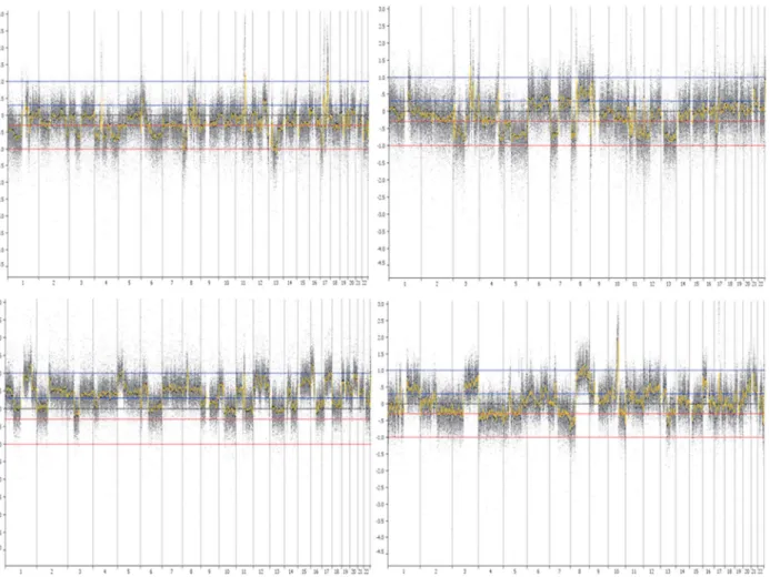
regions and low-level copy number changes involving whole or parts of chromosomes. A de-tailed list of CNAs is shown in Table A inS1 File. Among the 28 invasive cases with array-CGH data, 5 cases showed“simple”genomic changes characterized by very few copy number changes other than gain of 1q and loss of 16q; 5 cases displayed a more“complex”profile with extensive low-copy number changes but with no high-level amplifications. The majority of CCSS cases (N = 18) displayed amplifications in multiple regions (Table 3, Table A inS1 File). Among the amplifiers, the majority (N = 16) showed a“complex-amplifier”profile which is characterized by the presence of both low copy number changes and high-level amplifications
(Fig. 1). The fourin situcases showed similar CNA profiles (1 simple, 3 amplifier), and we
combined them with invasive cases in subsequent analyses. The most frequent low-copy num-ber changes were the gains of 1q, 3q, 6p, 8q, 16p, and 17q, and losses of 3p, 6q, 8p, 9p, 10q, 11q, 16q and 17p. The most frequent amplifications were 17q12 (containing HER2, n = 12 [38%]), 17q21–24 (n = 10 [31%]), 11q13 (containing CCND1, n = 7 [22%]), 8p11.2 (n = 7 [22%]), 8p12 (containing FGFR1, n = 5 [16%]), and 8q24 (containing MYC, n = 5 [16%]). Among 7 cases with HER2 IHC data available, results from IHC and arrayCGH were consistent for all but one sample (Table 3).
Among 28 cases with ER status available, 15 cases (54%) had luminal tumors, defined as positive for ER by IHC and negative for HER2 amplification; 4 cases (14%) were double posi-tive (ER+ and HER2amp); 6 cases (21%) were ER- and HER2amp; and 3 cases (11%) were dou-ble negative (ER-negative and HER2amp-negative) tumors. The frequency of high-level amplifications was significantly lower in ER+ tumors (53% vs. 100% in ER-, p = 0.01). The dif-ference remained significant when taking out the four in situ cases. All nine cases with a simple or complex CGH profile with the absence of amplifications had luminal tumors (ER status was undetermined in one“simple”and one“complex”case). The remaining six luminal tumors were amplifiers, harboring both amplifications (mostly in CCND1) and low-copy changes such as 16p gain and 16q loss. Among ten HER2-amplified tumors with known ER status, ER+ (N = 4) and ER- tumors (N = 6) showed similar CNAs in other chromosome regions. The three cases with double-negative tumors all had“complex-amplifier”CNA profiles.
Discussion
In this study, we characterized molecular and genetic changes in tumor tissue collected from CCSS breast cancer cases, the majority of whom were treated with chest radiation for a prior tumor when they were under 20 years of age. We found that breast cancers in the majority of these cases were positive for ER (68%, n = 19 out of 22), highly proliferative as indicated by Ki-67 staining (77%, n = 17 out of 22), and frequently had high-level amplifications (66%). The frequency of HER2 amplification appeared to be particularly higher in this irradiated series of cases (38%, n = 12) compared to breast cancer among young women in the general population [14–17].
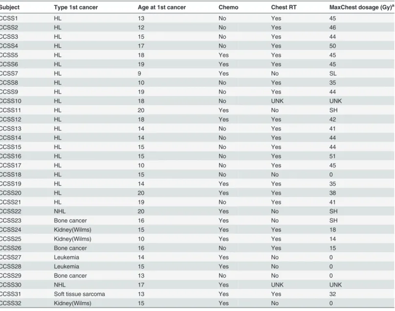
Table 1. Patient characteristics and treatment of CCSS cases included in this study.
Subject Type 1st cancer Age at 1st cancer Chemo Chest RT MaxChest dosage (Gy)a
CCSS1 HL 13 No Yes 45
CCSS2 HL 12 No Yes 46
CCSS3 HL 15 No Yes 44
CCSS4 HL 17 No Yes 50
CCSS5 HL 18 Yes Yes 45
CCSS6 HL 19 Yes Yes 45
CCSS7 HL 9 Yes No SL
CCSS8 HL 10 No Yes 35
CCSS9 HL 19 No Yes 44
CCSS10 HL 18 No UNK UNK
CCSS11 HL 20 Yes No SH
CCSS12 HL 18 Yes Yes 42
CCSS13 HL 14 No Yes 41
CCSS14 HL 14 No Yes 44
CCSS15 HL 15 No Yes 44
CCSS16 HL 15 No Yes 51
CCSS17 HL 10 No Yes 45
CCSS18 HL 15 No No 0
CCSS19 HL 14 Yes Yes 35
CCSS20 HL 20 Yes Yes 38
CCSS21 HL 19 No Yes 41
CCSS22 NHL 20 Yes No SH
CCSS23 Bone cancer 16 Yes No SH
CCSS24 Kidney(Wilms) 15 Yes Yes 18
CCSS25 Kidney(Wilms) 10 Yes Yes 14
CCSS26 Bone cancer 16 No Yes 15
CCSS27 Leukemia 14 Yes No 0
CCSS28 Leukemia 15 Yes No 0
CCSS29 Bone cancer 13 No No 0
CCSS30 NHL 17 Yes UNK UNK
CCSS31 Soft tissue sarcoma 13 Yes Yes 32
CCSS32 Kidney(Wilms) 15 Yes No 0
Abbreviations: HL = Hodgkin lymphoma, NHL = non-Hodgkin lymphoma, RT = radiotherapy, UNK = unknown.
a0 = no direct treatment to the chest, UNK = unknown dose,>0 and<70 Gy = direct treatment to the chest, SL = (low scatter) patient had RT, but not to the chest or an adjacent body region, SH = (high scatter) patient had RT that included arm, neck or abdomen but not chest.
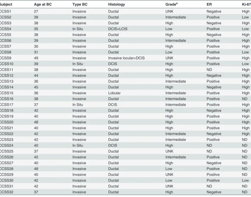
Genomic regions with low-copy gains and losses and high-level gene amplifications among CCSS cases were similar to those reported for breast cancers in the general population, with the most frequent gains on chromosomes 1q, 3q, 6p, 8q, 16p, and 17q; losses on 3p, 6q, 8p, 9p, 10q, 11q, 16q, and 17p; and high-level amplifications of 8p 11.2–12, 8q24, 11q13, 17q12, and 17q21–24. Previous array-CGH studies classified breast tumors into three genomic subtypes (simplex, complex, and amplifier) and found a significant correlation between these genomic subtypes and molecular subtypes defined by gene expression profiles [18,19]. Using a similar classification scheme, we found that a majority of CCSS cases demonstrated a“ complex-ampli-fier”profile, characterized by the presence of extensive low copy number changes and recurrent amplifications. High-level amplifications have been associated with short telomere length, Table 2. Breast tumor characteristics of CCSS cases included in this study.
Subject Age at BC Type BC Histology Gradea ER Ki-67
CCSS1 27 Invasive Ductal UNK Negative High
CCSS2 39 Invasive Ductal Intermediate Positive Low
CCSS3 38 Invasive Ductal High Negative High
CCSS4 35 In Situ DCIS+LCIS Low Positive Low
CCSS5 38 Invasive Ductal High Negative High
CCSS6 39 Invasive Ductal Intermediate Positive High
CCSS7 30 Invasive Ductal High Positive High
CCSS8 31 Invasive Ductal Low Positive Low
CCSS9 49 Invasive Invasive locular+DCIS UNK Positive High
CCSS10 39 In Situ DCIS High Positive Low
CCSS11 38 Invasive Ductal High ND High
CCSS12 44 Invasive Ductal High Negative High
CCSS13 36 Invasive Ductal Intermediate Positive High
CCSS14 45 Invasive Ductal High Negative High
CCSS15 36 Invasive Lobular Intermediate Positive High
CCSS16 38 Invasive Ductal Intermediate Positive ND
CCSS17 37 In Situ DCIS Intermediate Positive High
CCSS18 42 Invasive Ductal High Negative High
CCSS19 40 Invasive Ductal High Positive High
CCSS20 48 Invasive Ductal High Positive High
CCSS21 40 Invasive Ductal High Positive High
CCSS22 42 Invasive Ductal Intermediate Negative High
CCSS23 42 Invasive Ductal Intermediate Positive ND
CCSS24 40 In Situ DCIS High ND ND
CCSS25 37 Invasive Ductal UNK ND ND
CCSS26 42 Invasive Ductal Intermediate Positive ND
CCSS27 40 Invasive Ductal High Negative ND
CCSS28 48 Invasive Ductal Low Positive ND
CCSS29 40 Invasive Ductal UNK Positive ND
CCSS30 42 Invasive Ductal Low Positive Low
CCSS31 42 Invasive Ductal UNK ND ND
CCSS32 37 Invasive Ductal High Negative ND
Abbreviations: BC = breast cancer, DCIS = ductal cell in situ, ER = estrogen receptor, ND = not determined, UNK = unknown. aGrade: low = well differentiated or grade I, intermediate = moderately differentiated or grade II, high = poorly differentiated or grade III.
indicating high genomic instability, and poor prognosis independent of tumor grade and nodal status [18,19]. The majority of the CCSS cases (17 out of 22) in our study demonstrated prolif-eration as indicated by Ki-67 staining, suggesting that radiation may induce a highly prolifer-ative subtype of breast cancer through gene amplifications. It is possible that the high frequency of amplifications may be a characteristic of young breast cancer cases instead of being driven by radiation exposure since breast cancers in CCSS survivors occurred at young ages. However, the frequency of HER2 amplification in CCSS cases (38%) was still higher Table 3. ArrayCGH profile and molecular subtypes of CCSS cases included in this study.
Subject CGH subtypea Recurrent CNAs Subtypeb
CCSS1 Amplifier HER2 amp, CCND1 amp, 1p loss, 3p loss, 5p loss, 6q loss, 8p loss, 9p loss, 17p loss ER-HER2+
CCSS2 Amplifier CCND1 amp, 16p gain, 8p loss, 16q loss Luminal
CCSS3 Amplifier 8p11.2amp, 8q21–23amp, 12q12amp, 19q12–13.2amp, 10q loss ER-HER2-c
CCSS4 Simple 16q loss Luminal
CCSS5 Amplifier FGFR1 amp, 8q gain, 3p loss, 8p loss,17p loss
ER-HER2-CCSS6 Amplifier FGFR1 amp, CCND1 amp, 1q gain, 6p gain, 16p gain Luminal
CCSS7 Amplifier HER2 amp, ZNF217 amp, 1q gain, 8q gain, 16p gain, 3p loss, 8p loss ER+HER2+c
CCSS8 Amplifier MYC amp, ZNF217 amp, 16p gain, 3p loss, 16q loss Luminal
CCSS9 Amplifier HER2 amp, 6q loss, 16q loss ER+HER2+c
CCSS10 Amplifier CCND1 amp, 1q gain, 8q gain, 16p gain, 6q loss, 8p loss, 17p loss Luminal
CCSS11 Simple 1q gain, 3q gain, 10q gain ND
CCSS12 Amplifier CCND1 amp, 16p gain ER-HER2-c
CCSS13 Simple 16q loss Luminal
CCSS14 Amplifier HER2 amp, 1q gain, 6q loss, 8p loss, 9p loss, 10q loss ER-HER2+
CCSS15 Complex 1q gain, 6p gain, 17q gain, 3p loss, 6q loss, 13q loss, 17p loss Luminal
CCSS16 Amplifier MYC amp, FGFR1 amp, 10q loss, 16q loss Luminal
CCSS17 Amplifier HER2 amp ER+HER2+
CCSS18 Amplifier HER2 amp, 3q gain, 3p loss, 9p loss ER-HER2+
CCSS19 Complex 1q gain, 17q gain, 1p loss, 6q loss, 13q loss, 16q loss, 17p loss Luminal
CCSS20 Simple 1q gain, 18q loss Luminalc
CCSS21 Amplifier HER2 amp, CCND1 amp, FGFR1 amp, MYC amp, 8q gain, 16p gain, 8p loss, 16q loss, 17p loss ER+HER2+
CCSS22 Amplifier HER2 amp ER-HER2+
CCSS23 Amplifier CCND1 amp, 1q gain, 16p gain Luminal
CCSS24 Amplifier HER2 amp, 9p loss, 10q loss ND
CCSS25 Amplifier FGFR1 amp, HER2 amp ND
CCSS26 Simple 1p loss, 10q loss Luminalc
CCSS27 Amplifier HER2 amp, MYC amp, ZNF217 amp, 5p gain, 8q gain, 9q gain, 20q gain, 8p loss ER-HER2+c
CCSS28 Simple 8q gain Luminal
CCSS29 Complex 3q gain, 8q gain, 9q gain, 17q gain, 1p loss, 3p loss, 6q loss, 9p loss, 10q loss, 16q loss Luminal
CCSS30 Complex 10p gain, 16p gain, 20p gain, 10q loss, 16q loss Luminal
CCSS31 Complex 1q gain, 1p loss, 3p loss, 6q loss, 9p loss, 11q loss ND
CCSS32 Amplifier HER2 amp, MYC amp, 8p loss, 9p loss, 17p loss, 11p gain, 17q gain ER-HER2+
aArray-CGH subtype: simple: very few copy number changes other than gain of 1q and loss of 16q; complex; extensive low copy number changes but with no high-level amplifications; amplifiers: high-level amplifications usually accompanied by low copy number changes.
bER positivity was determined by immunohistochemistry; HER2 positivity was de
fined by the presence of amplification of 17q12 region (log2 ratio>1)
based on aCGH data.
cHER2 IHC status was available. IHC and CGH data were concordant for all but one sample (CCSS20), for which IHC was positive and ampli
fication was negative.
compared to young breast cancer (<45 years) in the general population (16%−25%) based on a literature search [14–17]. In addition, we compared the frequency of the most common ampli-fications to those in breast cancer cases diagnosed before 45 years of age in The Cancer Ge-nome Atlas (TCGA) Research Network (http://cancergenome.nih.gov/) [20] as well as in three published studies with accessible age and array-CGH data [21–23], and we found that the fre-quency of most amplifications, in particular, regions containing HER2 and possibly CCND1, still appeared higher compared to breast cancer in the general population (Table B inS1 File). Since the vast majority of cases in our study received high-dose radiation and very few cases had no radiation or radiation in other body areas, our study did not have the power in evaluat-ing the radiation dosage in relation to HER2 amplification frequency. However, our data are consistent with the high frequency of HER2 amplification observed in breast cancers among atomic-bomb survivors [10,24], supporting the view that HER2 amplification may be an im-portant mechanism in radiation-associated breast carcinogenesis.
A gene expression profiling analysis of breast tumors from 22 patients who developed breast cancer after HL suggested that radiation-associated tumors were associated with a higher fre-quency of basal-like tumors [9]. Similarly, a more recent study found that breast cancer pa-tients after radiation therapy were more likely to have triple-negative tumors [11]. Using Fig 1. Array-CGH images of CCSS cases displaying“complex-amplifier”genomic profiles.Test and reference DNA were labeled with different dyes and co-hybridized to Agilent CGH arrays containing 180,000 probes tiled across the genome. The chromosome number is shown at the bottom of the figure. Y axis shows the log2 ratios. Low-level copy number gains and losses were defined as absolute log2 ratios larger than 0.3. Amplifications were defined as log2 ratios larger than 1.
immunohistochemical ER status and HER2 amplification, we classified breast cancers into four subtypes, luminal, ER+/HER2+, ER-HER2+, and double negative. The frequency of ER+ tu-mors in our study (68%, n = 19 out of 28) is slightly higher compared to breast cancer among young cases in the general population (50–60%) [15,17], but is consistent with what was re-ported by Broeks et al. in the study of breast cancer after HL [9]. However, in contrast to the enrichment of basal-like or triple-negative tumors observed in previous studies, our results showed that breast tumors from CCSS cases were preferentially of two subtypes: luminal or HER2+, which is in line with what was observed by Castiglioni in breast tumors developed in women irradiated for HL within 4 years of menarche [25]. Our data is also consistent with findings from previous reports that radiation-associated breast cancers were characterized by more proliferative and aggressive features [9,24]. Since each individual study is unavoidably limited by the small sample size, a collaborative effort combining data from multiple such stud-ies is needed to more accurately characterize the distribution of molecular subtypes in radia-tion-associated breast tumors.
As expected, all cases without amplifications had luminal tumors in our study; however, lu-minal tumors also demonstrated extensive heterogeneity in genomic changes. In addition to the simple subtype, 5 luminal cases displayed a complex profile, and the remaining 6 luminal cases harbored high-level amplifications (4 had amplifications in CCND1). Our data are con-sistent with previous findings that the majority of CCND1-amplified tumors were ER+ [26]. Recently, a large-scale genomic and transcriptomic analysis of 2,000 breast tumors revealed a high-risk 11q13/14cis-acting luminal subtype that was associated with high mortality [8]. This
subgroup exhibited high-level amplification in CCND1 as well as alterations in several key cell cycle-related genes, which may be the drivers for the development of this tumor subtype and play a role in its aggressiveness. The higher frequency of ER-positive staining and focal amplifi-cations suggest that breast tumors in CCSS cases may be enriched for a high-risk luminal sub-type [27].
The major limitations of our study include small sample size, particularly in the non-irradi-ated group. There were only nine cases without chest radiation and, among them, 4 cases had radiation exposure but not directly to the chest. Therefore, we were not able to conduct a direct
“case-control”comparison within the cohort or to model copy number changes against quanti-tative radiation dosage because of small sample size. However, our study population is unique; most cases had high-dose radiation at early ages and developed breast cancer when they were young. In addition, we characterized genome-wide CNAs as well as ER expression and prolifer-ation status in these cases, which is lacking in the radiprolifer-ation-associated human breast
cancer literature.
In conclusion, data from our study are consistent with previous findings that radiation-asso-ciated breast cancer might have a distinct pathogenesis, characterized by high frequency of ge-nomic amplifications and high degree of proliferation. Future studies with large numbers of exposed and non-exposed cases are needed to validate these findings to determine the etiologic link between radiation exposure, early age onset, gene amplifications, and highly proliferative breast cancers.
Supporting Information
S1 File. Table A: Copy number changes in breast tumors in 32 CCSS cases.Table B: Fre-quency of amplifications in HER2, CCND1, FGFR1, and MYC in young breast cancer cases (age<45 years) in TCGA and published studies.
Acknowledgments
We thank the survivors who have contributed to the Childhood Cancer Survivor Study, the data abstractors, and the survey interviewers; the investigators of the institutions that partici-pated in the CCSS.
Author Contributions
Conceived and designed the experiments: XRY JKK KM PDI PM. Performed the experiments: JKK. Analyzed the data: XRY LSB HB YW SRD. Contributed reagents/materials/analysis tools: SH LCS JN MS REW LLR SB PDI. Wrote the paper: XRY PM.
References
1. Land CE, Tokunaga M, Koyama K, Soda M, Preston DL, et al. (2003) Incidence of female breast cancer among atomic bomb survivors, Hiroshima and Nagasaki, 1950–1990. Radiat Res 160: 707–717. PMID:14640793
2. Preston DL, Ron E, Tokuoka S, Funamoto S, Nishi N, et al. (2007) Solid cancer incidence in atomic bomb survivors: 1958–1998. Radiat Res 168: 1–64. PMID:17722996
3. Tokunaga M, Land CE, Tokuoka S, Nishimori I, Soda M, et al. (1994) Incidence of female breast cancer among atomic bomb survivors, 1950–1985. Radiat Res 138: 209–223. PMID:8183991
4. Clemons M, Loijens L, Goss P (2000) Breast cancer risk following irradiation for Hodgkin's disease. Cancer Treat Rev 26: 291–302. PMID:10913384
5. Kenney LB, Yasui Y, Inskip PD, Hammond S, Neglia JP, et al. (2004) Breast cancer after childhood cancer: a report from the Childhood Cancer Survivor Study. Ann Intern Med 141: 590–597. PMID:
15492338
6. Inskip PD, Robison LL, Stovall M, Smith SA, Hammond S, et al. (2009) Radiation dose and breast can-cer risk in the childhood cancan-cer survivor study. J Clin Oncol 27: 3901–3907. doi:10.1200/JCO.2008. 20.7738PMID:19620485
7. Moskowitz CS, Chou J.F., Wolden S.L., Bernstein J.L., Malhotra J., Friedman D.N., Mubdi N.Z., Hen-derson T.O., Leisenring W.M., Stovall M.,Hammond S., Boice J.D., Hudson M.M., Diller L., Bhatia S., Neglia J.P., Begg C.B., Robison L.L., Oeffinger K.C. (2012) New insights into the risk of breast cancer in childhood cancer survivors treated with chest radiation: A report from the Childhood Cancer Survivor Study (CCSS) and the Women's Environmental Cancer and Radiation Epidemiology (WECARE) Study. J Clin Oncol 30: CRA9513.
8. Curtis C, Shah SP, Chin SF, Turashvili G, Rueda OM, et al. (2012) The genomic and transcriptomic ar-chitecture of 2,000 breast tumours reveals novel subgroups. Nature 486: 346–352. doi:10.1038/ nature10983PMID:22522925
9. Broeks A, Braaf LM, Wessels LF, van de Vijver M, De Bruin ML, et al. (2010) Radiation-associated breast tumors display a distinct gene expression profile. Int J Radiat Oncol Biol Phys 76: 540–547. doi:
10.1016/j.ijrobp.2009.09.004PMID:20117289
10. Miura S, Nakashima M, Ito M, Kondo H, Meirmanov S, et al. (2008) Significance of HER2 and C-MYC oncogene amplifications in breast cancer in atomic bomb survivors: associations with radiation expo-sure and histologic grade. Cancer 112: 2143–2151. doi:10.1002/cncr.23414PMID:18348306
11. Horst KC, Hancock SL, Ognibene G, Chen C, Advani RH, et al. (2014) Histologic subtypes of breast cancer following radiotherapy for Hodgkin lymphoma. Ann Oncol 25: 848–851. doi:10.1093/annonc/ mdu017PMID:24608191
12. Robison LL, Mertens AC, Boice JD, Breslow NE, Donaldson SS, et al. (2002) Study design and cohort characteristics of the Childhood Cancer Survivor Study: a multi-institutional collaborative project. Med Pediatr Oncol 38: 229–239. PMID:11920786
13. Stovall M, Weathers R, Kasper C, Smith SA, Travis L, et al. (2006) Dose reconstruction for therapeutic and diagnostic radiation exposures: use in epidemiological studies. Radiat Res 166: 141–157. PMID:
16808603
14. Pronzato P, Mustacchi G, De Matteis A, Di Costanzo F, Rulli E, et al. (2011) Biological characteristics and medical treatment of breast cancer in young women-a featured population: results from the NORA study. Int J Breast Cancer 2011: 534256. doi:10.4061/2011/534256PMID:22332011
16. Choi DH, Shin DB, Lee MH, Lee DW, Dhandapani D, et al. (2003) A comparison of five immunohisto-chemical biomarkers and HER-2/neu gene amplification by fluorescence in situ hybridization in white and Korean patients with early-onset breast carcinoma. Cancer 98: 1587–1595. PMID:14534873
17. Keegan TH, DeRouen MC, Press DJ, Kurian AW, Clarke CA (2012) Occurrence of breast cancer sub-types in adolescent and young adult women. Breast Cancer Res 14: R55. PMID:22452927
18. Fridlyand J, Snijders AM, Ylstra B, Li H, Olshen A, et al. (2006) Breast tumor copy number aberration phenotypes and genomic instability. BMC Cancer 6: 96. PMID:16620391
19. Chin K, DeVries S, Fridlyand J, Spellman PT, Roydasgupta R, et al. (2006) Genomic and transcriptional aberrations linked to breast cancer pathophysiologies. Cancer Cell 10: 529–541. PMID:17157792
20. (2012) Comprehensive molecular portraits of human breast tumours. Nature 490: 61–70. doi:10.1038/ nature11412PMID:23000897
21. Andre F, Job B, Dessen P, Tordai A, Michiels S, et al. (2009) Molecular characterization of breast can-cer with high-resolution oligonucleotide comparative genomic hybridization array. Clin Cancan-cer Res 15: 441–451. doi:10.1158/1078-0432.CCR-08-1791PMID:19147748
22. Russnes HG, Vollan HK, Lingjaerde OC, Krasnitz A, Lundin P, et al. (2010) Genomic architecture char-acterizes tumor progression paths and fate in breast cancer patients. Sci Transl Med 2: 38ra47. doi:
10.1126/scitranslmed.3000611PMID:20592421
23. Jonsson G, Staaf J, Vallon-Christersson J, Ringner M, Holm K, et al. (2010) Genomic subtypes of breast cancer identified by array-comparative genomic hybridization display distinct molecular and clini-cal characteristics. Breast Cancer Res 12: R42. doi:10.1186/bcr2596PMID:20576095
24. Oikawa M, Yoshiura K, Kondo H, Miura S, Nagayasu T, et al. (2011) Significance of genomic instability in breast cancer in atomic bomb survivors: analysis of microarray-comparative genomic hybridization. Radiat Oncol 6: 168. doi:10.1186/1748-717X-6-168PMID:22152285
25. Castiglioni F, Terenziani M, Carcangiu ML, Miliano R, Aiello P, et al. (2007) Radiation effects on devel-opment of HER2-positive breast carcinomas. Clin Cancer Res 13: 46–51. PMID:17200337
26. Holm K, Staaf J, Jonsson G, Vallon-Christersson J, Gunnarsson H, et al. (2012) Characterisation of am-plification patterns and target genes at chromosome 11q13 in CCND1-amplified sporadic and familial breast tumours. Breast Cancer Res Treat 133: 583–594. doi:10.1007/s10549-011-1817-3PMID:
22002566
27. Kwei KA, Kung Y, Salari K, Holcomb IN, Pollack JR (2010) Genomic instability in breast cancer: patho-genesis and clinical implications. Mol Oncol 4: 255–266. doi:10.1016/j.molonc.2010.04.001PMID: