Human Pregnancies
Aloysius Ssemaganda1, Lindsay Kindinger2, Philip Bergin3, Leslie Nielsen1, Juliet Mpendo1, Ali Ssetaala1, Noah Kiwanuka1,6, Markus Munder5, Tiong Ghee Teoh2, Pascale Kropf4*, Ingrid Mu¨ller4*
1Uganda Virus Research Institute - International AIDS Vaccine Initiative, Entebbe, Uganda,2Department of Obstetrics and Gynecology, St. Mary’s Hospital, London, United Kingdom,3International AIDS Vaccine Initiative Human Immunology Laboratory, Faculty of Medicine, Imperial College London, London, United Kingdom, 4Section of Immunology, Department of Medicine, Faculty of Medicine, Imperial College London, London, United Kingdom,5Third Department of Medicine, Hematology, Oncology and Pneumology, University Medical Center, Mainz, Germany,6School of Public Health, College of Health Sciences, Makerere University, Kampala, Uganda
Abstract
We have previously shown that in successful pregnancies increased arginase activity is a mechanism that contributes to the suppression of the maternal immune system. We identified the main type of arginase-expressing cells as a population of activated low-density granulocytes (LDGs) in peripheral blood mononuclear cells and in term placentae. In the present study, we analyzed the phenotype of LDGs and compared it to the phenotype of normal density granulocytes (NDGs) in maternal peripheral blood, placental biopsies and cord blood. Our data reveal that only LDGs but no NDGs could be detected in placental biopsies. Phenotypically, NDGs and LDGs from both maternal and cord blood expressed different levels of maturation, activation and degranulation markers. NDGs from the maternal and cord blood were phenotypically similar, while maternal, cord and placental LDGs showed different expression levels of CD66b. LDGs present in cord blood expressed higher levels of arginase compared to maternal and placental LDGs. In summary, our results show that in maternal and cord blood, two phenotypically different populations of neutrophils can be identified, whereas in term placentae, only activated neutrophils are present.
Citation:Ssemaganda A, Kindinger L, Bergin P, Nielsen L, Mpendo J, et al. (2014) Characterization of Neutrophil Subsets in Healthy Human Pregnancies. PLoS ONE 9(2): e85696. doi:10.1371/journal.pone.0085696
Editor:Ana Claudia Zenclussen, Medical Faculty, Otto-von-Guericke University Magdeburg, Medical Faculty, Germany ReceivedSeptember 27, 2013;AcceptedDecember 6, 2013;PublishedFebruary 13, 2014
Copyright:ß2014 Ssemaganda et al. This is an open-access article distributed under the terms of the Creative Commons Attribution License, which permits unrestricted use, distribution, and reproduction in any medium, provided the original author and source are credited.
Funding:This work was supported in part by funds received from the HIV Trust (A. Ssemaganda, HIVVRT13-015), Imperial College (IM), The Wellcome Trust (07664/Z/05/Z, PK) and was made possible in part by the generous support of the American people through the United States Agency for International Development (USAID). The contents are the responsibilities of the study authors and do not necessarily reflect the views of USAID, the NIH or the United States Government. The funders had no role in study design, data collection and analysis, decision to publish, or preparation of the manuscript.
Competing Interests:The authors have declared that no competing interests exist. * E-mail: i.muller@imperial.ac.uk (IM); p.kropf@imperial.ac.uk (PK)
Introduction
Transient modulation of innate and adaptive maternal immu-nity during pregnancy contributes to the creation of an immunosuppressive state allowing implantation and growth of the fetus. Although there is bidirectional communication and migration of fetal and maternal cells throughout pregnancy the paternal antigens expressed by the fetus are not attacked and rejected by the maternal immune system [1]. It is generally accepted that in successful healthy pregnancies multiple mecha-nisms provided by both the mother and the fetus contribute to the development and maintenance of immune tolerance and immune privilege [2]. In normal pregnancies there is an increased systemic inflammation, enhanced number of polymorphonuclear cells (PMN), a low Th1/Th2 balance, a decrease in peripheral NK cells, and an increased number of regulatory T cells [3,4,5]. Although major progress has been made in the understanding of immune mechanisms that prevent rejection of the fetus, the generation of this immunosuppressive state is not fully elucidated [6].
Indoleamine 2,3 dioxygenase (IDO), a tryptophan-catabolizing enzyme expressed by both the maternal decidua and fetal trophoblast and catabolites of the tryptophan metabolism such
as kynurenine and picolinic acid can inhibit lymphocyte activation and have been shown to contribute to the Th2 bias and to tolerance induction and maintenance in pregnancy [7].
distin-guishes this population from the remaining granulocytes that co-purify with the erythrocyte fraction following density gradient centrifugation and thus have been named normal-density granu-locytes (NDGs).
In the present study we analyzed the phenotype and frequency of normal and low density granulocytes obtained from maternal peripheral blood, placental biopsies and cord blood.
Materials and Methods
Ethics Statement
This study protocol was approved by the NHS NRES Committee South Central Oxford A (REC reference 12/SC/ 0721; IRAS project ID 1181020).
Subjects and Samples
All individuals gave written, informed consent before partici-pation. Pregnant women (n = 7; mean age 34years, mean BMI 26) were recruited at the time of elective caesarean section; the mean gestational age at delivery was 38 weeks and 6 days. Exclusion criteria included any major complication of pregnancy or intercurrent illness, such as pre-eclampsia, pre- or post-term labor (,37 weeks or.42 weeks), intra-uterine growth retardation, viral, bacterial or parasitic infections.
Ten ml of maternal peripheral blood and of cord blood were collected in EDTA tubes and PBMCs were isolated by density gradient centrifugation on Histopaque 1077 (Sigma). Neutrophils were isolated from the erythrocyte fraction by dextran sulphate sedimentation [12].
Placentae were harvested directly after parturition and up to five small biopsies were taken through the full thickness of the
placenta. Single cell suspensions were obtained by homogenizing biopsies in PBS on cell dissociation sieves and purified from debris by Histopaque 1077 density gradient centrifugation. The placental cells (PlaC) obtained were washed and resuspended in PBS. All experiments were performed on fresh cells, immediately after processing.
Flow Cytometry
The following antibodies purchased from Biolegend were used: CD66bFITC (involved in respiratory burst, adhesion molecule, present in the membrane of specific granules); CD15PE(involved in cell-cell interactions, phagocytosis, stimulation of degranulation,
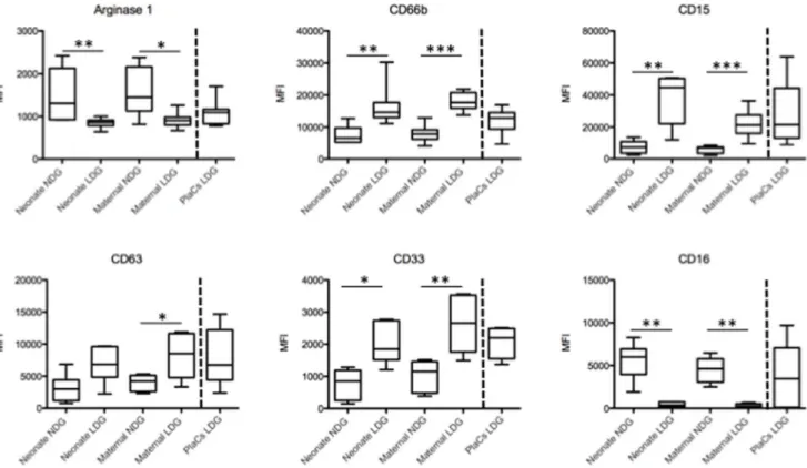
Figure 1. Phenotypic analysis of LDGs and NDGs.LDGs and NDGs were isolated as described in materials and methods (n = 7) and the expression levels of arginase, CD66b, CD15, CD63, CD33 and CD16 were determined by flow cytometry. Statistical significance was determined by a two-tailed Mann-Whitney test. Box = interquartile range and median; whiskers = range.
doi:10.1371/journal.pone.0085696.g001
Table 1.Expression levels of arginase and phenotypic markers of NDGs and LDGs in cord blood.
CORD BLOOD NDGs LDGs pvalues
Arginase 1 13126249 860642 0.0047
CD66b 658861153 1460562414 0.0023
CD15 1089661664 4472066046 0.0023
CD63 30046880 682061017 0.0513
CD33 8516216 18516296 0.0159
CD16 60066871 3646136 0.0043
NDGs and LDGs were isolated from cord blood (n = 7) as described in materials and methods. Expression levels (MFI) of phenotypic markers were determined by flow cytometry. Statistical significance was determined by a two-tailed Mann-Whitney test.
and respiratory burst); CD63PE-Cy7 (marker for release of azurophilic granules); CD33PE-Cy5 (marker of immature neutro-phils, adhesion molecule); CD16AF700(Marker of mature granu-locytes, involved in degranulation). In addition, Zenon Pacific blue conjugated Arginase-1 (Hycult Biotechnology/Invitrogen) was used to detect intracellular arginase 1. The Live/Dead Fixable Near-IR dye (Invitrogen) was used to distinguish live and dead cells.
Cells isolated from cord blood, PlaCs and PBMC were washed and incubated with 20ml FcR blocking reagent for 5 minutes and
stained for 20 minutes with cell surface markers at room temperature. Cells were washed with FACS medium and fixed with cold 4% formaldehyde on ice for 20 minutes.
For intracellular staining, 0.5% saponin was used to permea-bilize the cells for 20 minutes at room temperature and then stained with arginase 1 Pacific blue conjugated antibody for a further 20 minutes. The cells were washed and analyzed immediately with an LSRII flow cytometer (BD Bioscience). The results were analyzed using FlowJo v9.6.2 (Tree Star, Ashland, OR).
Statistical Analyses
Data were evaluated for statistical differences using a two-tailed Mann-Whitney test and a Kruskal-Wallis test when appropriate (GraphPad PRISM version 5); differences were considered significant at p,0.05. Results are expressed as median6SEM. Results
Phenotype and Frequency of Neutrophil Subpopulations in Maternal Blood, Cord Blood and Term Placentae
We have previously shown that the cells expressing arginase in the PBMCs and placentae of pregnant women are a population of low-density granulocytes (LDGs) that co-purify with PBMCs following density gradient centrifugation [8,13]. This difference in density distinguishes this population from the remaining granulocytes that co-purify with the erythocyte fraction following density gradient centrifugation and thus have been named normal-density granulocytes (NDGs).
Here we first determined whether both neutrophil subpopula-tions, LDGs and NDGs, were detectable in the maternal and cord blood and in the term placentae. Whereas both LDGs and NDGs were isolated from maternal and cord blood, NDGs could not be isolated from term placentae despite using different methods such as Histopaque 1119 density gradient centrifugation or isolation by
dextran sedimentation from the erythrocyte fraction of Histopaque 1077 gradients.
First we compared the expression levels of arginase, activation and maturation markers by LDGs and NDGs in maternal and cord blood since we had previously shown that the expression levels of arginase, CD15, CD16, CD33, CD63 and CD66b are modulated on LDGs [12]. The expression levels on placental LDGs is shown throughout Figure 1 (after the broken line) for comparison. As shown in Figure 1 and Tables 1 and 2, NDGs present in both maternal and cord blood express significantly more arginase than LDGs; the reduced expression of CD63 on NDGs is in agreement with the higher arginase levels and indicate that these cells are less activated and have not degranulated arginase-containing CD63+ azurophilic granules. The lower arginase expression by LDGs in cord blood, maternal blood and in the placenta corresponds with increased CD63 expression and indicate that these cells had been activated and had degranulated
Table 2.Expression levels of arginase and phenotypic markers of NDGs and LDGs in maternal peripheral blood.
MATERNAL BLOOD NDGs LDGs pvalues
Arginase 1 14526216 906669 0.0262
CD66b 783661021 1768261072 0.0006
CD15 68206914 2120763198 0.0006
CD63 42276505 851561259 0.0350
CD33 11546198 26586378 0.0043
CD16 46376531 247686 0.0012
NDGs and LDGs were isolated from maternal blood (n = 7) as described in materials and methods. Expression levels (MFI) of phenotypic markers were determined by flow cytometry. Statistical significance was determined by a two-tailed Mann-Whitney test.
doi:10.1371/journal.pone.0085696.t002
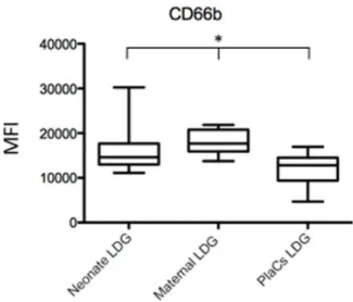
Figure 2. Comparison of the phenotype of LDGs in neonate and maternal blood and placentae. LDGs were isolated as described in materials and methods (n = 7) and the expression levels of CD66b was determined by flow cytometry. Statistical significance was determined by a kruskal-Wallis test. Box = interquartile range and median; whiskers = range.
doi:10.1371/journal.pone.0085696.g002
Table 3.Comparison of expression levels of arginase and phenotypic markers of LDGs in placentae, cord and maternal blood.
LDGs Cord blood Maternal blood Placentae pvalues
Arginase 1 860642 906669 10976118 0.2801 CD66b 1460562414 1768261072 1281861499 0.0233 CD15 4472066046 2120763198 2147467353 0.1961 CD63 682061017 851561259 673861686 0.6977 CD33 18516296 26586378 21996220 0.4565 CD16 3646136 247686 347261438 0.2368
LDGs were isolated from maternal and cord blood (n = 7) as described in materials and methods. Expression levels (MFI) of phenotypic markers were determined by flow cytometry. Statistical significance was determined by a two-tailed Mann-Whitney test.
arginase+ azurophilic granules. Furthermore, the increased expression of CD66b by LDGs from all three sources also indicates that LDGs are more activated than NDGs.
The expression levels CD33, a marker of immature neutrophils, were significantly higher in LDGs present in placentae, maternal and cord blood and indicate that LDGs might be a heterogenous population containing both mature and immature neutrophils. CD15 and CD16 are expressed by mature neutrophils and the increased expression of CD15 on LDGs from the three compartments analyzed could be due to upregulation of this molecule in response to activation and degranulation [14].
In conclusion, these results show that LDGs are phenotypically different from NDGs in both maternal and cord blood.
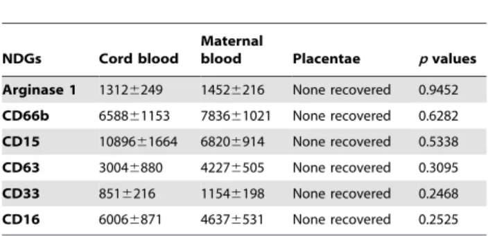
Next, we determined whether there are phenotypic differences in LDGs from the three compartments. As shown in Figure 1 and Table 3, the phenotype of LDGs in placentae, maternal and cord blood was similar, except for the expression levels of CD66b (Figure 2), which was lower in the placentae, as compared to maternal and cord blood. Results in Table 4 show that the phenotype of NDGs is similar in maternal and cord blood; however, NDG could not be isolated from placentae.
Finally, we determined the frequency of LDGs and our results show that there are significantly more LDGs in cord blood as compared to the placentae or maternal blood (Figure 3 and Table 5).
Discussion
Modulation of both innate and adaptive immune responses is crucial in successful pregnancy since the symbiosis between mother and fetus is not due to immunological ignorance but rather to suppression of maternal immune responses. Arginase-mediated L-arginine catabolism is a well-established mechanism of T cell suppression [10,15,16]. We showed previously that arginase activity is significantly increased in the placenta and in maternal blood and that arginase-induced L-arginine catabolism is one of the pathways contributing to suppression of maternal T cells responses in healthy human pregnancies [8]. Furthermore, we demonstrated recently that arginase activity and L-arginine levels return to physiological levels after birth [17].
In the present study we further characterized the phenotype of LDGs and compared it to normal density granulocytes (NDGs) isolated from placental biopsies, maternal and cord blood. At parturition both subsets of neutrophils are present in peripheral
maternal blood and in cord blood, however, from term placentae only LDGs could be isolated and despite using different methods it was not possible to isolate normal density granulocytes from term placentae. After density gradient centrifugation of blood, NDGs sediment with erythrocytes whereas LDGs co-purify with the PBMC fraction suggesting that their density is lower [13]. They also differ phenotypically: NDGs from maternal and cord blood express significantly higher cell surface levels of CD16 and significantly more intracellular arginase as compared to LDGs. The cell surface levels of CD63 were significantly lower in NDGs than in LDGs. In human neutrophils arginase is contained in azurophilic [18] and gelatinous granules and upregulation of CD63 coincides with the release of azurophilic granules [19]. Thus, our results showing that CD63 is increased in LDGs suggest that these cells have been activated and have degranulated. Furthermore, increased expression levels of CD15 have also been associated with degranulation [14]. Indeed, the increased cell surface expression of CD66b, CD15, and CD63 on LDGs support this conclusion.
The frequency of LDGs was highest in cord blood, however, the phenotype of LDGs in maternal blood, term placentae and cord blood was similar and the phenotype of NDGs in maternal and
Table 4.Comparison of expression levels of arginase and phenotypic markers of NDGs in placentae, cord and maternal blood.
NDGs Cord blood
Maternal
blood Placentae pvalues
Arginase 1 13126249 14526216 None recovered 0.9452 CD66b 658861153 783661021 None recovered 0.6282 CD15 1089661664 68206914 None recovered 0.5338 CD63 30046880 42276505 None recovered 0.3095 CD33 8516216 11546198 None recovered 0.2468 CD16 60066871 46376531 None recovered 0.2525
NDGs were isolated from maternal and cord blood (n = 7) as described in materials and methods. Expression levels (MFI) of phenotypic markers were determined by flow cytometry. Statistical significance was determined by a two-tailed Mann-Whitney test.
doi:10.1371/journal.pone.0085696.t004
Figure 3. Percentage of LDGs in neonatal and maternal blood and placentae. LDGs were isolated as described in materials and methods (n = 7) and the percentage of CD15+ arginase+ cells was determined by flow cytometry. The percentage of LDGs present in the peripheral blood of healthy controls was 0.2460.3 [12] Statistical significance was determined by a kruskal-Wallis test. Box = interquartile range and median; whiskers = range.
doi:10.1371/journal.pone.0085696.g003
Table 5.Percent of Arginase1+CD15+LDGs present in placentae, cord blood and maternal blood.
Arginase1+CD15+
LDGs Cord blood
Maternal
blood placentae pvalueip
Arginase 1 5.6661.24 1.72360.95 2.1460.53 0.0237
LDGs were isolated from maternal and cord blood and placentae (n = 7) as described in materials and methods. The percentage of CD15+arginase+cells was determined by flow cytometry. Statistical significance was determined by a Kruskal-Wallis test.
cord blood was also not significantly different. CD15+
neutrophils in the PBMC fraction have not only been observed in pregnancy (present study and [8]. It was also reported in a range of conditions including HIV [12], visceral leishmaniasis [20], cancer [15,16,21], trauma [22], and systemic lupus erythromatosus [23]. All of these conditions are frequently accompanied by various degrees of immune suppression.
Pregnancy has been associated with a shift in Th1/Th2 balance, reduced type 1responsiveness [3,4] and enhanced innate immu-nity. Indeed, the number of neutrophils increases in pregnancy and these cells undergo functional and metabolic changes [24,25,26]. Delayed apoptosis of neutrophils in normal pregnancy promotes inflammation to persist and may contribute to pregnan-cy-associated neutrophilia and pregnancy-induced inflammatory changes in neutrophils in the peripheral blood are akin to those of sepsis [27]. The function of neutrophils in pregnancy has to be very tightly controlled since enhanced inflammatory responses have been linked to pregnancy complications such as preeclamp-sia.
Neutrophils home to inflammatory sites, they can phagocytose pathogens, degranulate and release web-like structures, the neutrophil extracellular traps (NETs) that are implicated in inflammation and immunity [28]. Placental micro-debris has been shown to activate neutrophils and induce the formation of NETs in a dose-dependent manner [29]. NETs have been suggested to contribute to placental hypoxia in patients with preeclampsia [30]; however, the interplay between micro-debris, neutrophil activation and NETs in successful human pregnancies remains to be determined.
Arginase-mediated L-arginine catabolism is not only impacting on maternal immune responses [8]; amino acids such as L-arginine are also important for the developing fetus. Fetal growth is critically dependent on placental nutrient transport; specialized transporters can pass them to the fetus [31]. Placental amino-acid uptake takes place on the microvillous membrane of the syncytiotrophoblast and the efflux to the fetus is mediated by transporters on the basal membrane. The mammalian target of rapamycin (mTOR) pathway in the placenta regulates amino acid transporters such as L-arginine, and it has been proposed that the placental mTOR pathway constitutes a link between maternal nutrients and fetal growth [32].
The signals resulting in activation, degranulation of neutrophils and their development into LDGs in pregnancy remain to be determined and further work is required to identify the pathways leading to the generation of LDGs.
Acknowledgments
The authors would like to thank Dr. G.P. Taylor for critical reading of the manuscript and all the midwives for their outstanding help in sample collection.
Author Contributions
Conceived and designed the experiments: PK IM. Performed the experiments: A. Ssemaganda PK. Analyzed the data: A. Ssemaganda PK. Contributed reagents/materials/analysis tools: LK TGT. Wrote the paper: A. Ssemaganda LK PB LN JM A. Ssetaala NK MM PK IM.
References
1. Mold JE, Michaelsson J, Burt TD, Muench MO, Beckerman KP, et al. (2008) Maternal alloantigens promote the development of tolerogenic fetal regulatory T cells in utero. Science 322: 1562–1565.
2. Hunt JS, Petroff MG, McIntire RH, Ober C (2005) HLA-G and immune tolerance in pregnancy. FASEB J 19: 681–693.
3. Saito S, Nakashima A, Shima T, Ito M (2010) Th1/Th2/Th17 and regulatory T-cell paradigm in pregnancy. Am J Reprod Immunol 63: 601–610. 4. Mor G, Cardenas I (2010) The immune system in pregnancy: a unique
complexity. Am J Reprod Immunol 63: 425–433.
5. Watanabe M, Iwatani Y, Kaneda T, Hidaka Y, Mitsuda N, et al. (1997) Changes in T, B, and NK lymphocyte subsets during and after normal pregnancy. Am J Reprod Immunol 37: 368–377.
6. Chen SJ, Liu YL, Sytwu HK (2012) Immunologic regulation in pregnancy: from mechanism to therapeutic strategy for immunomodulation. Clin Dev Immunol 2012: 258391.
7. Mellor AL, Munn D (2004) Policing pregnancy: Tregs help keep the peace. Trends Immunol 25: 563–565.
8. Kropf P, Baud D, Marshall SE, Munder M, Mosley A, et al. (2007) Arginase activity mediates reversible T cell hyporesponsiveness in human pregnancy. Eur J Immunol 37: 935–945.
9. Ishikawa T, Harada T, Koi H, Kubota T, Azuma H, et al. (2007) Identification of arginase in human placental villi. Placenta 28: 133–138.
10. Bronte V, Zanovello P (2005) Regulation of immune responses by L-arginine metabolism. Nat Rev Immunol 5: 641–654.
11. Rodriguez PC, Ochoa AC (2008) Arginine regulation by myeloid derived suppressor cells and tolerance in cancer: mechanisms and therapeutic perspectives. Immunol Rev 222: 180–191.
12. Cloke T, Munder M, Taylor G, Muller I, Kropf P (2012) Characterization of a novel population of low-density granulocytes associated with disease severity in HIV-1 infection. PLoS One 7: e48939.
13. Schmielau J, Finn OJ (2001) Activated granulocytes and granulocyte-derived hydrogen peroxide are the underlying mechanism of suppression of t-cell function in advanced cancer patients. Cancer Res 61: 4756–4760.
14. Nakayama F, Nishihara S, Iwasaki H, Kudo T, Okubo R, et al. (2001) CD15 expression in mature granulocytes is determined by alpha 1,3-fucosyltransferase IX, but in promyelocytes and monocytes by alpha 1,3-fucosyltransferase IV. J Biol Chem 276: 16100–16106.
15. Munder M (2009) Arginase: an emerging key player in the mammalian immune system. Br J Pharmacol 158: 638–651.
16. Mu¨ller I, Munder M, Kropf P, Ha¨nsch GM (2009) Polymorphonuclear neutrophils and T lymphocytes: strange bedfellows or brothers in arms? Trends Immunol 30: 522–530.
17. Bhanot U, Cloke T, Munder M, Teoh TG, Kropf P, et al. (2013) Arginase-mediated L-arginine catabolism normalizes post partum. submitted for publication.
18. Munder M, Mollinedo F, Calafat J, Canchado J, Gil-Lamaignere C, et al. (2005) Arginase I is constitutively expressed in human granulocytes and participates in fungicidal activity. Blood 105: 2549–2556.
19. Kuijpers TW, Tool AT, van der Schoot CE, Ginsel LA, Onderwater JJ, et al. (1991) Membrane surface antigen expression on neutrophils: a reappraisal of the use of surface markers for neutrophil activation. Blood 78: 1105–1111. 20. Abebe T, Takele Y, Weldegebreal T, Cloke T, Closs E, et al. (2013) Arginase
activity - a marker of disease status in patients with visceral leishmaniasis in ethiopia. PLoS Negl Trop Dis 7: e2134.
21. Raber P, Ochoa AC, Rodriguez PC (2012) Metabolism of L-arginine by myeloid-derived suppressor cells in cancer: Mechanisms of T cell suppression and therapeutic perspectives. Immunol Invest 41: 614–634.
22. Bryk JA PP, Zenati MS, Munera V, Pribis JP, Ochoa JB (2010) Nature of myeloid cells expressing arginase 1 in peripheral blood after trauma. J Trauma 68: 843–852.
23. Denny MF, Yalavarthi S, Zhao W, Thacker SG, Anderson M, et al (2010) A distincy subset of proinflammatory neutrophils isolated from patients with systemic lupus erythrematosus induces vascular damage and synthesizes type I TFNs. J Immunol 184: 3284–3297.
24. Taniguchi K, Nagata H, Katsuki T, Nakashima C, Onodera R, et al. (2004) Significance of human neutrophil antigen-2a (NB1) expression and neutrophil number in pregnancy. Transfusion 44: 581–585.
25. Kindzelskii AL, Huang JB, Chaiworapongsa T, Fahmy RM, Kim YM, et al. (2002) Pregnancy alters glucose-6-phosphate dehydrogenase trafficking, cell metabolism, and oxidant release of maternal neutrophils. J Clin Invest 110: 1801–1811.
26. Kindzelskii A CA, Espinoza J, Maeda N, Aratani Y, Romero R, et al. (2006) Myeloid peroxidase accumulates at the neutrophil surface and enhances cell metabolism and oxidant release during pregnancy. Eur J Immunol 36: 1619– 1628.
27. Sacks GP, Studena K, Sargent K, Redman CW (1998) Normal pregnancy and preeclampsia both produce inflammatory changes in peripheral blood leukocytes akin to those of sepsis. Am J Obstet Gynecol 179: 80–86.
28. Branzk N PV (2013) Molecular mechanisms regulating NETosis in infection and disease. Semin Immunopathol 35: 513–530.
30. Hahn S, Giaglis S, Hoesli I, Hasler P (2012) Neutrophil NETs in reproduction: from infertility to preeclampsia and the possibility of fetal loss. Front Immunol 3: 362.
31. Grillo MA, Lanza A, Colombatto S (2008) Transport of amino acids through the placenta and their role. Amino Acids 34: 517–523.