J of Evidence Based Med & Hlthcare, pISSN- 2349-2562, eISSN- 2349-2570/ Vol. 2/Issue 52/Nov. 30, 2015 Page 8653
COMPARATIVE STUDY OF CYTOLOGIC AND COLPOSCOPIC FINDINGS IN PRECLINICAL
CERVICAL CANCER
Penagaluru Radha1, Yaragani Padma2, Mrudula Priyanka3
1Associate Professor, Department of Obstetrics & Gynaecology, Kakatiya Medical College, Warangal, Telangana. 2Associate Professor, Department of Obstetrics & Gynaecology, Kakatiya Medical College, Warangal, Telangana. 3Post Graduate, Department of Obstetrics & Gynaecology, Kakatiya Medical College, Warangal, Telangana.
ABSTRACT
BACKGROUND
The cytologic diagnosis of cervical smears has become a very important screening test for the detection of pre-invasive and invasive cervical epithelial abnormalities.
MATERIALS AND METHODS
It is a prospective study conducted for a period of 1 year in 100 women who fulfilled the inclusion criteria. Colposcopy, PAP smear and biopsy were done.
RESULTS
Majority 70.5% i.e., (12/17) of CIN occurred in the age group of 30-49 years. Among the 9 women who took OCP, 12% (2/17) had CIN. Incidence of CIN in the permanently sterilized group was 59% (10/17) and among IUCD user was 5.9% (1/17). Among women who were diagnosed to have CIN, 70.5% (12/17) complained of excessive vaginal discharge 11.7% (2/17) of women had post-coital bleeding. PAP smear had a sensitivity of 29% and a specificity of 88% which was attributed to the high number of false, negative smears. Colposcopy showed a sensitivity of 82% and a specificity of 81%. Sensitivity was more than pap smear but specificity was less than pap smear. Accuracy of Colposcopy was found to be 82% which was comparatively more accurate than pap smear (78%).
CONCLUSIONS
COLPOSCOPY offers an excellent tool in evaluating cervical lesions. It is an easy and perspective method and its importance lies in teaching, diagnosis and management of cervical lesions, both neoplastic and non-neoplastic.
KEYWORDS: Colposcopy, PAP smear, CIN.
HOW TO CITE THIS ARTICLE: Penagaluru Radha, Yaragani Padma, Mrudula Priyanka. “Comparative Study of Cytologic and Colposcopic findings in Preclinical Cervical Cancer”. Journal of Evidence based Medicine and Healthcare; Volume 2, Issue 52, November 30, 2015; Page: 8653-8660, DOI: 10.18410/jebmh/2015/1199
INTRODUCTION: Cancer of cervix continues to be the most common genital cancer encountered in India accounting for 80% of all female genital tract cancers. The concept of pre-invasive disease of cervix, which denotes changes that are confined to the cervical epithelial cells, was introduced in 1947. Early detection of pre-invasive disease and treatment of CIN, has the potential to improve the outcome of patients.1
Invasive cancer of cervix is considered to be a preventable condition, since it is associated with a long pre-invasive stage (CIN), making it amenable to screening and treatment. The easy accessibility of the cervix to clinical examination, application of cytologic and tissue sampling procedures have led to extensive screening programme for early detection and treatment of the disease.
Carcinoma cervix is the 2nd most common cancer of the genital tract cancer in females, next to carcinoma of breast. Approximately 500,000 new cases are estimated across the world every year. In India, the incidence of cervical cancer varies from 20 to 35/100,000 women between the age group of 35 to 64 years while in developed countries it is as low as 1-8/100,000 women. In India approximately 529000 new cases of cervical cancer are identified across the world every year and 275000 deaths/year.2
As invasive squamous cell carcinoma of the cervix is preceded by an Intraepithelial stage (CIN), which evolves over many years. There is an enormous opportunity for screening with the prospect of detection of cancer at the pre-clinical stage. The avoidable cancer deaths predominantly reflect the limitation of our cancer screening programmes which can be effectively done by a Pap smear, Colposcopy and cervical biopsies.
Pap smear cytological examination and Colposcopy are the principal methods used in the early detection of cancer of the uterine cervix. Their use depends on how closely their findings correlate with each other and with histology. To detect this widely prevalent cancer at an early stage, the
Submission 08-11-2015, Peer Review 09-11-2015 Acceptance 23-11-2015, Published 28-11-2015. Corresponding Author:
Dr. Pengaluru Radha, Associate Professor Department of Obstetrics & Gynaecology Kakatiya Medical College, Warangal, Telangana. E-mail: [email protected]
J of Evidence Based Med & Hlthcare, pISSN- 2349-2562, eISSN- 2349-2570/ Vol. 2/Issue 52/Nov. 30, 2015 Page 8654
simplest test has been a Pap smear. Reporting of Pap smears is done by using The Bethesda System 2001 prior to which many classification systems were developed. Traditionally the gold standard for assessing the performance of Pap smear has been histopathology.3 Although Papanicolaou smear
represents the most effective technique to prevent and detect precancerous lesions of uterine cervix. Its false negative results are still a reason of concern to the pathologist and gynaecologist. Hence histopathology control is one of the best ways to assess the accuracy of cytology diagnosis.
Traditionally, the tissue result has been the gold standard against which the accuracy of cytopathology is measured. Because sampling error and lesion regression account for many not correlating cytologic-histologic samples, there is a growing acceptance of consensual peer review of the Pap test as the accuracy standard. Inconsistencies between histopathologic and cytopathologic reporting terminologies and lack of standardized histopathologic criteria with poor inter and intra observer reproducibility rates are additional reasons for this diminishing confidence in the tissue result of the gold standard. The cytologic categories, Atypical Squamous Cells of Undetermined Significance (ASCUS) and the newly described squamous lesions, atypical immature metaplasia, papillary immature metaplasia, and transitional cell metaplasias exemplify these controversies and challenges. When cervical cytology has been applied in mass screening, there has been a reduction in the frequency and death rates from cervical carcinoma, the histogenesis and progression of cervical carcinoma is well documented. It is possible to prevent the development of invasive carcinoma by identifying and treating pre-invasive lesions. The efficacy of cervical smear study was established by George N. Papanicolaou in 1928.4
It has been known that the squamocolumnar junction of cervix is the site of predilection for carcinoma of the cervix. In an effort to study early malignant changes in the squamous cells thrown off specifically from this focus, the spatula cytology technique was developed and this is a means of collecting the cells before their exfoliation. There is evidence of falling mortality in most countries with well developed screening programmes. Tragically in many of the third world countries where the incidence of cervical cancer is much higher, the resources for comprehensive population screening are simply not available.5
Cytological examination of material from the female genital tract is not confined to the diagnosis of precancerous changes in the cervix. Reactive and infective conditions of the cervix and vagina can also be recognized. With an increase in the incidence of HIV infection there is an increase in the incidence of HPV infection of cervix. HPV infection is a known precursor of pre-neoplastic and neoplastic lesions of the cervix. Hence, there is a need to study various neoplastic and non-neoplastic lesions of cervix by cervical smear study and correlate with histopathological findings when needed.
The main aim of this study is to assess the accuracy of pap smear cytology and colposcopic examination as screening procedures in preclinical cervical cancer.
MATERIALS AND METHODS: This is a prospective clinical study of 100 women attending the Gynaecology OPD of CKM Government Maternity Hospital, Warangal. Study Period is two years from 2013-2014. A total of 100 cases were considered for the study and patients were subjected to Pap smear, Colposcopy and colposcopic directed cervical biopsy after taking informed consent. Relevant Obstetrics & Gynaecology history was taken and recorded.
Inclusion Criteria:
1. Women of age between 20-65 years presenting to the gynaec OPD with/without symptoms.
2. Women with symptoms like vaginal discharge, postcoital bleeding, postmenopausal bleeding, intermenstrual bleeding and persistent Leucorrhoea not responding to antibiotics.
3. Women with normal looking cervix but symptomatic. 4. Women with cervical lesions like polyps, erosion,
hypertrophied cervix, cervix with nabothian cyst. 5. Women with clinical evidence of acute pelvic infection.
Exclusion Criteria:
1. Women with vaginal bleeding at the time of examination.
2. Women who had been previously treated for carcinoma cervix
3. Pregnant women.
4. Women with frank lesions.
5. Women with previous cervical surgery.
PAP SMEAR: Place the patient in dorsal position, labia
aparted and the cusco’s self-retaining speculum gently introduced without the use of lubricant or jelly. Cervix is exposed; the squamocolumnar junction was scraped with
Ayre’s spatula by rotating the spatula all around. The scrapings were evenly spread onto glass slide, and immediately fixed by dipping the slide in the jar containing equal parts of 95% alcohol and ether.
Smear is classified as per The Bethesda 2001 classification system.
Cervical cytology correlated with Colposcopy and colposcopic directed biopsy.
J of Evidence Based Med & Hlthcare, pISSN- 2349-2562, eISSN- 2349-2570/ Vol. 2/Issue 52/Nov. 30, 2015 Page 8655
OS was moistened with normal saline. This increases the transparency of Cervix and provides a clearer view of vascular pattern when visualized with green filter. 5% acetic acid was applied on the cervix for 2 minutes. After 45 seconds the epithelial changes were noted and recorded. Biopsy was taken from abnormal areas.
Favourable period for colposcopic examination is 8-10th day of a cycle as the external OS is widely open during this time and abundance of watery secretions serve as a good refractory medium and facilitate examination of end cervix.
If the upper limit of TZ is not visible, the examination may be rescheduled and the patient is instructed to take Ethinylestradiol 50 µg/ dl given/5-7 days prior to examination which causes canal dilatation. In 15%, unsatisfactory colposcopic findings are seen, where Endocervical curettings are taken.
2002, International Federation of cervical pathology and Colposcopy (IFCPC) devised new colposcopic terminology (Barcelona).
Correlation between cervical cytology and histology findings and statistical analysis to assess the sensitivity and specificity. Results of pap smear cytology and colposcopic examination of each patient will be correlated with each other and with final histological diagnosis.
J of Evidence Based Med & Hlthcare, pISSN- 2349-2562, eISSN- 2349-2570/ Vol. 2/Issue 52/Nov. 30, 2015 Page 8656 RESULTS: Patients were subjected to Pap smear and
cervical biopsy.
Age In Years Total cases CIN cases
No. % No. %
20-29 13 3 1 6
30-39 38 38 6 35
40-49 31 31 6 35
50-59 18 18 4 24
Education
Illiterate 32 32 11 61
1st to 10th std 54 54 6 33
Degree 14 14 1 6
Table 1: Demographic distribution
Out of 100 patients 31% were in the age group of 40-49 years, Incidence of CIN was found to increase as age increases. In The mean age was 37.8 years.
Among 100 women 54% had primary /high school education higher.Among the illiterates 61% had CIN, This showed that there is higher incidence of CIN among illiterates than literates.
Income per month
Total cases
CIN
cases %
Less than 1000 77 15 19.5
1001-150 16 2 12.5
1501-2000 7 - -
Duration of marriage
<5yrs 13 1 7.6
5 to 10 years 35 4 11.4
11 to 20 years 32 7 21.8
>20 years 20 5 25
Contraception
Barrier 5 - -
OCP 9 2 11
IUCD 17 1 5
Permanent 39 10 59
Nil 30 4 25
Parity
1 8 1 1
2 34 7 2
3 38 6 3
>5 20 3 15
Table 2: Details in the study
Among 100 women studied, 32% were married for 11-20 years. There was high incidence of CIN, when the duration of exposure of sexual intercourse. CIN 39% had permanently sterilized, among them 59% (10/39) had CIN. 30% of women did not practice any form of contraception, among CIN.
Complaints Total Cases N=100
CIN Cases
N=17 %
White discharge 56 12 21.4
PCB 7 2 28.5
Int.M.B 11 1 9.09
PMB 5 2 40.00
Loss of wt 5 - -
Others 16 - -
Total 100 17
Table 3: Chief Complaints
The common complaints were white discharge & bleeding per vagina which was either post coital, intermenstrual or of post menopausal type.
PAp Smear Findings Total Cases N=100
Normal 5
Inflammatory atypia 80
Mild dysplasia 10
Moderate dysplasia 3
Severe dysplasia 2
Invasive cancer -
Coloscopic appearance
Normal 3
Erosion cervix 31
Inflammatory changes 16
Polyps 5
Leukoplakia 2
AW areas 17
Punctate pattern 8
Mosaic pattern 4
Atypical vessels -
Unsatisfactory 14
J of Evidence Based Med & Hlthcare, pISSN- 2349-2562, eISSN- 2349-2570/ Vol. 2/Issue 52/Nov. 30, 2015 Page 8657 HPE findings
Chronic cervicitis 46 Chronic cervicitis +erosion 28
Erosion cervix 2
Epithelial hyperplasia 2
Polyp (benign) 5
Mild dysplasia 8
Moderate dysplasia 5
Severe dysplasia 4
Table 3: PAP smear, Colposcopic and HPE findings of cervix
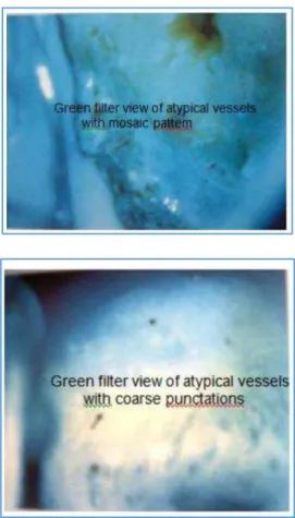
PAP smear was taken for all patients. 5% of smear were found to be normal, 80% showed inflammatory atypia, 10% showed mild dysplasia, 3% showed moderate dysplasia and 2% showed severe dysplasia. Among the 100 cases studied, 29% 29(/100) were diagnosed as colposcopically abnormal. Among the abnormal cases, AW areas were diagnosed in 17%, punctate pattern of vessels was seen in 8 % of women and mosaic pattern of vessels was diagnosed in 4% of women.
All 100 cases were subjected to colposcopically directed biopsy. Majority of cases, 46% had chronic cervicitis.
Total cases
CIN cases N=17 % Clinical appearance of cervix
Atrophy 2 - -
Congestion 16 2 16.9
Erosion cervix 59 10 12.5 Hypertrophy+congestion 6 2 33.4 Hypertrophy+erosion 12 3 25
Polyps 5 - -
Total 100 17 -
Lugol’s Iodine Application
Positive 24 - -
Partial positive 46 5 11
Negative 30 12 40
Total 100 17 -
AW area within TZ
Flat AW area with sharp
margins 17 6 35.2%
Dense, opaque AW area with sharp margin with punctate / mosaic pattern.
12 11 91.6%
Total 29 17
Table 4: Appearance of Cervix
Erosion of cervix was found in majority 59% of patients. Among women with erosion cervix 12.5% (10/59) had CIN.
Mature squamous epithelium stained deep brown with
lugol’s iodine, called Iodine positivity was found in 24% of
cases. Among them none had CIN. Iodine partial positivity was found to be 46% speckled or variegated appearance with an area of slight AW area change that might be due to Immature metaplasia, regenerating epithelium or CIN 1.
PAP smear
No. Normal HPE
No. % Mild dysplasia Moderate dysplasia Severe dysplasia Total
No % No. % No. % No. %
Normal/
inflammatory atypia 83 71 87.9 5 6 4 4.8 3 3.6 12 14.5
Mild dysplasia 10 8 80 2 20 - - - - 2 20
Moderate dusplasia 3 1 33.3 - - 2 66.7 - - 2 66.7
Severe dysplasia 4 1 25 - - - - 3 75 3 75
Total dysplasia smears 17 10 66.7 2 11.8 2 11.8 3 17.6 7 41.2
Table 5: Comparision of PAP Smear With HPE Result
Abnormal colposcopic findings No. of cases % HPE
CIN1 CIN2 CIN3 Invasive CA
AW epithelium 17 58.6 5 1 - -
Punctate 8 27.5 3 3 1 -
Mosiac 4 13.7 - 1 3 -
Atypical vessels - - - -
Total 29 100 8 5 4 -
J of Evidence Based Med & Hlthcare, pISSN- 2349-2562, eISSN- 2349-2570/ Vol. 2/Issue 52/Nov. 30, 2015 Page 8658
Test True
Positive
False Positive
True Negative
False
Negative Sensitivity Specificity PPV NPV
Pap smear 5 10 73 12 29.4 87.9 33.3 85
Colpo-scopy 14 15 68 13 82 81 48.2 96
Table 7: Sensitivity And Specificity In Pap Smear And Colposcopy
Accuracy for pap smear= 78%. Accuracy for Colposcopy= 82%.
DISCUSSION: The cytologic diagnosis of cervical smears has become a very important screening test for the detection of pre-invasive and invasive cervical epithelial abnormalities. Screening of female population for cervical Neoplasia is a simple, inexpensive and reliable method which greatly reduces the mortality and morbidity associated with carcinoma cervix, if detected in its pre-invasive stage.
Cervical cytodiagnosis has been the subject of several investigations evaluating the efficacy of this method as a diagnostic test. The present study was conducted to know the accuracy of cervical cytology in diagnosis of cervical Neoplasia. An attempt has been made to compare the various parameters in the study with the results obtained by different workers.
In the present study screening was done in 100 women with abnormal symptoms like excessive white discharge, post coital bleeding, post menopausal bleeding etc. women with unhealthy cervix, and women with dysplastic smears, with Colposcopy and its results were correlated with pap smear and biopsy to determine the sensitivity and specificity of these methods in detecting CIN. Regarding age distribution, high incidence of CIN was found among age group of 30-49 years with mean age of 41 years, which was seen in 19% of cases. Kushtagi and Fernandez,6 in their study showed the
prevalence of CIN was higher in women over 30 years. Vaidya, et al 7 showed in his study that CIN was more
prevalent in the age group of >35 years. Shalini, et al8
showed the mean age of patients with cancer cervix was 41 vs 32 in patients with benign pathology in cervix.
Regarding parity, our study showed increased incidence of CIN among multiparous women. 20.5% were para 2, 15.7% were para 3 and 15% were para 4 or more.
Similar study by Shalini, et al8 showed the mean parity
was 4.2 in patients with invasive cancer. Kushtagi and Fernandez6 showed the prevalence of CIN was significantly
higher in parity of more than 4. Vaidya7 showed more positive
cases of CIN were found with parity more than 4. This might be attributed to hormonal and nutritional changes that occur in pregnancy, immunosuppression during pregnancy and cervical trauma during delivery (Becker, et al and Adadevoh, et al).9
Regarding the literacy, CIN was more prevalent among the illiterates, in our study, 61% (11 out of 32) of CIN was found among the illiterates. This was attributed to lack of awareness of symptoms and failure to seek medical care. Socio-economic status had always been playing an epidemiological role in genesis of dysplasia. In our study, the incidence of CIN was found to be higher among the low income group (19.5%) Vaidya7 had showed that low
socio-economic status had a definite role in the development of Dyskaryosis. In his study 80% of CIN I and 50% of CIN II were from the low income group. Poor personal hygiene, poor living conditions, unstable marriages, and early age at first intercourse are factors associated with both low socio-economic conditions and cervical cancer.
Duration of marriage and duration of exposure to sexual intercourse had a distinct role in genesis of cervical dysplasia. In our study, the incidence of CIN was 22% in women married for 11-20 years and 25% among women who were married for > 20 years. Kushtagi, et al6 had demonstrated
the severity of underlying CIN increased with increase in the duration of marital life and hence the increase in the duration of sexual intercourse.
Increasing number of sexual partners had the effect of increasing the risk of developing CIN and invasive disease. Sex with high risk males was also another risk factor for the development of CIN.
The relationship between oral contraceptives and development of CIN had been investigated by Research IARC- International agency for Research in Cancer and they concluded that the use of OCP increased the risk of CIN upto 4 fold after 5 or more years among the HPV DNA positive women. In our study, we found that none of the women who practiced barrier contraception had CIN. Out of 17% of IUCD users, the incidence of CIN was 5% (1/17). Out of 39% of women who had undergone sterilization permanently, the incidence of CIN was 59% (10/39).
Prospective studies by Stern, et al10 in Los Angeles
suggested an increased risk of progression of cervical dysplasia among the users of hormonal contraceptive. Vaidya, et al7 in their study showed 40% of risk of CIN 1 in
J of Evidence Based Med & Hlthcare, pISSN- 2349-2562, eISSN- 2349-2570/ Vol. 2/Issue 52/Nov. 30, 2015 Page 8659
Among the complaints, majority of women (56%) complained of excessive white discharge 21.4% (12/56), playing a role in contributing to the development of CIN, was also proved to be a risk factor in the study conducted by Vaidya, et al7. In their study, 24% had vaginal discharge.
Postcoital bleeding was found in 7% (7/100) of cases. Among them CIN was found in 28.6% (2/7). Shalini R., Amitha S.,8 in their study showed the relationship of post
coital bleeding and CIN. In their study, among the women who had postcoital bleeding, 85.5% had benign findings, 5.6% had HPV and CIN 1,3.6% had CIN 2 and 3 and 55% had invasive cancer.
There was no correlation between the duration of bleeding and pathology. Among those with intermenstrual bleeding, 9.09% (1/11) had CIN. Among those with post menopausal bleeding 40% (2/5) had CIN.
Regarding the clinical appearances of cervix, the most common finding was erosion of cervix where the squamous epithelium of ectocervix was replaced by the columnar epithelium of endocervix. Erosion was seen in 59% (59/100), rest of the patients showed congestion in 16%. Hypertrophy with congestion seen in 6%, hypertrophy with erosion was seen in 12% and polyp was found in 5% of cases.
CIN was found in 16.9% (2/16) in women who showed congestion, 12.5% (10/59) in women who showed erosion and 33.3% in women with hypertrophy + congestion and 25% in women with hypertrophy + erosion.
5% acetic acid application produces suspicious areas in 29% (29/100) cases. Among them, AW areas without any vascular pattern was found in 17.5%, punctate pattern was seen in 4% and 2% showed mosaic pattern. Among those with AW areas, 35.2% (6/17) were found to be CIN positive. Among those with dense opaque acetowhite areas, 91% (11/12) had CIN. Londhe M. Seshadri in their study showed VIAM had a sensitivity of 72.4% and a specificity of 54% and a false negative rate of 15.2%.
Lugol’s iodine application produced iodine positivity in 24%. Among them, none had CIN. CIN was found in 29.4% (5/17) in partial iodine positivity and 70.5% (12/17) in iodine negativity.
Pap smear was taken for all cases. It showed mild dysplasia in 10%, moderate dysplasia in 3% and severe dysplasia in 2%. Pap smear correctly estimated CIN in 78% and underestimated in 10% and overestimated in 12% (false positivity).
Sensitivity of Pap smear was found to be very low - 29% compared to its specificity which was 88%. This attributed to the high number of false negative smears.
Authors Sensitivity Specitivity
Londhe M, George S,
Seshadri I 13.2% 96.3%
Shalini R, Amith S, Neera
M. A.8 56% 90%
Basu PS and
Sankaranarayanan12 29.5% 92.3%
Pete I, Toth V, Bosze P13 47% 77%
Sukhpreet Singh14 20% 91.25%
Present study 29.4% 87.9%
Table 8: Sensitivity and specificity of pap smear by various authors
This data suggests that with Colposcopy as a screened tool, the rate of false negative cytology could be significantly reduced. Colposcopy enhanced cervical screening particularly in women with otherwise negative smears.
Correlation between cytology and HPE was poor as far as mild dysplasia were concerned. But the correlation was good for moderate and severe dysplastic lesions. Correlation between colposcopic findings and biopsy showed a good correlation for higher grade lesions (CIN 2 and CIN 3). Sensitivity was found to be 83% and specificity was 81%. This showed a high sensitivity and a low specificity when compared to pap smear. Low specificity when compared to Pap smear was due to the high incidence of unsuspected AW epithelium which might be due to inflammation, immature metaplasia, and latent HPV infections. Out of 17 cases which showed AW areas without any vascular pattern only 5 were confirmed on biopsy.
Authors Sensitivity Specificity
Pete I, Toth V, Bosze P13 87% 15%
Olaniyan B Meta analysis15 87-99% 26-87%
Massad L S Collins Y C 16 89% 52%
Kier Kegaard, et al17 67% -
Sukhpreet L Singh, et al14 95% 63.5%
Present study 82% 81%
Table 9: Sensitivity and specificity of Colposcopy by various authors
Colposcopy and biopsy were positive in 14 out of 17 (82.4%) cases while Pap smear and biopsy were positive in only 5 out of 17 (29.4%) cases. This indicated the usefulness of Colposcopy in diagnosing lesions missed by pap smear.
Olaniyan, et al15 did a meta analysis of eight longitudinal
studies and compared the correlation of Colposcopy impression with biopsy results. Colposcopy accuracy was found to be 89% which agreed exactly with histology in 61% of cases. In the present study, the accuracy of colposcopic impression was found to be 82%.
CONCLUSION: Colposcope in general has a role in the evaluation of women with abnormal pap smears, unhealthy cervix, and seems to be more accurate in detecting CIN. Hence, primary Colposcopy can be incorporated into genitourinary tract screening at first visit.
J of Evidence Based Med & Hlthcare, pISSN- 2349-2562, eISSN- 2349-2570/ Vol. 2/Issue 52/Nov. 30, 2015 Page 8660 REFERENCES:
1. Papanicolaou GN, Traut AF: The diagnostic value of vaginal smears in carcinoma of the cervix. Am J Obstet Gynecol 1941;42:192-206.
2. GLOBOCAN 2008 (IARC), Section of Cancer Information (8/10/2012). (WHO, 2009); GLOBOCAN 2002 database, IARC.
3. Albert SO, Oguntayo OA, Samaila MAO. Comparative study of visual inspection of the cervix using acetic acid and papanicolaou smears for cervical cancer screening. Ecancer medical science 2012; 6:262. 4. Gan, Tel Aviv, Israel Colposcopic Terminology of the
International Federation for Cervical Pathology and Colposcopy Obstetrics and Gynaecology 2012; 120(1):166-72.
5. Dasari P. A grossly abnormal cervix: Evidence for using Colposcopy in the absence of a squamous intraepithelial lesion by the conventional
Papanicolau’s test. Journal of Gynaecologic Surgery. 2011 March; 27(1) 2.
6. Kushtagi P, Fernandez. P. Significance of persistent inflammatory, cervical smears in sexually active women of reproductive age. J of Obst & Gynaecol. of India 2002; 52(1):124-6.
7. Vaidya, Olaniyan OB. Validity of Colposcopy in the diagnosis of early cervical neoplasia a review – African Journal of Reproductive Health 2002, 6:59-69. 8. Shalini R, Amitha S, Neera MA. How alarming is post coital bleeding—A cytologic, colposcopic and histological evaluation. Gynaecol Obstet Invest. 1998, 45(3):205-8.
9. Adadevoh SW, Forkouh BK. Cervical cancer screening. International Journal of Gynaecology and Obstretics; 1993; 43(1):63-4.
10. Stern E. Epidemiology of dysplasias, Obstet gynaecol survey 1969, 24:711-23.
11. Duggan, et al, Natural history of CIN lesions, European J Gynaecol Onco 1988, 19(4)338-44. 12. Basu PS, Shankaranarayanan R, Mandal R, Roy C, Das
P. Visual inspection of cervix with acetic acid and cytology in the early detection of cervical neoplasia in Kolkata-India. International Journal of Cancer 2005; Sept-Oct 13(6):626-632.
13. Pete I, Toth. V, Bosze. P. The Value of Colposcopy in screening of cervical carcinoma. European Journal of Gynaecol Oncology 1998, 19(2):120-2.
14. Sukhpreet L Singh, Nayana A Dastur, Murari S. Nanavatti; Comparison of Colposcopy and Pap smear: Sensitivity, specificity and predictive values. BHJ No. 4, Vol. 42, Oct 2000.
15. Olaniyan OB. Validity of Colposcopy in the diagnosis of early cervicalneoplasia a review. African Journal of Reproductive Health 2002,6:59-69 11.
16. Massad LS, Jeronimo J, Katki HA, Schiffman M. The accuracy of the colposcopic grading for the detection of high-grade cervical intraepithelial neoplasia. J Low Genit Tract Dis. 2009 Jul;13(3):137-44.