Recebido em 29.11.2001. / Received in November, 29thof 2001.
Aprovado pelo Conselho Consultivo e aceito para publicação em 04.06.2002. / Approved by the Consultive Council and accepted for publication in June, 04t hof 2002. * Trabalho realizado no Serviço de Dermatologia do Hospital das Clinicas da Universidade Federal de Minas Gerais. / Work done at Dermatology Service of “Dermatologia do Hospital das Clinicas da Universidade Federal de Minas Gerais".
1Residente em Dermatologia do Hospital das Clínicas - UFMG. / Dermatology Resident at the Hospital das Clínicas - Federal University of Minas Gerais (HC-UFMG).. 2Graduando em Medicina (12o Período) da Faculdade de Medicina - UFMG. / 12th period medical student, Faculty of Medicine - UFMG.
3Professor Assistente de Dermatologia do Departamento de Clínica Médica da Faculdade de Medicina da UFMG, Mestre em Medicina, Coordenador do Ambulatório de Hanseníase do HC - UFMG. / Assistant professor of Dermatology, Dept. of Clinical Medicine, Faculty of Medicine - UFMG, Masters in Medicine, Coordinator of the Leprosy Ambulatory HC - UFMG. 4Professor Adjunto de Dermatologia do Departamento de Clínica Médica da Faculdade de Medicina da UFMG, Doutor em Medicina, Coordenador do Serviço de Dermatologia do HC-
UFMG. / Adjunct Professor of Dermatology, Dept. of Clinical Medicine, Faculty of Medicine - UFMG, Ph.D. Medicine, Coordinator of Dermatology service, HC- UFMG.
©2003by Anais Brasileiros de Dermatologia
Linfoma não Hodgkin simulando hanseníase
virchowiana
*
Non-Hodgkin's Lymphoma simulating Lepromatous
Leprosy
*
Vanessa Barreto Rocha
1Saôny Victor de Carvalho
2Marcelo Grossi Araújo
3Antônio Carlos Martins Guedes
4Resumo: Os autores relatam caso de linfoma não Hodgkin em paciente do sexo feminino, de 28 anos, ressaltando o diagnóstico diferencial com formas multibacilares de hanseníase. Além de achados clínicos passíveis de confusão, a histologia mostrava, de modo não usual, infiltrado inflamatório mononuclear peri-neural e perianexial.
Palavras-chave: diagnóstico; diagnóstico diferencial; hanseníase; linfoma não Hodgkin.
Summary:The authors report a case of a 28-year-old woman with non-Hodgkin's lymphoma, first diagnosed as multibacillary leprosy. The differential diagnosis is discussed, with emphasis on leprosy, since there were similarities in the clinical aspects. Furthermore the histopathological findings were unusual, displaying perineural and periannexal inflammatory infiltrate composed of mononuclear cells.
Key-words: diagnosis; diagnosis, differential; leprosy; lymphoma, non-Hodgkin.
INTRODUÇÃO
Os linfomas não Hodgkin representam um grupo he-terogêneo de neoplasias com diferenças na apresentação clínica, histologia e no curso clínico. Os linfomas cuja mani-festação clínica inicial se dá na pele são freqüentemente de diagnóstico difícil e têm comportamento clínico enigmático. O exame histopatológico deve distinguir os linfo-mas de várias entidades benignas que cursam com infiltra-do inflamatório linfomatóide. Dessas podem ser citainfiltra-dos linfocitoma cútis, infiltração linfocítica de Jessner, pitiríase liquenóide aguda, hiperplasia linfóide cutânea e, neste
caso, de modo não usual, hanseníase.1,2Do ponto de vista
clínico, o polimorfismo das lesões cutâneas coloca várias
INTRODUCTION
Non-Hodgkin's lymphomas represent a heteroge-neous group of neoplasias with differing clinical presenta-tion, histology and clinical course. Those lymphomas with initial clinical manifestation in the skin are frequently diffi-cult to diagnose and present an enigmatic clinical behavior.
The histopathological exam should distinguish between the lymphomas of various benign entities which course with an inflammatory lymphomatoid infiltrate. These include cutaneous lymphocytoma, Jessner's lymphocytic infiltration, acute lichenoid pthiriasis, cutaneous lymphoid hyperplasia and, as in this case though unusually, leprosy.1,2
diagnos-possibilidades diagnósticas e, quando ocorre infiltração da pele e surgem placas e nódulos, a possibilidade de hanseníase multibacilar deve ser considerada.
RELATO DO CASO
Paciente do sexo feminino, de 28 anos, servente escolar, natural de Ubaporanga e residente em Caratinga, MG, foi atendida pela primeira vez no Serviço de
Dermatologia do HC-UFMGem setembro de 2000. Relatava
ser hígida até fevereiro, quando iniciou com lesões hiper-crômicas assintomáticas no abdômen e dorso que, após biopsiadas, foram diagnosticadas como hanseníase. Foi
tratada com poliquimioterapia esquema II da OMS(PQT II)
durante seis meses, evoluindo com anemia importante, atribuída à hemólise por dapsona. Apresentou, também, edema poliarticular, piora das lesões da pele e surgimento de nódulos cervicais, interpretados como quadro reacional, para o qual foi iniciada prednisona 1mg/kg/dia.
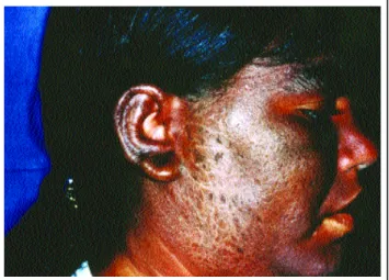
Ao exame clínico apresentava-se com estado geral comprometido, placas ictiósicas e várias lesões escleroder-miformes disseminadas; a face mostrava-se infiltrada bem como, de modo marcante, os pavilhões auriculares, não se notando madarose (Figuras 1 e 2). Não apresentava neurite ou espessamento neural nem alteração de sensibilidade. Apresentava, ainda, linfadenomegalia muito importante em várias cadeias, com linfonodos endurecidos, conflu-entes, aderidos. O exame abdominal revelou fígado a 8cm do rebordo costal direito e tamanho do baço aumentado (Boyd III).
Exames complementares: adenomegalia hilar impor-tante na radiografia de tórax, hipoalbuminemia (2,8g/dl), anemia importante (Hb 8,2g/dl), normocítica
normocrômi-ca, leucocitose (12500 células/mm3) com linfocitose (77%
= 9625) e presença de blastos no sangue periférico. A
tic possibilities and when associated with infiltration of the skin with onset of plaques and nodules, the possibility of multibacillary leprosy should be considered.
CASE REPORT
Female patient, 28 years old, school worker, born in Ubaporanga and resident in Caratinga, Minas Gerais State, was attended for the first time at the Dermatology Service of
HC-UFMG [Hospital das Clínicas, Federal University of Minas Gerais] in September, 2000. She was reportedly healthy until February, when she noticed the onset of asymp-tomatic hyperchromic lesions in the abdomen and back. After biopsy these were diagnosed as leprosy. She was treated with polychemotharapy (PQT) according to the WHOregimen II (PQT II) for six months, coursing with important anemia, attributed to dapsone induced hemolysis. She also presented polyarticular edema, aggravation of the skin lesions and onset of cervical nodules, this was interpreted as a reaction-al picture, for which 1mg/kg/day prednisone was initiated.
At clinical exam she presented with a poor general state, ichthyosiform plaques and several sclerodermiform disseminated lesions; her face was infiltrated and also the auricular pavilions to marked degree, madarosis was not apparent (Figures 1 and 2). She did not present neuritis, neural thickening nor sensitivity alterations. Furthermore, she presented very important enlarged lymph nodes in sev-eral chains, with hardened, confluent and adhered lymph nodes. Abdominal exam revealed liver 8cm from the right costal arch and enlarged spleen (Boyd III).
Complementary exams: important hilar ade-nomegaly in the chest x-ray, hypoalbuminemia (2.8g/dl), significant anemia (Hb 8.2g/dl) which was both normocyt-ic and normochromnormocyt-ic, leukocytosis (12500 cells/mm3) with
lymphocytosis (77% = 9625) and presence of blasts in the
Figura 1: Placas ictiósicas e esclerodermiformes na hemiface direita, com infiltração difusa, principalmente nos pavilhões
auriculares. / Figure 1: Icteroid and sclerodermatous plaques
in right hemifacial region, with diffuse infiltration, specially in the auricles.
Figura 2: Placas ictiósicas e esclerodermiformes no antebraço
direito. / Figure 2: Icteroid and sclerodermatous plaques in right
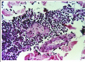
Figure 3: Histological skin sec-tions, stained with hema-toxylin and eosin (100x), showing dense, inflammatory, perivascular and periadnexal infiltrate, comprising lymphoid cells, with discrete cellular atypia and some mitotic figures, the majority of lymphocytes were well differentiated.
Figura 3: Cortes histológicos de pele corados pela hematoxilina-eosina, em aumento de 100x, mostrando denso infiltrado
inflamatório perivascular e perianexial, constituído por células linfóides, com atipias celulares discretas, algumas figu-ras de mitose, sendo a maioria dos linfócitos bem
diferenciados.
peripheral blood. The test for alcohol-acid-resistant bacilli (Baar) in the lesions, ear lobes and elbows was negative.
Biopsies were performed in five sites of the skin and histology showed a dense infiltrate comprised of lymphoid cells, with discreet cellular atypia and some mitotic figures, the majority of which well-differentiated lymphocytes, arranged particularly in a perivascular manner, which were at times confluent, occupying the entire dermis and hypodermis (Figure 3). The diagnosis of non-Hodgkin's lymphoma was reached. Biopsy of the lymph node presented subversion of its architec-ture by a monotonous mass of atypical cells with evident nucle-oli and high mitotic index - compatible with non-Hodgkin's lymphoma. CD3immunohistochemistry was slightly positive,
suggestive of diffuse T cell lymphoma (Figures 4 and 5). Myelogram showed a medulla with 84.4% hypercel-lular infiltration by blasts and with characteristics of acute lymphoblastic leukemia (ALL - L2).
Following the diagnosis of leukemid lymphoblastic non-Hodgkin's lymphoma, clinical evaluation and negative laboratory findings for leprosy, the PQTwas suspended. Chemotherapy was initiated this time using the DOP regi-men (daunoblastine, vincristine and prednisone) + L-asparaginase, and the patient presented considerable regression of the lesions (Figure 6) and her general state improved, but she coursed with medullar recurrence and was submitted to bone marrow transplant (compatible sis -ter). In the second week post transplant she presented graft versus host disease involving the skin, liver, gastrointestinal tract and lung. Approximately thirty days later she died despite the use of various immunosuppressive therapies.
DISCUSSION
The diagnosis of cutaneous lymphoma is frequently a challenge in dermatology.3,4,5
Lymphoblastic lymphoma is a disease with a high degree of malignancy that occurs most frequently in chil-dren.4 Sooner or later acute lymphocytic-leukemia (ALL)
occurs due to the proliferation of totipotent lymphoid cells, as in this case. In children there is usually an associated thymic tumor. The prognostic for these lymphomas is poor
pesquisa de bacilos álcool-ácidorresistentes (Baar) nas lesões, lóbulos de orelha e cotovelos foi negativa.
Foram feitas biópsia de cinco locais da pele, e a his -tologia mostrou denso infiltrado constituído por células lin-fóides, com atipias celulares discretas, algumas figuras de mitose, em sua maioria linfócitos bem diferenciados, dis -postos especialmente de maneira perivascular, por vezes confluentes, ocupando toda a derme e hipoderme (Figura 3). Firmou-se o diagnóstico de linfoma não Hodgkin. A biópsia de linfonodo mostrou subversão de sua arquitetura por massa monótona de células atípicas de nucléolos evi-dentes e alto índice mitótico - compatível com linfoma não Hodgkin com imuno-histoquímica CD3 fracamente positi-va, sugestiva de linfoma difuso de células T (Figuras 4 e 5).
O mielograma mostrou medula hipercelular infiltra-da por 84,4% de blastos, com características de leucemia
linfoblástica aguda (LLA- L2).
Com o diagnóstico de linfoma não Hodgkin lin-foblástico leucemizado e com avaliação clínica e
laborato-rial negativas para hanseníase, suspendeu-se a PQT. Foi
ini-ciada quimioterapia, com esquema DOP (daunoblastina,
vincristina e prednisona) + L-asparginase, e a paciente apre-sentou regressão importante das lesões (Figura 6) e melho-ra do estado gemelho-ral, mas evoluiu com recidiva medular e foi submetida a transplante de medula óssea (irmã compatível). Na segunda semana depois do transplante apresentou
doença enxerto versushospedeiro envolvendo pele, fígado,
trato gastrointestinal e pulmão, evoluindo para o óbito cerca de 30 dias após, apesar dos vários esquemas imunossupres -sores utilizados.
DISCUSSÃO
O diagnóstico do linfoma cutâneo é,
freqüente-mente, um desafio em dermatologia.3,4,5
O linfoma linfoblástico é doença de alto grau de
malignidade que ocorre mais freqüentemente em crianças.4
Devido à proliferação de células linfóides totipotentes, cedo
ou tarde ocorre leucemização (LLA), como neste caso. Em
Figure 6: Patient 60 days after first cycle of chemotherapy, with significant regression of the cutaneous lesions.
poucos meses há metástase a distância, leucemização e curso letal.
As lesões da pele ocorrem raramente, parecem ser secundárias e se manifestam com nódulos e infiltração de extremidades, em particular no abdômen e couro cabeludo. A histologia dos linfomas revela infiltrado infla-matório monomórfico de linfoblastos, que neste caso se
mostrou com características não usuais,6 com linfócitos
perivasculares, envolvendo pequenos nervos, de modo semelhante ao da hanseníase.
As formas multibacilares de hanseníase (vir-chowiana, dimorfa) cursam com comprometimento cutâneo importante. Caracterizam-se pela presença de manchas, pla-cas, infiltrações nas extremidades e madarose. As alterações de sensibilidade e o espessamento de troncos nervosos são achados neurológicos importantes para o diagnóstico da hanseníase. O exame histopatológico da pele mostra alter-ações que são bem estabelecidas para as formas dimorfa e virchowiana. Já as lesões
precoces e a forma indeter-minada podem trazer dificul-dades para o diagnóstico, mesmo para patologistas
experimentados.7 Nas lesões
precoces o infiltrado mono-nuclear envolvendo e desor-ganizando a estrutura
ner-and the course is subacute. Within a few months there is dis-tant metastasis, leukemogenesis and lethal course.
Skin lesions rarely occur and appear to be secon-dary. They present with nodules and infiltration of the extremities, especially in the abdomen and scalp.
The histology of the lymphomas reveals a monomor-phic inflammatory infiltration of lymphoblasts, which in this case presented unusual characteristics,6with
perivas-cular lymphocytes, involving minor nerves, in a similar manner to leprosy.
The multibacillary forms of leprosy (lepromatous and dimorphous) course with important cutaneous involve-ment. They are characterized by the presence of stains, plaques, infiltration in the extremities and madarosis. Sensitivity alterations and thickening of the nerve branches are important neurological findings for the diagnosis of le-prosy. Histopathological exam of the skin reveals altera-tions that are very well-established for the dimorphous and lepromatous forms. While the precocious lesions and indeterminate form can cause difficulty in the diag-nosis, even among experi-enced pathologists.7 In
pre-cocious lesions the mononu-clear infiltration involving and disorganizing the
nerv-Figura 4: Imunohistoquímica de cortes histológicos de pele, em aumento de 100X, mostrando-se CD3 com marcação fraca-mente positiva, com infiltrado inflamatório linfóide, com
pre-sença de atipia. / Figure 4: Immunohistochemistry of
histologi-cal skin sections (100X) showing CD3 with weakly positive marking, lymphoid inflammatory infiltrate and the presence
of atypia.
Figura 5: Detalhe da imunohistoquímica, mostrando infiltrado perineural, sem agressão ao nervo.
Figure 5: Immunohistochemical detail, showing perineural infil-tration, without aggression to the nerve.
9. World Health Organization. Leprosy - Global situation. Weekly Epidemiological Records [on line], 2000; 75 (n. 28): 226-231. Disponível na Internet: <http:www.who.int/wer>
10. Croft, R. et al. Case Report: Cutaneous Lymphoma and
Borderline Leprosy simulating Lepromatous Leprosy. Lepr Rev 1996; 67: 145-147.
11. Balanchandran, C. et al. Cutaneous Lymphoma masquerading
as Lepromatous Leprosy. Int Lepr 1990; 58: 115-161. REFERÊNCIAS / REFERENCES
1. Garvin, AJ. et al. An Autopsy Study of Histologic Progression
in Non-Hodgkin's Lymphomas. Cancer 1983; 52: 393-398.
2. Burke, JS. et al. Cutaneous Malignant Lymphomas. Cancer;
1981: 47: 300-310.
3. Braun-Falco, O. et al. Dermatology. Berlin: Springer-Verlag,
1991: 1082-1101.
4. Linfoma não-Hodgkin. In: Wintrobe, M. N. et al Wintrobe
Hematologia Clínica.1a edição. São Paulo: Manole, 1998:
5. Nathwani, BN. et al. Malignant Lymphoma, Lymphoblastic.
Cancer 1976; 38: 964-983.
6. Derringer, GA. et alExtranodal spread of anaplastic large cell
(CD30+) Lymphoma presenting as a cutaneous perivascular infil-trate. J Cutan Pathol 1996; 23: 323-327.
7. Fine, P. E. M. et al. Comparability among histopathologists in
the diagnosis and classification of lesions suspected of leprosy in Malawi. J Leprosy 1986; 54: 614-625.
8. Job, CK. Pathology of Leprosy. In: Hastings, R.C. & Opromolla, D.V.A. Leprosy. 2ª edição. Edinburgh: Churchill Livinsgtone, 1994: 193-224.
ENDEREÇO PARA CORRESPONDÊNCIA: / MAILINGADDRESS: Marcelo Grossi Araújo
Rua Ouro Fino, 215/801 Cruzeiro Belo Horizonte MG 30310-110 Tel/Fax: (31) 3241-3096 / 3226-3066 E-mail: mgrossi@medicina.ufmg.br
vosa deve levar à suspeita de hanseníase.8
Deve ser ressaltado o fato de que a baciloscopia no caso relatado foi negativa desde o início, assim como a pesquisa de bacilos nos fragmentos de biópsia corados pelo Wade. O quadro clínico acima descrito e os exames real-izados permitiram afastar o diagnóstico de hanseníase.
Sabendo-se que o Brasil é o segundo país tanto na incidência anual de hanseníase (42.055 casos registrados em 1999 - 2,59/10.000) como na prevalência (78.068 casos
registrados em 1999 - 4,3/10.000hab),9 e, sabendo-se da
necessidade de diagnóstico e tratamento precoces dessa doença e da raridade das manifestações cutâneas primárias nos linfomas, salienta-se a dificuldade do diagnóstico dife-rencial que casos como o relatado podem trazer para o
tra-balho de campo.10,11
q
ous structure should lead to the suspicion of leprosy.8
The fact should be emphasized that bacilloscopy in the reported case was negative since onset, as well as tests for bacilli in the biopsy specimens prepared with Wade-Fite stain. The above described clinical picture and exams enabled the diagnosis of leprosy to be discarded.
Since it is known that Brazil is the second country in terms of the annual incidence of leprosy (42,055 cases reg-istered in 1999 - 2.59/10,000) and prevalence (78,068 cases registered in 1999 - 4.3/10,000),9and given the need
for precocious treatment and diagnosis of this disease together with the rarity of primary cutaneous manifesta-tions in the lymphomas; the authors underscore the diffi-culty in the differential diagnosis that cases such as this can cause for field work.10,11