PONTIFÍCIA UNIVERSIDADE CATÓLICA DO RIO GRANDE DO SUL
FACULDADE DE ODONTOLOGIA
PROGRAMA DE PÓS-GRADUAÇÃO EM ODONTOLOGIA
ÁREA DE CONCENTRAÇÃO EM ESTOMATOLOGIA CLÍNICA
EXPRESSÃO IMUNOISTOQUÍMICA DAS ENZIMAS DNA METILTRANSFERASES 1, 3a E 3b EM LEUCOPLASIAS E CARCINOMAS DE CÉLULAS
ESCAMOSAS BUCAIS
FILIPE IVAN DANIEL
FILIPE IVAN DANIEL
EXPRESSÃO IMUNOISTOQUÍMICA DAS ENZIMAS
DNA METILTRANSFERASES 1, 3a E 3b EM LEUCOPLASIAS E CARCINOMAS DE CÉLULAS ESCAMOSAS BUCAIS
Tese apresentada à Faculdade de Odontologia da Pontifícia Universidade Católica do Rio Grande do Sul, como parte dos requisitos para a obtenção do título de Doutor em Odontologia, área de concentração em Estomatologia Clínica.
Orientadora: Profa. Dra. Fernanda Gonçalves Salum
FILIPE IVAN DANIEL
EXPRESSÃO IMUNOISTOQUÍMICA DAS ENZIMAS
DNA METILTRANSFERASES 1, 3a E 3b EM LEUCOPLASIAS E CARCINOMAS DE CÉLULAS ESCAMOSAS BUCAIS
Tese apresentada à Faculdade de Odontologia da Pontifícia Universidade Católica do Rio Grande do Sul, como parte dos requisitos para a obtenção do título de Doutor em Odontologia, área de concentração em Estomatologia Clínica.
Aprovada em 13 de novembro de 2009.
BANCA EXAMINADORA:
_________________________________________ Profa. Dra. Elena Riet Correa Rivero – UFSC _________________________________________ Profa. Dra. Sandra Beatriz Chaves Tarquinio – UFPel
_________________________________________ Profa. Dra. Márcia Gaiger de Oliveira – UFRGS _________________________________________
Dedico esta Tese à Inah, minha esposa, que através de seu
amor incondicional, uniu seus sonhos aos meus
AGRADECIMENTOS ESPECIAIS
Aos meus pais, Ivan e Isabel, minha avó Laura, minha irmã Viviane e meu cunhado Fernando, pelo eterno apoio e carinho.
Aos meus sogros, Sr. Afonso e Sra. Ana Maria, pelo exemplo e incentivo.
AGRADECIMENTOS
Ao Serviço de Estomatologia e Prevenção do Câncer
Bucomaxilofacial do Hospital São Lucas da PUC/RS, pelo acolhimento e pelo grande aprendizado obtido durante estes três anos de curso.
Às professoras Dra. Liliane Soares Yurgel, Dra. Karen Cherubini e Dra. Maria Antônia de Figueiredo, pela experiência e ensinamentos passados.
À professora Dra. Fernanda Gonçalves Salum, pela orientação.
Às minhas colegas de curso, Márcia e Renata, por gerarem um ambiente de amizade e cooperação.
À professora Dra. Maria Martha Campos, pelo seu exemplo de
humildade e conhecimento, dignos de serem seguidos.
Aos demais professores do Programa de Pós-graduação em
Odontologia da PUC/RS. À Pontifícia Universidade Católica do Rio Grande do Sul (PUCRS) pela oportunidade de realizar este curso.
A atual coordenação do Programa de Pós-graduação em Odontologia da PUC/RS, na pessoa do professor Dr. José Antônio Poli de Figueiredo, e à anterior coordenação, na pessoa da professora Dra. Nilza Pereira da Costa, pela possibilidade de ingressar e concluir este curso.
Aos funcionários da secretaria do Programa de Pós-graduação em Odontologia da PUC/RS, Ana Lúcia, Carlos Eduardo, Davenir e Marcos, pela atenção dispensada durante a realização deste curso.
Aos funcionários do Laboratório de Anatomia Patológica do Hospital São Lucas da PUC/RS, em especial ao Tiago Giuliani, pela perseverança e dedicação na execução da imunoistoquímica.
Ao Ambulatório de Estomatologia do Hospital Universitário da UFSC, em especial às professoras Sônia Lückmann Fabro e Maria Inês Meurer pelo indispensável incentivo e apoio.
Ao Conselho Nacional de Desenvolvimento Científico e Tecnológico (CNPq), pela possibilidade de concluir o doutorado como bolsista.
A Deus, pela vida e saúde indispensável durante esta trajetória.
“Senhor, dê
-me serenidade para aceitar as coisas que não posso mudar,
coragem para mudar as coisas que posso e sabedoria para distinguir
umas das outras.”
RESUMO
Objetivos: Neste estudo foi investigada a expressão das enzimas DNA metiltransferases (DNMTs) 1, 3a e 3b em carcinomas escamocelulares (CEC) e leucoplasias bucais, relacionando-a com a graduação histopatológica e presença de displasia epitelial, respectivamente, bem como com os parâmetros clínicos dos pacientes.
Metodologia: A técnica de imunoistoquímica utilizando anticorpos DNMT1, anti-DNMT3a e anti-DNMT3b (diluição de 1:700) foi realizada para detectar a expressão das três DNMTs em 21 amostras de leucoplasias com diagnóstico histopatológico de acantose e/ou hiperceratose (leucoplasia sem displasia), 16 leucoplasias com diagnóstico histopatológico de displasia epitelial, 20 CEC bem diferenciados (grau I), 20 CEC moderadamente diferenciados (grau II) e 20 CEC pobremente diferenciados (grau III). Vinte amostras de tecidos orais não tumorais foram utilizadas como controles. O material, incluído em parafina, foi obtido do arquivo do Laboratório de Patologia do Serviço de Estomatologia e Prevenção do Câncer Bucomaxilofacial do Hospital São Lucas da PUCRS. A análise estatística foi realizada por meio da Análise de Variância (ANOVA), teste Student-Newman-Keuls, correlação de Pearson e teste t. Resultados: A incidência da imunorreatividade nuclear para DNMT3a nos grupos de CEC (39,9%) foi significativamente superior a do grupo-controle (22,6%) (p<0,05),
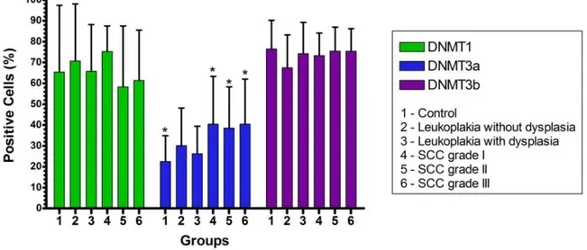
mas não diferiu dos grupos de leucoplasias (28,2%). Para a DNMT1 e DNMT3b não houve diferença estatisticamente significativa entre os grupos de CEC (65% e 74,7%), de leucoplasias (68,3% e 70,9%) e controle (65,4% e 76,5%). A expressão imunoistoquímica das enzimas DNMTs não exibiu correlação com a idade dos pacientes nem associação com gênero e consumo de chimarrão. Houve uma significativa associação entre DNMT3a e uso de álcool (p = 0,01) e uma associação
inversa entre DNMT1 e tabagismo (p = 0,048).
DESCRITORES1:
Metilação de DNA, Metiltransfease, Câncer bucal, Carcinoma de células escamosas, leucoplasia, imunoistoquímica.
1
ABSTRACT
Objectives: This study investigated the expression of DNA methyltransferase (DNMT) 1, 3a, and 3b enzymes in oral squamous cell carcinoma (SCC) and leukoplakia, their relationship with histopathologic graduation and dysplasia, respectively, and with clinical parameters.
Study design: Immunohistochemistry using antibodies anti-DNMT1, anti-DNMT3a, and anti-DNMT3b (dilution of 1:700, Imgenex, San Diego, USA) was carried out to detect the expression of the 3 DNMTs proteins in 21 oral leukoplakias with histopathologic diagnosis of acanthosis and/or hyperkeratosis (leukoplakia without dysplasia), 16 leukoplakias with histopathologic diagnosis of epithelial dysplasia, 20 oral well differentiated SCC (grade I), 20 oral moderately differentiated SCC (grade II), and 20 oral poorly differentiated SCC (grade III). Twenty samples of non-tumor oral tissues were obtained as control. The tissues were embedded in paraffin and obtained from the pathologic archive of Division of Stomatology and Prevention of Bucomaxillofacial Cancer, São Lucas Hospital, PUCRS. Statistical analysis was done using Analysis of Variance (ANOVA), Student-Newmann-Keuls test, Pearson correlation and t test.
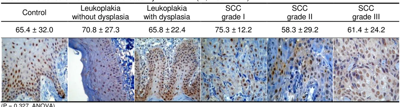
Results: The incidence of nuclear DNMT3a immunoreactivity in oral SCC groups (39.9%) was significantly higher than in control (22.6%) (P<0.05), but not when compared to oral leukoplakias groups (28.2%). For DNMT1 and DNMT3b, there were no statistically significant difference between oral SCC groups (65% and 74.7%), oral leukoplakia groups (68.3% and 70.9%) and control (65.4% and 76.5%). DNMT protein expression exhibited no correlation with age and no association with gender and mate. There were a significant association between the DNMT3a and alcohol use (P = 0.01), and an inverse association between DNMT1 and smoking (P = 0.048).
DESCRIPTORS2
DNA methyltransferase, DNMT, oral cancer, squamous cell carcinoma, oral leukoplakia, immunohistochemistry.
2
LISTA DE ILUSTRAÇÕES
Artigo de Revisão
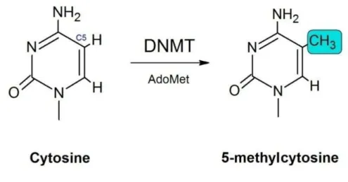
Figure 1 Structures of cytosine before and after the transfer of a methyl group from the cofactor AdoMet, catalyzed by DNA methyltransferases …... 30
Figure 2 Genetic and epigenetic events that can block the expression of tumor suppressor genes in cancer. When only one allele is hit either by genetic or epigenetic alteration, the other can still express the protein that controls cell growth (first hit). However, if the other allele is inactivated (second hit), gene expression will be blocked, contributing to the
development of cancer. LOH: Loss of heterozygosity ...………. 32
Figure 3 Mechanism of transcriptional silencing by DNA methylation. (A) Promoter of gene in a transcriptionally active state. Chromatin in this phase is occupied by spaced nucleosomes composed of acetylated histone complexes and with tails of histone H3 methylated at lysine 4, which configures the euchromatin, making the region accessible to components of the transcription machinery. (B) DNA methyltransferase adds the methyl group to the cytosine of CpG islands. (C) The methylated CpG sites attract methyl-binding proteins such as MeCP2 which, in turn, attract Sin3A and HDAC to the region. Chromatin structure is modified, with deacetylated histone and methylated lysine 9 of histone H3, configuring the heterochromatin. Once these changes have occurred, the transcription factors are repelled and transcription is
blocked ..……… 35
Figure 4 Maintenance versus de novo methylation. (A) After semiconservative
DNA replication, the daughter strand is base-paired with one of the methylated parental strand. The enzyme DNMT1 is responsible for maintaining the methylation pattern by completing half-methylated sites.
(B) De novo methylation of unmethylated sequences, catalyzed by
DNMT3 family enzymes ..……….... 37
Artigo de Pesquisa
Figure 1 DNMT immunohistochemical expression in control, leukoplakia and squamous cell carcinoma groups. * Statistically significant difference between group 1 and groups 4, 5, and 6 (P<0.05, ANOVA,
Apêndices
Figuras 1 e 2 Hiperplasia fibroepitelial evidenciando epitélio com áreas de hiperceratose e hiperplasia irregular das projeções epiteliais (HE, ~40X e ~100X) ... 83
Figuras 3 e 4 Hiperplasia fibroepitelial. Marcação do anticorpo anti-DNMT1 no epitélio (núcleos corados em marrom), contra-corado com hematoxilina de Harris (núcleos corados em azul); ~200X ... 83
Figuras 5 e 6 Hiperplasia fibroepitelial. Marcação do anticorpo anti-DNMT3a no epitélio (núcleos corados em marrom), contra-corado com hematoxilina de Harris (núcleos corados em azul); ~200X ... 84
Figuras 7 e 8 Hiperplasia fibroepitelial. Marcação do anticorpo anti-DNMT3b (núcleos corados em marrom), contra-corado com hematoxilina de Harris (núcleos corados em azul); ~200X ... 84
Figuras 9 e 10 Leucoplasia sem displasia. Observa-se tecido epitelial apresentando hiperceratose e acantose (HE, ~100X e ~200X) ... 85
Figuras 11 e 12 Leucoplasia sem displasia. Marcação do anticorpo anti-DNMT1 (núcleos corados em marrom), contra-corado com hematoxilina de Harris (núcleos corados em azul); ~200X ... 85
Figuras 13 e 14 Leucoplasia sem displasia. Marcação do anticorpo anti-DNMT3a (núcleos corados em marrom), contra-corado com hematoxilina de Harris (núcleos corados em azul); ~200X ... 86
Figuras 15 e 16 Leucoplasia sem displasia. Marcação do anticorpo anti-DNMT3b ((núcleos corados em marrom), contra-corado com hematoxilina de Harris (núcleos corados em azul); ~200X ... 86
Figuras 17 e 18 Leucoplasia com displasia, demonstrando projeções epiteliais em forma de gota, aumento do número de figuras de mitose, pleomorfismo celular e nuclear e hipercromatismo nuclear (HE, ~100X e ~200X) ... 87
Figuras 19 e 20 Leucoplasia com displasia. Marcação do anticorpo anti-DNMT1 (núcleos corados em marrom), contra-corado com hematoxilina de Harris (núcleos corados em azul); ~200X ... 87
Figuras 21 e 22 Leucoplasia com displasia. Marcação do anticorpo anti-DNMT3a (núcleos corados em marrom), contra-corado com hematoxilina de Harris (núcleos corados em azul); ~200X ... 88
de Harris (núcleos corados em azul); ~200X ... 88
Figuras 25 e 26 CEC grau I. Observar proeminente ceratinização, poucas figuras mitóticas e mínimo pleomorfismo celular/nuclear (HE, ~100X e ~200X) ... 89
Figuras 27 e 28 CEC grau I. Marcação do anticorpo anti-DNMT1 (núcleos corados em marrom), contra-corado com hematoxilina de Harris (núcleos corados em azul); ~200X ... 89
Figuras 29 e 30 CEC grau I. Marcação do anticorpo anti-DNMT3a (núcleos corados em marrom), contra-corado com hematoxilina de Harris (núcleos corados em azul); ~200X ... 90
Figuras 31 e 32 CEC grau I. Marcação do anticorpo anti-DNMT3b (núcleos corados em marrom), contra-corado com hematoxilina de Harris (núcleos corados em azul); ~200X ... 90
Figuras 33 e 34 CEC grau II, com menor grau de ceratinização, maior pleomorfismo celular/nuclear e com mitoses mais numerosas (HE, ~100X e ~200X) ... 91
Figuras 35 e 36 CEC grau II. Marcação do anticorpo anti-DNMT1 (núcleos corados em marrom), contra-corado com hematoxilina de Harris (núcleos corados em azul); ~200X ... 91
Figuras 37 e 38 CEC grau II. Marcação do anticorpo anti-DNMT3a (núcleos corados em marrom), contra-corado com hematoxilina de Harris (núcleos corados em azul); ~200X ... 92
Figuras 39 e 40 CEC grau II. Marcação do anticorpo anti-DNMT3b (núcleos corados em marrom), contra-corado com hematoxilina de Harris (núcleos corados em azul); ~200X ... 92
Figuras 41 e 42 CEC grau III evidenciando numerosas mitoses atípicas, ceratinização rara e pleomorfismo celular/nuclear evidente (HE, ~100X e ~200X) ... 93
Figuras 43 e 44 CEC grau III. Marcação do anticorpo anti-DNMT1 (núcleos corados em marrom), contra-corado com hematoxilina de Harris (núcleos corados em azul); ~200X ... 93
Figuras 45 e 46 CEC grau III. Marcação do anticorpo anti-DNMT3a (núcleos corados em marrom), contra-corado com hematoxilina de Harris (núcleos corados em azul); ~200X ... 94
LISTA DE TABELAS
Artigo de Revisão
Table 1 Most studied hypermethylated promoters of genes implicated in
carcinogenesis ...……… 33
Table 2 Summary of DNA methyltransferase studies in human cancer tissues
and cell lines ..……… 40
Artigo de pesquisa
Table 1 Clinical characteristics of study subjects ………... 58
Table 2 Incidence of nuclear immunoreactivity for DNMT1 (%, mean ± SD) .... 59
Table 3 Incidence of nuclear immunoreactivity for DNMT3a (%, mean ± SD) .. 59
LISTA DE ABREVIATURAS
AdoMet S-adenosyl-L-methionine
ANOVA Analise de variância / Analysis of variance
APC Adenomatosis polyposis coli gene
C Citosina / Cytosine
CEC Carcinoma de células escamosas
CNPq Conselho Nacional de Desenvolvimento Científico e Tecnológico
CpG Dinucleotídeo Citosina-Guanina / Cytosine-Guanine dinucleotide
DAP-K Death-associated protein kinase
DNA Ácido desoxirribonucléico / deoxyribonucleic acid
DNMT DNA metiltransferase / DNA methyltransferase
EDTA Ethylenediamine tetraacetic acid
FHIT Fragile histidine triad gene
G Guanina / Guanine
H2O2 Hydrogen peroxide
HAC Histone acetylase
HDAC Histone deacetylase
HE Hematoxilina e eosina
IHC Immunohistochemistry
K-ras v-Ki-ras2 Kirsten rat sarcoma viral oncogene homolog
LOH Loss of heterozygosity
MBD Methyl-CpG-binding domain
MeCP2 Methyl-CpG-binding protein 2
MGMT Methylguanine-DNA methyltransferase
MLH1 DNA mismatch repair protein Mlh1
mRNA Ácido ribonucleic mensageiro / Messenger ribonucleic acid
NA Not available
NBH Northem blot hybridization
oC Graus Celsius
p Probabilidade / probability
p15 Cyclin-dependent kinase inhibitor 2B gene
p16 Cyclin-dependent kinase inhibitor 2A gene
p53 Tumor protein p53
PBS Phosphate-buffered saline
PCNA Proliferating cell nuclear antigen
PCR Reação em cadeia da polymerase
pH Potential of hydrogen
PUCRS Pontifícia Universidade Católica do Rio Grande do Sul
qRT-PCR Real-time polymerase chain reaction
r Coeficiente de Correlação de Pearson
RAR Retinoic acid receptor
RASSF1 Ras association domain family member 1 gene
Rb Retinoblastoma gene
RB1 Retinoblastoma 1 gene
SCC Squamous cell carcinoma
SD Standard deviation
SPSS Statistical Package for the Social Sciences
TpG Timine-guanine dinucleotide
TRD Transcriptional repression domain
W Watts
SUMÁRIO
1 INTRODUÇÃO ... 21
2 ARTIGO DE REVISÃO ... 27
ABSTRACT ………..……… 28
KEYWORDS ………. 28
INTRODUCTION ……….. 29
METHYLATION PROCESS AND CANCER ……….. 30
MECHANISMS OF SILENCING ……… 33
METHYLATION AND MUTATION ……… 36
DNA METHYLTRANSFERASES (DNMTs) ……… 36
DNMT1 ………... 36
DNMT3 FAMILY ………... 38
DNMT AND CANCER ………. 39
DNMT INHIBITORS ………. 41
CONCLUSION ……….. 41
ACKNOWLEDGMENTS ………. 42
REFERENCES ………. 42
3 ARTIGO DE PESQUISA ……… 52
ABSTRACT ……….. 53
KEYWORDS ………. 53
INTRODUCTION ……….. 55
MATERIALS AND METHODS ……….. 55
Immunohistochemistry ……….. 56
RESULTS ……….. 57
DISCUSSION ………. 61
CONCLUSION ……… 64
ACKNOWLEDGMENTS ………... 64
REFERENCES ………... 64
4 DISCUSSÃO GERAL .………. 70
5 CONCLUSÕES ………... 75
REFERÊNCIAS BIBLIOGRÁFICAS ………. 77
APÊNDICES ……… 81
APÊNDICE A – Ficha para coleta de dados ……… 82
APÊNDICE B – Fotomicrografias (grupo-controle) ……….. 83
APÊNDICE C –Fotomicrografias (leucoplasias sem displasia) …... 85
APÊNDICE D –Fotomicrografias (leucoplasias com displasia) ….. 87
APÊNDICE E –Fotomicrografias (CEC grau I) ……….. 89
APÊNDICE F – Fotomicrografias (CEC grau II) ………. 91
APÊNDICE G –Fotomicrografias (CEC grau III) ………... 93
ANEXOS ……….. 95
Anexo A – Carta de aprovação pela comissão Científica e de Ética da Faculdade de Odontologia – PUCRS ……… 96
1 INTRODUÇÃO
O carcinoma de células escamosas ou carcinoma escamocelular (CEC) é a neoplasia maligna mais comum da cavidade bucal, representando 90% a 95% de todos os cânceres dessa região1,2, com cerca de 14 mil novos casos no Brasil em 2008.3 Como toda neoplasia maligna, o CEC bucal é uma doença genética com modificações moleculares complexas, mais especificamente nos genes que regulam a proliferação celular, impedindo que os controles normais de crescimento sejam eficientes.4,5
Os fatores de risco mais comumente associados a essa neoplasia são o consumo crônico de cigarro e álcool, sendo este risco proporcional à quantidade e ao tempo de uso desses agentes.4,6,7 A interação sinérgica entre eles está comprovadamente envolvida na etiologia do CEC bucal e na evolução das lesões cancerizáveis6,8.
As lesões precursoras de câncer ou potencialmente malignas possuem algumas das características moleculares e fenotípicas similares às células neoplásicas, apresentando risco aumentado de transformação em câncer.9 A leucoplasia é o exemplo mais comum e, ainda que não necessariamente evolua para o carcinoma, pode apresentar uma ou mais modificações genéticas que se traduzem em diferentes graus de displasia epitelial. Entretanto, mesmo na ausência de displasia, elas podem já possuir características genéticas em comum às do CEC bucal4,7,10 e nestes casos a habilidade de identificar o risco de transformação maligna é limitada.4 Para Reibel (2003)11, as características clínicas e histopatológicas (presença ou ausência de displasia epitelial) ainda são os parâmetros mais importantes para a predição do desenvolvimento do CEC em tais lesões.
As estratégias terapêuticas do câncer bucal visam a sua cura, com a preservação e restauração das funções dos órgãos envolvidos, minimizando as seqüelas da doença e/ou tratamento. As modalidades atualmente disponíveis envolvem cirurgia, radioterapia e quimioterapia.12 A morbidade resultante de
tratamentos e a alta taxa de mortalidade do câncer de boca são atribuídos ao fato de grande parte das lesões serem diagnosticadas em estágio avançado.7
Os principais fatores prognósticos do câncer bucal ainda são o estadiamento clínico do tumor, a sua localização e a graduação histopatológica, embora, muitas vezes, estes fatores falhem na sua função preditiva. Diversas moléculas relacionadas à carcinogênese do CEC bucal, como aquelas envolvidas na regulação do ciclo celular (p53, p21, ciclina D1), apoptose (Bcl-2, Bax), angiogênese (Fator de crescimento endotelial vascular) e metástase (E-caderina, laminina, colágeno tipo IV) têm sido estudadas com o intuito encontrar um marcador biológico que possa auxiliar no diagnóstico precoce, no seguimento clínico dos pacientes e na escolha das medidas terapêuticas.13-15
Um melhor entendimento das alterações moleculares que ocorrem desde os estágios iniciais da carcinogênese bucal, da displasia epitelial, carcinoma in situ
até carcinoma invasivo, pode aumentar as possibilidades de se detectar o potencial de transformação maligna das lesões cancerizáveis, bem como diagnosticar precocemente o câncer. A descoberta de marcadores moleculares para diferenciar as leucoplasias que apresentem maior propensão a sofrer transformação maligna poderá auxiliar o profissional na conduta terapêutica frente a lesões precursoras.11
Entretanto, as evidências científicas disponíveis sobre esses marcadores
permanecem inconclusivas.8
O desenvolvimento do câncer envolve, basicamente, o acúmulo de mutações gênicas com perda do controle do crescimento celular.16,17 Duas classes de genes que sofrem mutações estão envolvidas no câncer: os proto-oncogenes e
os genes supressores tumorais.5 Os proto-oncogenes estão diretamente
relacionados com a regulação do crescimento celular e, quando alterados por mutações, translocações e amplificações, são chamados de oncogenes, pois promovem a proliferação das células independente de qualquer controle.16,18 Já os
genes supressores tumorais codificam proteínas que de alguma forma impedem ou reduzem o crescimento celular. A sua inativação contribui para o desenvolvimento do câncer, por resultar na diminuição desses inibidores do crescimento.5,6
informação genética fornece a sequência de aminoácidos para a síntese protéica, a epigenética informa quando, como e onde estas informações genéticas devem ser utilizadas.20 A alteração epigenética mais encontrada nas células de mamíferos é a
metilação de DNA, ou seja, a adição covalente de um radical metil para o carbono 5 do nucleotídeo citosina20,21, além de modificações das histonas22, que são proteínas
nucleares responsáveis pela condensação da cromatina.23 Essas alterações podem
ser transmitidas por meio da divisão celular às células filhas e contribuir para a inativação de genes supressores tumorais no câncer.24,25
A metilação de genes resulta no seu silenciamento, enquanto a não metilação permite a sua transcrição. Após a metilação, o produto do gene não é produzido, embora a seqüência do DNA seja mantida.19,22,26 Quando isto ocorre em
um gene supressor tumoral a célula perde a propriedade de transcrição de substâncias importantes para o controle do crescimento celular.20
A metilação ocorre nas ilhas CpG, que são sítios do DNA ricos em nucleotídeos Citosina (C) e Guanina (G), localizados dentro de regiões promotoras de quase metade de todos os genes dos mamíferos, que geralmente não estão metiladas. A metilação dessas regiões está associada com a perda de função do gene, por meio do bloqueio da transcrição, o que pode levar a uma proliferação celular descontrolada quando esta inativação ocorre em genes supressores tumorais.22,27 Essas modificações têm sido encontradas durante os estágios iniciais
da oncogênese.22,28
Neoplasias podem exibir dois tipos de defeitos no padrão de metilação: hipometilação global, que ocorre em regiões não reguladoras de genes, capaz de causar instabilidade/alterações estruturais nos cromossomos27,29 e hipermetilação de regiões promotoras ou reguladoras da expressão gênica, bloqueando a transcrição de genes supressores tumorais.28,30
e DNMT3b, por outro lado, como não são capazes de diferenciar entre nucleotídeos hemimetilados e não metilados, são consideradas responsáveis pela adição de
grupos metil ao DNA não previamente metilado, processo denominado metilação de
novo.28,33,34 Estas últimas são altamente expressas em células embrionárias e
neoplásicas, e pouco expressas após a diferenciação celular e em células somáticas
adultas.24,35-37 Embora as DNMT1 e DNMT3a/DNMT3b sejam consideradas
responsáveis pelas metilações de manutenção e de novo, respectivamente, é
provável que todas as três possuam ambas as funções durante a carcinogênese.20 A expressão aumentada das enzimas DNMTs leva a um padrão de metilação descontrolado no genoma e em muitos genes supressores de tumor.28
O padrão de metilação das células tumorais vem sendo estudado como marcador tanto para o diagnóstico precoce e determinação do prognóstico, quanto para avaliação da resposta à terapia antineoplásica.19 Por se tratar de uma alteração reversível, certas drogas inibidoras de DNA metiltransferase como a decitabina (5-aza-2’-deoxicitidina) têm sido testadas em linhagens celulares neoplásicas, mostrando efeito antiproliferativo38 e radiossensibilizador.39 Esta droga é capaz de reverter o estado de metilação, permitindo a re-expressão dos genes silenciados.40
O papel da metilação do DNA e a expressão das enzimas DNMTs são estudados tanto em tecidos tumorais quanto nas lesões precursoras de câncer de bexiga, colorretal, rins, fígado, pâncreas e colo de útero.28,32,36,41,42
Robertson et al. (1999)36 investigaram o padrão de expressão de mRNA de DNMT1, DNMT3a e DNMT3b em tecidos tumorais de bexiga, cólon, rim e pâncreas, bem como em tecidos normais adjacentes a essas lesões. Foi observado significativo aumento da expressão de DNMT3b nos tumores, enquanto DNMT1 e DNMT3a demonstraram uma moderada elevação da expressão e com uma freqüência menor.
tecidos pré-neoplásicos e neoplásicos foram avaliados. Os dados sugerem que a expressão da DNMT1 e DNMT3a tem importante papel nos estágios precoces da hepatocarcinogênese.
Em 48 pacientes com carcinoma colorretal, Zhu et al. (2007)28 verificaram
que a porcentagem de células mostrando imunorreatividade para DNMT1 nos tumores (75,3%) foi significativamente maior do que no tecido normal (39%).
Sawada et al. (2007)42 investigaram, por imunoistoquímica, a expressão de DNMT1 em 30 amostras de CEC cervical, 97 de lesões precursoras e 34 de epitélio normal, provenientes de 49 pacientes. A imunorreatividade foi significativamente mais elevada nos tumores, quando comparada com o tecido histologicamente normal. A expressividade da enzima foi superior nas lesões precursoras classificadas como de alto grau e nos carcinomas micro-invasivos, reduzindo nos carcinomas invasivos, sugerindo que o aumento da expressão da DNMT1 foi um evento precoce da oncogênese.
2 ARTIGO DE REVISÃO
THE ROLE OF EPIGENETIC TRANSCRIPTION REPRESSION AND
DNA METHYLTRANSFERASES IN CANCER
Authors:
Filipe Ivan Daniel
Fernanda Gonçalves Salum
Affiliation:
ABSTRACT
Epigenetic alterations such as DNA methylation have been implicated in the development and progression of various cancers. DNA methylation consists of the reversible addition of a methyl group to the carbon 5 position of cytosine in CpG dinucleotides, and is considered essential for normal embryonic development. However, global genomic hypomethylation and aberrant hypermethylation of regulatory regions of tumor suppressor genes have been associated with chromosomal instability and transcription repression, respectively, providing neoplastic cells with a selective advantage. DNA methyltransferases are the enzymes responsible for the addition of methyl groups to CpG dinucleotides, which together with histone modifiers, initiate the events necessary for transcription repression to occur. It has been demonstrated that increased expression of DNA methyltransferases may contribute to tumor progression through methylation-mediated gene inactivation in various human cancers. Given their importance, this article reviews the main epigenetic mechanisms for regulating transcription and its implications in cancer development.
KEYWORDS
INTRODUCTION
Heritable and reversible mechanisms known as epigenetic alterations do not require direct alterations of DNA sequences, but they can be responsible for modifying gene expression and are related to cancer development. 1-5 While the genetic information provides the sequence for protein synthesis, the epigenetic information provides instructions on how, where, and when the genetic information will be used.6
Several epigenetic mechanisms regulate gene expression: DNA methylation, modifications of histone proteins and functional noncoding RNAs.1 The major form present in mammalian cells is DNA methylation, which is the covalent addition of a methyl group to the carbon 5 position of cytosine predominantly in the CpG dinucleotide.6,7 This cytosine modification pattern can be transmitted through cell division and may contribute to gene inactivation in cancer.3,4
DNA methylation is essential for normal embryonic development and has a variety of important functions, such as the regulation of gene expression, control of cell differentiation and development, chromatin modification, mutation accumulation, silencing of endogenous retroviruses, preservation of chromosomal integrity, genomic imprinting control, and X chromosome inactivation.6,8-12 Initially discovered as a mechanism for control of development, it plays an important role in many tumor types.13 Genomic methylation patterns are frequently altered in tumor cells with global hypomethylation accompanying region-specific hypermethylation sites. When hypermethylation occurs within the promoter of a tumor suppressor gene, it can silence expression of the associated gene and provide the cell with a growth advantage in a manner akin to deletions or mutations.6,14,15
METHYLATION PROCESS AND CANCER
In mammalian cells, the DNA targets for modification through methylation are cytosine bases adjacent to guanine bases (CpG dinucleotides).16,17 Sequences of
CpG, when found at a high frequency in the genome, are referred to as CpG islands.18 Most of the 29,000 CpG islands found in the human genome are in the promoter regions of almost half of the genes and are generally unmethylated in normal cells.3,15,19
The modification of cytosine is catalyzed by the enzymes DNA methyltransferases (DNMTs) using S-adenosyl-L-methionine (AdoMet) as the methyl donor (figure 1).8,12,18 The methyl group of AdoMet is bound to a sulfonium ion which thermodynamically destabilizes the molecule and makes the relatively inert methylthiol of the methionine moiety very reactive toward activated carbon atoms8. The reaction involves DNMT-DNA binding, flipping the target cytosine out of the double helix, and formation of a transient covalent complex with the cytosine residue.17 DNMT adds a cysteine thiolate to the 6-carbon of the substrate cytosine followed by transfer of the methyl group to the 5-carbon.20
The distribution of methylated and non-methylated CpG dinucleotides is not random, but rather conforms to a pattern.16 Certain genomic sites, such as
pericentromeric regions, imprinted regions, and genes on the inactive X chromosome in females, are hypermethylated while other sites, such as CpG islands which are often associated with gene promoter regions, are hypomethylated.6,12,21
Cancers exhibit at least two types of methylation defects: hypomethylation characterized by a global loss of methylation and hypermethylation of CpG islands of regulatory regions of tumor suppressor genes.22,23
Hypomethylation of non-promoter regions of DNA (known as global hypomethylation) may cause genomic instability and structural changes in chromosomes in cancer, although the relationship between the two processes is not clear.15,24 Two resulting effects of losses of methylation in tumorigenesis have been proposed. First, weakening of transcriptional repression in normally silent regions of the genome could cause the potentially harmful expression of inserted viral genes and of normally silenced genes, such as imprinted genes and genes on the inactive X chromosome. Second, losses of methylation of nuclear structures other than genes could affect the functional stability of chromosomes, such as pericentromeric regions.25
Methylation of CpG islands in gene promoter regions may be involved in carcinogenesis as a result of three possible mechanisms: cytosine methylation facilitates gene mutation as 5-methylcytosine is deaminated to thymine,26,27 aberrant DNA methylation may be associated with allelic loss,6,28 and tumor suppressor genes may be inactivated by DNA hypermethylation.3,29
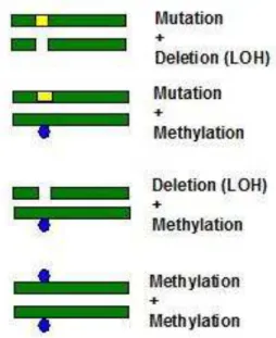
Fig. 2: Genetic and epigenetic events that can block the expression of tumor suppressor genes in cancer. When only one allele is hit either by genetic or epigenetic alteration, the other can still express the protein that controls cell growth (first hit). However, if the other allele is inactivated (second hit), gene expression will be blocked, contributing to the development of cancer. LOH: Loss of heterozygosity.
It is important to note that only methylation within or around the promoter region is associated with gene silencing. Dense methylation within the body of a gene, even within CpG islands, does not hinder transcription.25,32 Many tumor
suppressor genes associated with mutations in cancer have been found with promoter hypermethylation (table 1).
Table 1: Most studied hypermethylated promoters of genes implicated in carcinogenesis
Gene Tissue Reference
p15 Oral cancer Ogi et al.36 (cell cycle regulator) Hepatic cancer Oh et al.37
p16 Oral cancer Kulkarni, Saranath.38; Shaw et al.39; Ogi et al.36 (cell proliferation inhibitor) Saliva of oral cancer patients Rosas et al.40
Saliva ofleukoplakia patients López et al.33
Head and neck cancer Sanchez-Cespedes et al.41; Maruya et al.42 Hepatic cancer Oh et al.37
Colorectal cancer Eads et al.43; Renal cancer Arai et al.11 Lung cancer Lin et al.44
RASSF1 Nasopharyngeal cancer Fendri et al.45
(cell cycle regulator) Hepatic cancer Oh et al.37
Bladder cancer Friedrich et al.46; Abbosh et al.47
MLH1 Colorectal cancer Eads et al.43; Herman et al.48 (cell cycle regulator) Renal cancer Arai et al.11
MGMT Oral cancer Kulkarni; Saranath.38
(DNA repair) Saliva of oral cancer patients Rosas et al.40 Saliva of leukoplakia patients López et al.33
Head and neck cancer Sanchéz-Cespedes et al.41; Maruya et al.42 Bladder cancer Abbosh et al.47
Lung cancer Vallböhmer et al.49
FHIT Lung cancer Lin et al.44; Kim et al.50
(DNA replication regulator)
DAP-K Oral cancer Kulkarni; Saranath.38; Ogi et al.36
(proapoptotic protein) Saliva of oral cancer patients Rosas et al.40 Nasopharyngeal cancer Fendri et al.45
Head and neck cancer Sanchez-Cespedes et al.41; Maruya et al.42 Pancreatic cancer Dansranjavin et al.51
Renal cancer Christoph et al.52 Lung cancer Vallböhmer et al.49
APC Colorectal cancer Eads et al.43; Esteller et al.53 (cell adhesion) Lung cancer Vallböhmer et al.49
Cadherin Hepatic cancer Kanai et al.28; Oh et al.37 (cell adhesion) Pancreatic cancer Dansranjavin et al.51
Lung cancer Kim et al.50
RAR Nasopharyngeal cancer Fendri et al.45
(retinoic acid receptor) Head and neck cancer Maruya et al.42 Lung cancer Kim et al.50; Lin et al.44
MECHANISMS OF SILENCING
In higher eukaryotes, DNA methylation and histone modifications appear to be the main events responsible for the formation of transcriptionally active or inactive chromatin.54
factors.21,55 Many factors are known to bind CpG-containing sequences, and some of
these fail to bind when the CpG is methylated.21 However, it is unlikely to be a
widespread mechanism for transcriptional silencing, since most transcription factors do not have CpG dinucleotides within their DNA binding sites.6 Another possible
mechanism is that specific transcriptional repressors may recognize methyl-CpG and turn off transcription.55
Four proteins with a methyl-CpG-binding domain (MeCP2, MBD1, MBD2, and MBD3) recognize methylated DNA and are implicated in transcriptional repression. These proteins also have an affinity for histone modifying enzymes which cause chromatin condensation and gene silencing.56 MeCP2 contains both a
methyl-CpG-binding domain and a transcriptional repression domain (TRD), which can be tethered to another protein called Sin3A which interacts with histone deacetylase, a member of another transcriptional repression system.57
Histones, nuclear proteins that interact with DNA to form nucleosomes, besides being responsible for packing DNA within chromosomes, are also essential for transcription regulation.10,58 Histone modifications such as acetylation and
methylation may be read by the DNA methylation machinery, leading to either methylation of or failure to methylate a particular CpG dinucleotide.21 Histone
acetylation occurs at sites where transcription takes place, resulting in chromatin decondensation (euchromatin) to permit binding of transcription factors to DNA.18,58
Acetylation is controlled by histone acetylases (HACs) and histone deacetylases (HDACs).59 Deacetylation of these proteins (in particular H3 and H4) by HDAC leads
to a tighter nucleosomal packing and the formation of a compacted chromatin environment (heterochromatin) that inhibits transcription.3,10 Moreover, histone methylation is linked to euchromatic and heterochromatic states.59,60 Methylation of lysine 9 in the core histone H3 is associated with silenced genes,61 whereas methylation of lysine 4 in histone H3 is a feature of active genes.21
DNA methylation and histone modifications are intricately connected with each other.7,24 Thus, the methylation level is connected to the broad organization of
chromatin, with unmethylated DNA usually being part of euchromatin, whereas heavily methylated DNA is part of heterochromatin.8 HDAC may play an important
associated co-repressors and HDACs, resulting in tighter packaging of DNA and reduced access of transcription factors to their binding sites (figure 3).10,62,63
Fig. 3: Mechanism of transcriptional silencing by DNA methylation. (A) Promoter of gene in a transcriptionally active state. Chromatin in this phase is occupied by spaced nucleosomes composed of acetylated histone complexes and with tails of histone H3 methylated at lysine 4, which configures the euchromatin, making the region accessible to components of the transcription machinery. (B) DNA methyltransferase adds the methyl group to the cytosine of CpG islands. (C) The methylated CpG sites attract methyl-binding proteins such as MeCP2 which, in turn, attract Sin3A and HDAC to the region. Chromatin structure is modified, with deacetylated histone and methylated lysine 9 of histone H3, configuring the heterochromatin. Once these changes have occurred, the transcription factors are repelled and transcription is blocked.
METHYLATION AND MUTATION
Cytosine methylation can increase mutation rates because of the spontaneous hydrolytic deamination of methylated cytosine, which causes C T transition mutation.15,18,27 This phenomenon was used to explain the high incidence of CpG to TpG transition mutations observed in the p53 tumor suppressor gene.66
The epigenetic silencing of the DNA repair enzyme O6-MGMT (O6 -methylguanine-DNA methyltransferase) is another example of how abnormal methylation may lead to increased rates of mutation.67 The O6-MGMT protein
removes carcinogen-induced O6-methylguanine adducts from DNA, which produce G
A transition mutations if left unrepaired.15 Tumors with silenced O6-MGMT alleles seem to be predisposed to mutation in key genes, such as p5367 and K-ras.68
DNA METHYLTRANSFERASES (DNMTs)
Three DNA methyltransferases (DNMT1, DNMT3a and DNMT3b) are responsible for adding methyl groups to CpG dinucleotides.6,13
DNMT1
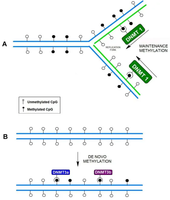
foci and, under experimental conditions, has up to 50-fold preference for hemimethylated DNA substrate.14,70
This enzyme can maintain CpG methylation after DNA replication by methylating the daughter DNA strand, using the methylation pattern of the parental strand as a template (figure 4A).8,16,71 Its inactivation produces global demethylation,
which is consistent with the fact that DNMT1 is required for maintenance methylation.71 The structural and mechanistic basis for the specificity of the enzyme for CpG sites as well as its preference for hemimethylated DNA is still unknown.8
Fig. 4: Maintenance versus de novo methylation. (A) After semiconservative DNA replication, the
daughter strand is base-paired with one of the methylated parental strand. The enzyme DNMT1 is responsible for maintaining the methylation pattern by completing half-methylated sites. (B) De novo
Three sequences located in the N-terminal increase the precision of maintenance methylation and give the enzyme direct access to the nuclear replication site: proliferating cell nuclear antigen (PCNA) binding domain,72 replication
foci targeting sequence73 and polybromo homology domain.17,74 PCNA is required for
DNA replication, and the DNMT1-PCNA interaction may allow the newly synthesized daughter strands to be rapidly remethylated before being packaged into chromatin.6,72 This tight association of the DNMT1 with the replication machinery allows DNMT1 to bind newly replicated and still naked DNA.17
Otherwise, some genes may make this interaction difficult with replicating foci. p21, a cell cycle regulator, can disrupt the DNMT-PCNA interaction, suggesting that p21 may negatively regulate methylation by blocking access of DNMT to PCNA,72 particularly during DNA damage, when p21 protein is induced.75 It was also demonstrated that p21 may inhibit DNMT1 gene expression.75 The retinoblastoma gene product, Rb, another cell cycle regulator protein, can bind to DNMT1 and inhibit its methyltransferase activity during DNA replication in the cell cycle.76 Loss of
functional Rb may grant DNMT1 free access to the genome which could allow for aberrant de novo methylation of CpG.6 These observations point to a complicated
network of connections between DNMT1 and several cellular proteins involved in gene regulation and epigenetic signaling during cell replication.54
DNMT3 FAMILY
Although DNMT1 is the major DNMT in humans, two other enzymes, DNMT3a and DNMT3b, have also been shown to possess DNMT activity.11 They
catalyze DNA methylation at CpG dinucleotides in unmethylated genomic sequences.29
Since DNMT3a and DNMT3b cannot differentiate between unmethylated and hemimethylated CpG sites, they obviously cannot copy a specific pattern of methylation or contribute to the maintenance of methylation pattern.8 Because they
show no preference for hemimethylated DNA, both enzymes appear to function as de
not associated with replication sites even during S-phase (figure 4B).17 This fact
suggests that these DNMTs utilize a different mechanism for accessing the densely packed chromatin and for interacting with their target sites which may involve auxiliary factors such as chromatin remodeling complexes.17
DNMT3a and DNMT3b are highly expressed in early embryonic cells, the
stage in which most programmed de novo methylation events occur, are
downregulated after differentiation and in adult somatic tissues, and are overexpressed in tumor cells.3,21,77-79 DNMT3b has been shown to play a crucial role
in incorporating de novo hypermethylation of promoter CpG islands, a possible
mechanism for tumor suppressor gene inactivation within human cancer cells.7,18 Another member of the DNMT3 family is DNMT3L, a regulatory factor for
the de novo methylation without methylation capacities. Its amino acid sequence is
very similar to that of DNMT3a and DNMT3b, but lacks the residues required for DNA methyltransferase activity in the C-terminal domain.7
DNMT AND CANCER
Although the DNMT1 and DNMT3 families have been considered, respectively, maintenance and de novo methyltransferases, it is likely that all three
DNMTs possess both functions in vivo, particularly during carcinogenesis.6
Excessive amounts of DNMT1, which cannot target replication foci, may participate in the de novo methylation of CpG islands that are not methylated in
normal cells,80 supporting the idea that DNMT can contribute to tumor progression
through CpG island methylation-mediated gene inactivation.81 Thus, its increased
expression may play an important role in the malignant progression of cancer, leading to aberrant methylation in many important tumor suppressor genes.22
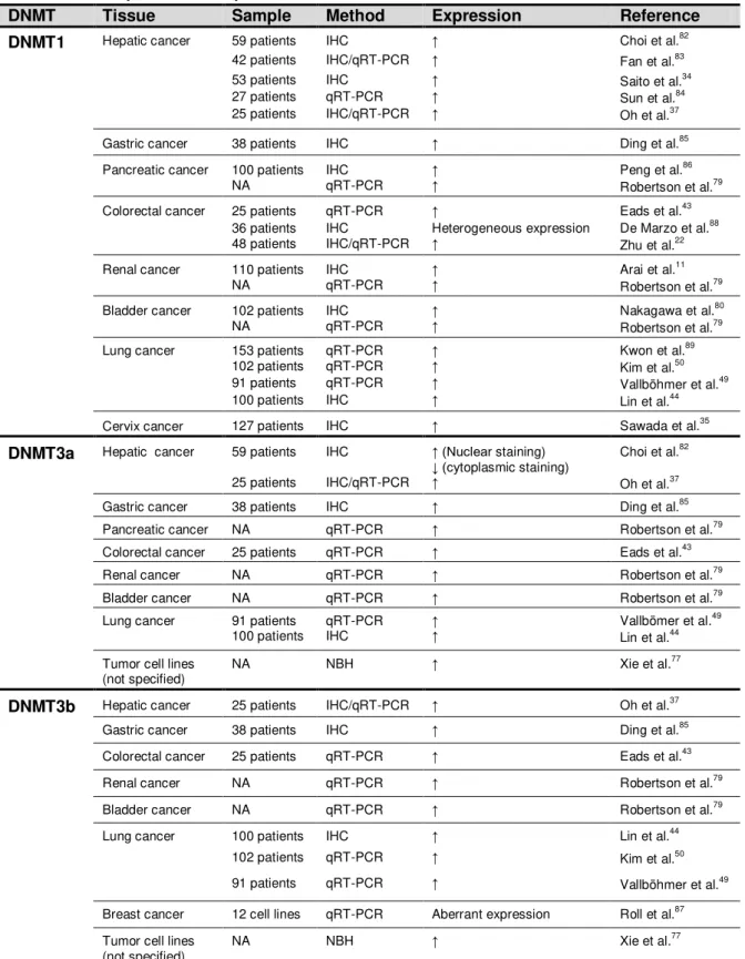
Table 2: Summary of DNA methyltransferase studies in human cancer tissues and cell lines
DNMT Tissue Sample Method Expression Reference
DNMT1 Hepatic cancer 59 patients IHC Choi et al.82
42 patients IHC/qRT-PCR Fan et al.83 53 patients IHC Saito et al.34 27 patients qRT-PCR Sun et al.84 25 patients IHC/qRT-PCR Oh et al.37 Gastric cancer 38 patients IHC Ding et al.85 Pancreatic cancer 100 patients IHC Peng et al.86
NA qRT-PCR Robertson et al.79 Colorectal cancer 25 patients qRT-PCR Eads et al.43
36 patients IHC Heterogeneous expression De Marzo et al.88 48 patients IHC/qRT-PCR Zhu et al.22 Renal cancer 110 patients IHC Arai et al.11
NA qRT-PCR Robertson et al.79 Bladder cancer 102 patients IHC Nakagawa et al.80 NA qRT-PCR Robertson et al.79 Lung cancer 153 patients qRT-PCR Kwon et al.89
102 patients qRT-PCR Kim et al.50 91 patients qRT-PCR Vallböhmer et al.49 100 patients IHC Lin et al.44 Cervix cancer 127 patients IHC Sawada et al.35 DNMT3a Hepatic cancer 59 patients IHC (Nuclear staining)
(cytoplasmic staining) Choi et al. 82
25 patients IHC/qRT-PCR Oh et al.37 Gastric cancer 38 patients IHC Ding et al.85 Pancreatic cancer NA qRT-PCR Robertson et al.79 Colorectal cancer 25 patients qRT-PCR Eads et al.43 Renal cancer NA qRT-PCR Robertson et al.79 Bladder cancer NA qRT-PCR Robertson et al.79 Lung cancer 91 patients qRT-PCR Vallbömer et al.49
100 patients IHC Lin et al.44 Tumor cell lines
(not specified) NA NBH Xie et al. 77
DNMT3b Hepatic cancer 25 patients IHC/qRT-PCR Oh et al.37
Gastric cancer 38 patients IHC Ding et al.85 Colorectal cancer 25 patients qRT-PCR Eads et al.43 Renal cancer NA qRT-PCR Robertson et al.79 Bladder cancer NA qRT-PCR Robertson et al.79 Lung cancer 100 patients IHC Lin et al.44
102 patients qRT-PCR Kim et al.50 91 patients qRT-PCR Vallböhmer et al.49 Breast cancer 12 cell lines qRT-PCR Aberrant expression Roll et al.87 Tumor cell lines
(not specified) NA NBH Xie et al. 77
NA: not available; IHC: immunohistochemistry; qRT-PCR: real-time PCR; NBH: Northern blot hybridization; : increased
DNMT INHIBITORS
In contrast to genetic alterations, epigenetic changes in cancer are potentially reversible, which has spurred the development of pharmacologic inhibitors of DNA methylation and histone deacetylation.90 Indeed, the reactivation of epigenetically silenced genes in cancer may have a profound antitumor effect, thereby being a rational target for therapy and prevention.25
DNA methylation inhibitors such as 5-aza-2’-deoxycytidine (Decitabine) can be utilized to reverse the effects of methylation, including the reduction of mutations at methylated CpG sites, reactivation of genes suppressed by hypermethylation and restoration of cell growth control.91 Treatment of cultured cells with this drug has been shown to cause cell growth inhibition, G2/M arrest, and cell apoptosis.78 The disadvantage of this demethylating agent is its myelosuppressive
effect, particularly when used at high doses.15
The combination of DNMTs and HDAC inhibitors may have an advantage in the treatment of cancer.25 The use of a histone deacetylase inhibitor such as trichostatin A and phenylbutyrate in combination with 5-aza-2’-deoxycytidine has resulted in a strong synergistic growth inhibition in both cell lines and tumor.92,93
CONCLUSION
Since it is a potentially reversible change, the epigenetic event represents new opportunities for the clinical management of cancer through the development of strategies to reverse gene silencing. Further, the associated molecular changes (such as DNMT/HDAC overexpression and gene promoter hypermethylation) may serve as markers for risk assessment, diagnosis and prognosis of cancer.
ACKNOWLEDGMENTS
This study was supported by the Brazilian Research Council CNPq.
REFERENCES
1. Foley DL, Craig JM, Morley R, Olsson CJ, Dwyer T, Smith K, et al. Prospects
for epigenetic epidemiology. Am J Epidemiol. 2009 Feb 15;169(4):389-400.
2. Egger G, Liang G, Aparicio A, Jones PA. Epigenetics in human disease and
prospects for epigenetic therapy. Nature. 2004 May 27;429(6990):457-63.
3. Jones PA, Laird PW. Cancer epigenetics comes of age. Nat Genet. 1999 Feb;21(2):163-7.
4. Shiah SG, Chang LC, Tai KY, Lee GH, Wu CW, Shieh YS. The involvement of
promoter methylation and DNA methyltransferase-1 in the regulation of EpCAM expression in oral squamous cell carcinoma. Oral Oncol. 2009 Jan;45(1):e1-8.
5. Stadler ME, Patel MR, Couch ME, Hayes DN. Molecular biology of head and
neck cancer: risks and pathways. Hematol Oncol Clin North Am. 2008 Dec;22(6):1099-124.
6. Robertson KD. DNA methylation, methyltransferases, and cancer. Oncogene.
7. Cheng X, Blumenthal RM. Mammalian DNA methyltransferases: a structural perspective. Structure. 2008 Mar;16(3):341-50.
8. Hermann A, Gowher H, Jeltsch A. Biochemistry and biology of mammalian DNA methyltransferases. Cell Mol Life Sci. 2004 Oct;61(19-20):2571-87.
9. Li E. Chromatin modification and epigenetic reprogramming in mammalian development. Nat Rev Genet. 2002 Sep;3(9):662-73.
10. Ng HH, Bird A. DNA methylation and chromatin modification. Curr Opin Genet
Dev. 1999 Apr;9(2):158-63.
11. Arai E, Kanai Y, Ushijima S, Fujimoto H, Mukai K, Hirohashi S. Regional DNA
hypermethylation and DNA methyltransferase (DNMT) 1 protein overexpression in both renal tumors and corresponding nontumorous renal tissues. Int J Cancer. 2006 Jul 15;119(2):288-96.
12. Bestor TH. The DNA methyltransferases of mammals. Hum Mol Genet. 2000
Oct;9(16):2395-402.
13. Ha PK, Califano JA. Promoter methylation and inactivation of tumour-suppressor genes in oral squamous-cell carcinoma. Lancet Oncol. 2006 Jan;7(1):77-82.
14. Valinluck V, Sowers LC. Endogenous cytosine damage products alter the site
selectivity of human DNA maintenance methyltransferase DNMT1. Cancer Res. 2007 Feb 1;67(3):946-50.
15. Jones PA, Baylin SB. The fundamental role of epigenetic events in cancer. Nat Rev Genet. 2002 Jun;3(6):415-28.
16. Bird A. DNA methylation de novo. Science. 1999 Dec 17;286(5448):2287-8.
17. Margot JB, Cardoso MC, Leonhardt H. Mammalian DNA methyltransferases show different subnuclear distributions. J Cell Biochem. 2001 Aug 21-Sep 5;83(3):373-9.
18. Momparler RL, Bovenzi V. DNA methylation and cancer. J Cell Physiol. 2000
19. Lander ES, Linton LM, Birren B, Nusbaum C, Zody MC, Baldwin J, et al. Initial sequencing and analysis of the human genome. Nature. 2001 Feb 15;409(6822):860-921.
20. Erlanson DA, Chen L, Verdine GL. DNA Methylation through a Locally Unpaired Intermediate. J Am Chem Soc. 1993;115(26):12583-4.
21. Bird A. DNA methylation patterns and epigenetic memory. Genes Dev. 2002 Jan 1;16(1):6-21.
22. Zhu YM, Huang Q, Lin J, Hu Y, Chen J, Lai MD. Expression of human DNA
methyltransferase 1 in colorectal cancer tissues and their corresponding distant normal tissues. Int J Colorectal Dis. 2007 Jun;22(6):661-6.
23. Rhee I, Jair KW, Yen RW, Lengauer C, Herman JG, Kinzler KW, et al. CpG
methylation is maintained in human cancer cells lacking DNMT1. Nature. 2000 Apr 27;404(6781):1003-7.
24. Feinberg AP, Tycko B. The history of cancer epigenetics. Nat Rev Cancer. 2004 Feb;4(2):143-53.
25. Herman JG, Baylin SB. Gene silencing in cancer in association with promoter
hypermethylation. N Engl J Med. 2003 Nov 20;349(21):2042-54.
26. Shen JC, Rideout WM, Jones PA. High frequency mutagenesis by a DNA methyltransferase. Cell. 1992 Dec 24;71(7):1073-80.
27. Cheng X, Roberts RJ. AdoMet-dependent methylation, DNA
methyltransferases and base flipping. Nucleic Acids Res. 2001 Sep 15;29(18):3784-95.
28. Kanai Y, Ushijima S, Tsuda H, Sakamoto M, Hirohashi S. Aberrant DNA methylation precedes loss of heterozygosity on chromosome 16 in chronic hepatitis and liver cirrhosis. Cancer Lett. 2000 Jan 1;148(1):73-80.
29. Ting AH, Jair KW, Suzuki H, Yen RW, Baylin SB, Schuebel KE. Mammalian DNA methyltransferase 1: inspiration for new directions. Cell Cycle. 2004 Aug;3(8):1024-6.
30. Esteller M, Fraga MF, Guo M, Garcia-Foncillas J, Hedenfalk I, Godwin AK, et
31. Baylin SB, Ohm JE. Epigenetic gene silencing in cancer - a mechanism for early oncogenic pathway addiction? Nat Rev Cancer. 2006 Feb;6(2):107-16.
32. Jones PA. The DNA methylation paradox. Trends Genet. 1999 Jan;15(1):34-7.
33. Lopez M, Aguirre JM, Cuevas N, Anzola M, Videgain J, Aguirregaviria J, et al. Gene promoter hypermethylation in oral rinses of leukoplakia patients--a diagnostic and/or prognostic tool? Eur J Cancer. 2003 Nov;39(16):2306-9.
34. Saito Y, Kanai Y, Nakagawa T, Sakamoto M, Saito H, Ishii H, et al. Increased protein expression of DNA methyltransferase (DNMT) 1 is significantly correlated with the malignant potential and poor prognosis of human hepatocellular carcinomas. Int J Cancer. 2003 Jul 1;105(4):527-32.
35. Sawada M, Kanai Y, Arai E, Ushijima S, Ojima H, Hirohashi S. Increased expression of DNA methyltransferase 1 (DNMT1) protein in uterine cervix squamous cell carcinoma and its precursor lesion. Cancer Lett. 2007 Jun 28;251(2):211-9.
36. Ogi K, Toyota M, Ohe-Toyota M, Tanaka N, Noguchi M, Sonoda T, et al. Aberrant methylation of multiple genes and clinicopathological features in oral squamous cell carcinoma. Clin Cancer Res. 2002 Oct;8(10):3164-71.
37. Oh BK, Kim H, Park HJ, Shim YH, Choi J, Park C, et al. DNA
methyltransferase expression and DNA methylation in human hepatocellular carcinoma and their clinicopathological correlation. Int J Mol Med. 2007 Jul;20(1):65-73.
38. Kulkarni V, Saranath D. Concurrent hypermethylation of multiple regulatory genes in chewing tobacco associated oral squamous cell carcinomas and adjacent normal tissues. Oral Oncol. 2004 Feb;40(2):145-53.
39. Shaw RJ, Hall GL, Woolgar JA, Lowe D, Rogers SN, Field JK, et al. Quantitative methylation analysis of resection margins and lymph nodes in oral squamous cell carcinoma. Br J Oral Maxillofac Surg. 2007 Dec;45(8):617-22.
40. Rosas SL, Koch W, da Costa Carvalho MG, Wu L, Califano J, Westra W, et al.
Promoter hypermethylation patterns of p16,
O6-methylguanine-DNA-methyltransferase, and death-associated protein kinase in tumors and saliva of head and neck cancer patients. Cancer Res. 2001 Feb 1;61(3):939-42.
42. Maruya S, Issa JP, Weber RS, Rosenthal DI, Haviland JC, Lotan R, et al. Differential methylation status of tumor-associated genes in head and neck squamous carcinoma: incidence and potential implications. Clin Cancer Res. 2004 Jun 1;10(11):3825-30.
43. Eads CA, Danenberg KD, Kawakami K, Saltz LB, Danenberg PV, Laird PW. CpG island hypermethylation in human colorectal tumors is not associated with DNA methyltransferase overexpression. Cancer Res. 1999 May 15;59(10):2302-6.
44. Lin RK, Hsu HS, Chang JW, Chen CY, Chen JT, Wang YC. Alteration of DNA
methyltransferases contributes to 5'CpG methylation and poor prognosis in lung cancer. Lung Cancer. 2007 Feb;55(2):205-13.
45. Fendri A, Masmoudi A, Khabir A, Sellami-Boudawara T, Daoud J, Frikha M, et
al. Inactivation of RASSF1A, RARbeta2 and DAP-kinase by promoter methylation correlates with lymph node metastasis in nasopharyngeal carcinoma. Cancer Biol Ther. 2009 Mar;8(5):444-51.
46. Friedrich MG, Weisenberger DJ, Cheng JC, Chandrasoma S, Siegmund KD,
Gonzalgo ML, et al. Detection of methylated apoptosis-associated genes in urine sediments of bladder cancer patients. Clin Cancer Res. 2004 Nov 15;10(22):7457-65.
47. Abbosh PH, Wang M, Eble JN, Lopez-Beltran A, Maclennan GT, Montironi R,
et al. Hypermethylation of tumor-suppressor gene CpG islands in small-cell carcinoma of the urinary bladder. Mod Pathol. 2008 Mar;21(3):355-62.
48. Herman JG, Umar A, Polyak K, Graff JR, Ahuja N, Issa JP, et al. Incidence and functional consequences of hMLH1 promoter hypermethylation in colorectal carcinoma. Proc Natl Acad Sci U S A. 1998 Jun 9;95(12):6870-5.
49. Vallbohmer D, Brabender J, Yang D, Schneider PM, Metzger R, Danenberg KD, et al. DNA methyltransferases messenger RNA expression and aberrant methylation of CpG islands in non-small-cell lung cancer: association and prognostic value. Clin Lung Cancer. 2006 Jul;8(1):39-44.
50. Kim H, Kwon YM, Kim JS, Han J, Shim YM, Park J, et al. Elevated mRNA levels of DNA methyltransferase-1 as an independent prognostic factor in primary nonsmall cell lung cancer. Cancer. 2006 Sep 1;107(5):1042-9.
52. Christoph F, Hinz S, Kempkensteffen C, Schostak M, Schrader M, Miller K. mRNA expression profiles of methylated APAF-1 and DAPK-1 tumor suppressor genes uncover clear cell renal cell carcinomas with aggressive phenotype. J Urol. 2007 Dec;178(6):2655-9.
53. Esteller M, Sparks A, Toyota M, Sanchez-Cespedes M, Capella G, Peinado MA, et al. Analysis of adenomatous polyposis coli promoter hypermethylation in human cancer. Cancer Res. 2000 Aug 15;60(16):4366-71.
54. Margot JB, Ehrenhofer-Murray AE, Leonhardt H. Interactions within the mammalian DNA methyltransferase family. BMC Mol Biol. 2003 May 30;4:7.
55. Kass SU, Pruss D, Wolffe AP. How does DNA methylation repress
transcription? Trends Genet. 1997 Nov;13(11):444-9.
56. Klose RJ, Bird AP. Genomic DNA methylation: the mark and its mediators. Trends Biochem Sci. 2006 Feb;31(2):89-97.
57. Bird AP, Wolffe AP. Methylation-induced repression--belts, braces, and chromatin. Cell. 1999 Nov 24;99(5):451-4.
58. Wolffe AP. Histone deacetylase: a regulator of transcription. Science. 1996 Apr 19;272(5260):371-2.
59. Tamaru H, Selker EU. A histone H3 methyltransferase controls DNA
methylation in Neurospora crassa. Nature. 2001 Nov 15;414(6861):277-83.
60. Jenuwein T, Allis CD. Translating the histone code. Science. 2001 Aug 10;293(5532):1074-80.
61. Nakayama J, Rice JC, Strahl BD, Allis CD, Grewal SI. Role of histone H3 lysine 9 methylation in epigenetic control of heterochromatin assembly. Science. 2001 Apr 6;292(5514):110-3.
62. Jones PL, Veenstra GJ, Wade PA, Vermaak D, Kass SU, Landsberger N, et
al. Methylated DNA and MeCP2 recruit histone deacetylase to repress transcription. Nat Genet. 1998 Jun;19(2):187-91.
64. Cameron EE, Bachman KE, Myohanen S, Herman JG, Baylin SB. Synergy of demethylation and histone deacetylase inhibition in the re-expression of genes silenced in cancer. Nat Genet. 1999 Jan;21(1):103-7.
65. Suzuki H, Gabrielson E, Chen W, Anbazhagan R, van Engeland M,
Weijenberg MP, et al. A genomic screen for genes upregulated by demethylation and histone deacetylase inhibition in human colorectal cancer. Nat Genet. 2002 Jun;31(2):141-9.
66. Rideout WM, Coetzee GA, Olumi AF, Jones PA. 5-Methylcytosine as an endogenous mutagen in the human LDL receptor and p53 genes. Science. 1990 Sep 14;249(4974):1288-90.
67. Esteller M, Risques RA, Toyota M, Capella G, Moreno V, Peinado MA, et al.
Promoter hypermethylation of the DNA repair gene O(6)-methylguanine-DNA methyltransferase is associated with the presence of G:C to A:T transition mutations in p53 in human colorectal tumorigenesis. Cancer Res. 2001 Jun 15;61(12):4689-92.
68. Esteller M, Toyota M, Sanchez-Cespedes M, Capella G, Peinado MA, Watkins
DN, et al. Inactivation of the DNA repair gene O6-methylguanine-DNA methyltransferase by promoter hypermethylation is associated with G to A mutations in K-ras in colorectal tumorigenesis. Cancer Res. 2000 May 1;60(9):2368-71.
69. Hsieh CL. Dynamics of DNA methylation pattern. Curr Opin Genet Dev. 2000
Apr;10(2):224-8.
70. Fatemi M, Hermann A, Pradhan S, Jeltsch A. The activity of the murine DNA
methyltransferase Dnmt1 is controlled by interaction of the catalytic domain with the N-terminal part of the enzyme leading to an allosteric activation of the enzyme after binding to methylated DNA. J Mol Biol. 2001 Jun 22;309(5):1189-99.
71. Okano M, Bell DW, Haber DA, Li E. DNA methyltransferases Dnmt3a and Dnmt3b are essential for de novo methylation and mammalian development. Cell. 1999 Oct 29;99(3):247-57.
72. Chuang LS, Ian HI, Koh TW, Ng HH, Xu G, Li BF. Human DNA-(cytosine-5)
methyltransferase-PCNA complex as a target for p21WAF1. Science. 1997 Sep 26;277(5334):1996-2000.
74. Liu Y, Oakeley EJ, Sun L, Jost JP. Multiple domains are involved in the targeting of the mouse DNA methyltransferase to the DNA replication foci. Nucleic Acids Res. 1998 Feb 15;26(4):1038-45.
75. Tan HH, Porter AG. p21(WAF1) negatively regulates DNMT1 expression in mammalian cells. Biochem Biophys Res Commun. 2009 Apr 24;382(1):171-6.
76. Pradhan S, Kim GD. The retinoblastoma gene product interacts with
maintenance human DNA (cytosine-5) methyltransferase and modulates its activity. Embo J. 2002 Feb 15;21(4):779-88.
77. Xie S, Wang Z, Okano M, Nogami M, Li Y, He WW, et al. Cloning, expression
and chromosome locations of the human DNMT3 gene family. Gene. 1999 Aug 5;236(1):87-95.
78. Cui M, Wen Z, Chen J, Yang Z, Zhang H. 5-Aza-2'-deoxycytidine is a potent
inhibitor of DNA methyltransferase 3B and induces apoptosis in human endometrial cancer cell lines with the up-regulation of hMLH1. Med Oncol. 2009 Mar 21.
79. Robertson KD, Uzvolgyi E, Liang G, Talmadge C, Sumegi J, Gonzales FA, et
al. The human DNA methyltransferases (DNMTs) 1, 3a and 3b: coordinate mRNA expression in normal tissues and overexpression in tumors. Nucleic Acids Res. 1999 Jun 1;27(11):2291-8.
80. Nakagawa T, Kanai Y, Saito Y, Kitamura T, Kakizoe T, Hirohashi S. Increased
DNA methyltransferase 1 protein expression in human transitional cell carcinoma of the bladder. J Urol. 2003 Dec;170(6 Pt 1):2463-6.
81. Vertino PM, Yen RW, Gao J, Baylin SB. De novo methylation of CpG island
sequences in human fibroblasts overexpressing DNA
(cytosine-5-)-methyltransferase. Mol Cell Biol. 1996 Aug;16(8):4555-65.
82. Choi MS, Shim YH, Hwa JY, Lee SK, Ro JY, Kim JS, et al. Expression of DNA
methyltransferases in multistep hepatocarcinogenesis. Hum Pathol. 2003 Jan;34(1):11-7.
83. Fan H, Zhao ZJ, Cheng J, Su XW, Wu QX, Shan YF. Overexpression of DNA
methyltransferase 1 and its biological significance in primary hepatocellular carcinoma. World J Gastroenterol. 2009 Apr 28;15(16):2020-6.
84. Sun L, Hui AM, Kanai Y, Sakamoto M, Hirohashi S. Increased DNA