DOUGLAS FERREIRA PARREIRA
MICOBIOTA FITOPATOGÊNICA ASSOCIADA À PLANTA INVASORA
Tibouchina herbacea
Dissertação apresentada à Universidade Federal de Viçosa, como parte das exigências do Programa de Pós-Graduação em Fitopatologia, para obtenção do título de Magister Scientiae.
VIÇOSA
DOUGLAS FERREIRA PARREIRA
MICOBIOTA FITOPATOGÊNICA ASSOCIADA À PLANTA INVASORA
Tibouchina herbacea
Dissertação apresentada à Universidade Federal de Viçosa, como parte das exigências do Programa de Pós-Graduação em Fitopatologia, para obtenção do título de Magister Scientiae.
APROVADA: 28 de julho 2008
______________________________ _________________________________ Dr. Dartanha José Soares Prof. Maria Catarina Megumi Kasuya (Co-orientador)
_______________________________ _______________________________ Prof. Leandro Grassi Freitas Dr. Bruno Sérgio Vieira
__________________________________ Prof. Robert Weingart Barreto
Aos meus pais, Cosme Damião e Maria Aparecida, minha base de apoio.
AGRADECIMENTOS
A Deus por ter me abençoado e permitido que chegasse tão longe
A meus pais, pela criação amorosa, apoio e companheirismo ao longo de toda minha vida
A meu avô Luis Parreira Coelho, pelo carinho e pelo exemplo de vida. Aos meus irmãos Vanessa e Elias.
A meu cunhado Francisco e a minha sobrinha Ingred pelo carinho.
Ao meu grande amigo e companheiro de inúmeras jornadas Mohamad Hammoud
À minha namorada Wania Neves pelo apoio, auxílio e colaboração durante essa empreitada
À Universidade Federal de Viçosa, pela assistência estudantil que na forma de gratuidade de alojamento e de bolsa atividade permitiu-me graduar-me como engenheiro agrônomo, e, ao Departamento de Fitopatologia pela oportunidade de realizar o curso de Mestrado
Ao Prof. Robert Weingart Barreto pela orientação e ensinamentos
Ao Dr. Dartanhã José Soares pelo apoio e acompanhamento durante meu aprendizado em micologia
Ao Prof. Olinto Liparini Pereira pelos ensinamentos transmitidos e pela colaboração
A todos meus colegas de alojamento e de república, por preencherem o vazio causado pela distancia da família
Aos colegas e amigos do Departamento de Fitopatologia, e da Clínica de Doença de Plantas, em especial a Paulinho e Hélvio, pelo companheirismo nas noites mal dormidas de estudo durante o mestrado
A CAPES, pela bolsa concedida durante o curso
BIOGRAFIA
Douglas Ferreira Parreira é filho de Cosme Damião Parreira e Maria Aparecida Ferreira Parreira, nascido em Três Pontas, MG.
Em marco de 2007 concluiu o curso de Agronomia pela Universidade Federal de Viçosa, em Viçosa, MG.
SUMÁRIO
RESUMO ... v
ABSTRACT ... vii
INTRODUÇÃO GERAL ... 01
REFERÊNCIAS BIBLIOGRÁFICAS... 05
ARTIGO : Parreira D. F.; Barreto R. W.; Soares D. J. and Pereira O. L. (2008). The mycobiota of the invasive weed Tibouchina herbacea (normas da revista Fungal Diversity)... 09
RESUMO
PARREIRA, Douglas Ferreira, M.Sc., Universidade Federal de Viçosa, julho de 2008. MICOBIOTA FITOPATOGÊNICA ASSOCIADA À PLANTA INVASORA
Tibouchina herbacea. Orientador: Robert Weingart Barreto. Co-orientadores:
Dartanhã José Soares e Olinto Liparini Ferreira.
uma futura utilização de fungos fitopatogênicos no controle de T. herbacea. 16 espécies de fungos foram obtidas, sendo: seis hifomicetos – Cladosporium, Passalora,
Cercospora apii e três espécies do gênero Pseudocercospora; quatro coelomicetos – Septoria, Hainesia, Chaetophiophoma e Pestalotiopsis; e seis ascomicetos – Asteridiella, Mollisia, Asterina, Perisporiopsis, Gnomonia, Leptosphaeria. Foram
reconhecidas como taxa novos para a ciência e são aqui descritos: Cladosporium
tibouchinensis, Mollisia tibouchinae, Passalora tibouchinae, Pseudocercospora subsinematosa, Pseudocercospora tibouchinensis, Pseudocercospora tibouchinicola
and Septoria tibouchinensis. Dentre as espécies de fungos encontradas no presente trabalho, três parecem ter potencial para uso em programas de controle biológico por causarem doenças severas em T. herbacea: S. tibouchinensis, P. tibouchinae e M. tibouchinae. Embora a especificidade destes fungos não tenha ainda sido testada, os
ABSTRACT
PARREIRA, Douglas Ferreira, M.Sc., Universidade Federal de Viçosa, July, 2008. THE MYCOBIOTA OF THE INVASIVE WEED Tibouchina herbacea. Advisor: Robert Weingart Barreto. Co-advisors: Dartanhã José Soares and Olinto Liparini Ferreira.
Weed plant invasions, in natural as well as in anthropic modified environments, results in significant economical losses or ecosystem disequilibrium. Some ecosystems are more susceptible than others to biological invasions, once they have disharmonic fauna and flora (with empty ecological niches or niches occupied by low adapted species to the ecological functions required), due to its geographic isolation, as in oceanic islands (Hawaii, French Polynesia, Fernando de Noronha and other archipelagos), or due to its separation form the continental bulk during the process of continental derivation in a remote past, as in the case of Australian continent. In such places, the introduction of exotic vegetal or animal species may be a disaster for the ecosystem. A exemple is the introduction of Tibouchina herbacea in the Hawaiian archipelago resulted in environmental invasion. This plant belongs to the
Melastomataceae family and it is native that South America. A biological program
step for the development of a future biological control program using phytopatogenic fungi to control the weed plant Tibouchina herbacea. In present work 16 fungi species were found: 6 Hyphomycetes – Cladosporium, Passalora, Cercospora and three
Pseudocercosporas; 4 Coelomycetes – Septoria, Hainesia, Chaetophiophoma, Pestalotiopsis ;and 6 Ascomycetes – Asteridiella, Mollisia, Asterina, Perisporiopsis, Gnomonia, Leptosphaeria. There are recognized as new to science and described here: Cladosporium tibouchinensis, Mollisia tibouchinae, Passalora tibouchinae, Pseudocercospora subsinematosa, Pseudocercospora tibouchinensis, Pseudocercospora tibouchinicola and Septoria tibouchinensis Among the species of
fungi founded in this study, three have potential for use in biological control programs causing severe disease in T. herbacea: S. tibouchinensis, P. tibouchinae and M.
tibouchinae. Although the specificity of these fungi has not yet been tested, the first two
INTRODUÇÃO GERAL
As invasões causadas por plantas tanto em ambientes naturais como em
ambientes modificados pela ação antrópica resultam em grandes perdas econômicas e
no desequilíbrio de ecossistemas. Em ambientes de exploração agrícola plantas
daninhas podem competir diretamente com a cultura, podem dificultar a colheita
mecânica ou podem servir como hospedeiros alternativos de pragas e patógenos.
Alguns ecossistemas são mais suscetíveis a invasões biológicas, pois têm fauna e
flora endêmicas desarmônicas (com nichos ecológicos vazios ou ocupados por espécies
pouco adaptadas às funções ecológicas ali desempenhadas) devido ao seu surgimento
em isolamento geográfico, como as ilhas oceânicas dos arquipélagos Havaiano,
Polinésia Francesa, Fernando de Noronha e outros, ou à sua separação da massa
continental no processo de deriva dos continentes em passado evolutivo remoto, como é
o caso do Continente Australiano. Nesses locais a introdução de uma espécie exótica
vegetal ou animal pode ser desastrosa para o ecossistema. Uma alternativa para a
remediação nos casos de invasões causadas por plantas pode ser o controle biológico,
que pode seguir duas estratégias diferentes, a clássica e a inundativa.
O controle biológico clássico ou estratégia inoculativa segue os princípios e
métodos aplicados pelos entomologistas há cerca de um século (CATE, 1990). O uso de
fitopatógenos neste caso é similar ao uso de insetos, envolvendo a importação de um
agente de biocontrole para uma área onde populações da planta daninha existem livres
de inimigos naturais, visando estabelecer um equilíbrio da população da planta alvo. No
caso de utilização de fitopatógenos a preferência tem sido por fungos fitopatogênicos
parasitas obrigatórios, provenientes do centro de origem da planta-alvo (MORTENSEN,
esta baseada na separação espacial da planta daninha e de seus inimigos naturais por um
período de tempo, onde a planta tende a perder a resistência ao patógeno, tendendo a se
tornarem mais vulnerável (CHARUDATTAN, 1990a; CHARUDATTAN, 1990b). O
patógeno inoculado é meramente liberado em uma infestação da planta relativamente
pequena quando comparada com a área total infestada. Dessa maneira uma pequena
dose de inóculo inicial é utilizada para que, eventualmente, uma grande população da
planta daninha seja afetada (CHARUDATTAN & DeLOACH, 1988; McRAE, 1988).
Porém o processo pode ser lento, pois depende do aumento gradual da doença que pode
levar alguns anos, mas que devido a seu baixo custo é apropriado para o controle de
infestações em grandes áreas não manejadas ou com baixo valor econômico (ALTMAN
et al, 1990; CHARUDATTAN, 1990b). O fungo fitopatogênico usado neste tipo de
estratégia tem que ter alta capacidade de disseminação, de maneira que isso permita
uma rápida dispersão na população alvo, sem que seja necessária a intervenção humana
intensiva e repetida. Dessa forma, os tipos de infestação mais apropriados para o
sucesso na aplicação da estratégia clássica de controle biológico são os que ocorrem em
áreas com pouco ou nenhum distúrbio antrópico como por exemplo: áreas de vegetação
natural, áreas de pastagens e cursos d’agua, dentre outros (MORTENSEN, 1986;
ADAMS, 1988; CHARUDATTAN, 1985; CHARUDATTAN & DeLOACH, 1988).
Outra característica importante para o fitopatógeno é a capacidade de se multiplicar e
manter a epidemia, implicando na permanência de inóculo de um ano para outro.
Comumente, aponta-se para as Uredinales, grupo de fungos que causa as doenças
conhecidas como ferrugens como o mais adequado para o uso em programas de controle
biológico clássico, devido à sua (comumente) elevada especificidade, fácil dispersão de
propágulos pelo vento e desenvolviemento de endemias após a liberação inicial
(MORTENSEN, 1986; TeBEEST et al, 1992). O controle biológico clássico é um
sistema que possui mecanismos de auto regulação, onde os níveis de doença aumentam
ou diminuem junto com a população da planta alvo (CHARUDATTAN, 1985;
WATSON, 1992). Uma vez que o agente de controle biológico é liberado e se
estabelece ele não pode ser mais contido, sendo necessário que ele tenha alta
especificidade pela planta a ser controlada (WATSON, 1991; WAAGE, 1992). O
exemplo mais amplamente conhecido de sucesso do controle biológico clássico
severamente infestadas, a população da planta foi reduzida, em menos de um ano e
meio, em 99%.
A estratégia inundativa ou de bio-herbicida tipicamente envolve o uso de fungos
fitopatogênicos endêmicos, já associados à planta alvo, que são produzidos em massa,
formulados e aplicados de modo semelhante aos utilizados com herbicidas químicos
onde a população da planta invasora encontra-se estabelecida. Consiste na produção de
propágulos infectivos do organismo em larga escala, sendo aplicados na população alvo.
Esse tipo de trabalho usualmente depende, para o seu sucesso, do desenvolvimento de
formulações e técnicas apropriadas de aplicação para que um controle adequado seja
obtido (TeBEEST et al, 1992). Existem diversos exemplos de produtos que se tornaram disponíveis comercialmente como resultado de estudos feitos nesta abordagem de
biocontrole e que foram objeto de discussão em diversas revisões publicadas. Um
exemplo é a publicação de EVANS et al (2001).
A primeira etapa de qualquer programa de controle biológico clássico, depois da
escolha da espécie de organismo que se pretende controlar, é a busca no centro de
origem do organismo por inimigos naturais. O Brasil é parte do centro de origem de
muitas plantas invasoras importantes em escala mundial. Aproveitando esta localização
privilegiada, diversos estudos prévios sobre a micobiota de plantas invasoras
selecionadas têm sido feitos (BARRETO, 1991; BARRETO & EVANS, 1994; 1995a;
1995b; 1996b, 1998; BARRETO et al. 1995; POMELA & BARRETO, 1997;
BARRETO et al. 1999; BARRETO & TORRES, 1999; BARRETO et al. 2000;
PEREIRA & BARRETO, 2000; PEREIRA et al. 2003; PEREIRA & BARRETO, 2005; VIEIRA & BARRETO, 2005; PEREIRA & BARRETO, 2006; SOARES &
BARRETO, 2006; POMELLA et al, 2007; PEREIRA et al, 2007; ROCHA et al, 2007; SEIXAS et al, 2007; SOARES & BARRETO, 2008)
A espécie de planta alvo deste trabalho, Tibouchina herbacea (DC.) Cogn. (Melastomataceae), foi introduzida no Havaí como ornamental e, devido à ausência de
inimigos naturais e condições edafo-climáticas favoráveis se dispersou rapidamente
pelas florestas nativas e regiões úmidas, sendo hoje encontrada nas ilhas Maui, Hawaii,
Molokai, Oahu e Lanai (ALMASI, 2000). Nesses locais forma populações densas,
sendo que o controle químico ou mecânico desta espécie são hoje considerados como
economicamente inviáveis. O controle biológico já foi reconhecido como a única
maneira de se mitigar este problema (WIKLER & SOUZA, 2005). Essa espécie quando
úmida, possui flores autógamas, não dependendo de polinizadores, e produz um grande
número de sementes de tamanho diminuto dispersas facilmente pelo vento. Nas regiões
de origem de T. herbacea, as plantas atingem no máximo 1,5 m de altura, sofrendo forte pressão de fitopatógenos e de insetos. No Havaí devido às condições edafoclimáticas
favoráveis e a ausência de inimigos naturais pode atingir 4,0 m de altura (ALMASI,
2000). Nas ilhas havaianas não existe nenhuma espécie pertencente à família
Melastomataceae. Todas as espécies desta família que ocorrem neste arquipélago ,
foram introduzidas pelo homem e se tornaram invasoras, sendo listadas como espécies
nocivas. Sua venda e transporte sendo inclusive consideradas atividades ilegais
(http://www.state.hi.us/dlnr/dofaw/hortweeds/species/tibher.htm). A espécie T. herbacea.é considerada como parte de um complexo altamente polimórfico, possuindo grande variação morfológica e genética, com ampla distribuição. Segundo uma das
autoridades brasileiras nesta família (com. pessoal R. Goldemberg) há uma clara
necessidade de uma revisão detalhada deste complexo para esclarecimento de sua
taxonomia e delimitação clara de cada taxon que o compõe. Deve-se levar em
consideração, as limitações impostas por este cenário imperfeito quanto à identidade das
plantas hospedeiras para os fungos estudados neste trabalho quando da utilização da
informação aqui gerada. Plantas herbáceas do gênero Tibouchina, com morfologia equivalente ou próxima da apresentada pelas plantas presentes como invasoras no
Havaí, e que estivessem doentes foram coletadas e incorporadas neste estudo, mesmo
quando alguma discrepância, por exemplo, número de pétalas nas flores, pilosidade nas
hastes ou outro aspecto se apresentasse como discrepante do padrão usual fosse
REFERÊNCIAS BIBLIOGRÁFICAS
ADAMS, E. B. Fungi in classical biocontrol of weeds. In Fungi in biological control systems, ed. M. N. Burge. Manchester University Press: Manchester 111-122. 1988.
ALMASI, K.N. A non-native perennial invades a native forest. Biological Invasions 2: 219-230. 2000.
ALTMAN, J., NEATE, S. & ROVIRA, A.D. Herbicide-pathogen interactions and mycoherbicides as alternative strategies for weed control. In Microbes and microbial products as herbicides, ed. R. E. Hoagland. American Chemical Society: Washington DC 240–259. 1990.
BARRETO, R.W. & EVANS, H.C. Fungal biocontrol of weeds and its potential role in ecosystem sustainability. In: Chapela H. I. & M. E. Palm (eds). Mycology in Sustainable Development: Expanding Concepts and Vanishing Borders. Parkway Publishers Inc: Boone. 1996a.
BARRETO, R.W. & EVANS, H.C. Fungal pathogens of Euphorbia heterophylla and E. hirta in Brazil and their potential as weed biocontrol agents. Mycopathologia 141: 21–36. 1998.
BARRETO, R.W. & EVANS, H.C. Fungal pathogens of weeds collected in the Brazilian tropics and subtropics and their biocontrol potential. In: Delfosse, E.S. & R.R. Scott (eds). Proceedings of the Eighth International Symposium on Biological Control of Weeds. DSIR-CSIRO: Melbourne. 1996b.
BARRETO, R.W. & EVANS, H.C. Mycobiota of the weed Cyperus rotundus in the state of Rio de Janeiro, with an elucidation of its associated Puccinia complex. Mycological Research 99: 407–419. 1995a.
BARRETO, R.W. & EVANS, H.C. The mycobiota of the weed Chromolaena odorata in southern Brazil with particular reference to fungal pathogens for biological control. Mycological Research 98: 1107–1116. 1994.
BARRETO, R.W. & EVANS, H.C. The mycobiota of the weed Mikania micrantha in southern Brazil with particular reference to fungal pathogens for biological control. Mycological Research 99: 343–352. 1995b.
BARRETO, R.W. & TORRES, A.N.L. Nimbya alternantherae and Cercospora
alternantherae: two new records of fungal pathogens on Alternanthera philoxeroides (alligatorweed) in Brazil. Australasian Plant Pathology 28: 103–107. 1999.
BARRETO, R.W., CHARUDATTAN, R., POMELLA, A.W.V. & HANADA, R.E. Biological control of neotropical aquatic weeds with fungi. Crop Protection 19: 697–703. 2000.
BARRETO, R.W., EVANS, H.C. & ELLISON, C.A. The mycobiota of the weed Lantana camara in Brazil, with particular reference to biological control. Mycological Research 99: 769–782. 1995.
BARRETO, R.W., EVANS, H.C. & HANADA, R.E. First record of Cercospora pistiae causing leaf spot of water lettuce (Pistia stratioites) in Brazil, with particular reference to weed biocontrol. Mycopathologia 144: 81–85. 1999.
CATE, J.R. Biological control of pests and diseases: integrating a diverse heritage. In New directions in biological control: Alternatives for suppressing agricultural pests and disease, ed. R. R. Baker. Alan R Liss: New York 23–43. 1990.
CHARUDATTAN, R. & DeLOACH, C.J. Management of pathogens and Insects for weed control in agroecosystems. In Weed Management in agroecosystems: ecological approaches, eds M. A. 59 Altieri and M. Liebman. CRC press. Boca Raton: Florida 245–264. 1988.
CHARUDATTAN, R. Pathogens with potential for weed control. In Microbes and microbial products as herbicides, ed. R. E. Hoagland. American Chemical Society: Washington DC 132–154. 1990a.
CHARUDATTAN, R. Prospects for biological control of weeds by plant pathogens. Fitopatologia Brasileira 15(1): 13–19. 1990b.
CHARUDATTAN, R. The mycoherbicide approach with plant pathogens. In: TeBeest, D.O. (Ed.). Microbial control of weeds. New York, Chapman & Hall 24–57. 1991.
CHARUDATTAN, R. The use of genetically altered strains of pathogens for weed control. In Biological control in agricultural IPM systems, eds M. A. Hoy and D. C. Herzog. Academic Press: London 347–372. 1985.
CULLEN, J.M. & HASAN, S. Pathogens for the control of weeds. Philosophical Transactions of the Royal Society of London 318: 213–224. 1988.
CULLEN, J.M., KABLE, P.F. & CATT, M. Epidemic spread of a rust imported for biological control. Nature 244: 462–464. 1973.
DANIEL, J.T., TEMPLETON, G.E., SMITH, R.J. & FOX, W.T. Biological control of northern jointvech in rice with an endemic fungal disease. Weed Science 21: 303– 307. 1973.
McRAE, C.F. Classical and inundative approaches to biological weed control compared. Plant Protection Quarterly 3(3): 124–127. 1988.
MORTENSEN, K. Biological control of weeds with plant pathogens. Canadian Journal of Plant Pathology 8: 229–231. 1986.
PEREIRA, J.M. & BARRETO, R.W. Additions to the mycobiota of the weed Lantana camara (Verbenaceae) in Southeastern Brazil. Mycopathologia 151: 71–80. 2000.
PEREIRA, O.L. & BARRETO, R.W. Pseudocercospora palicoureae sp. nov.
associated with the toxic rubiaceous weed Palicourea marcgravii in Brazil, with observations on its mycobiota. Fungal Diversity 23: 243–253. 2006.
PEREIRA, O.L. & BARRETO, R.W. The mycobiota of the weed Mitracarpushirtus in Minas Gerais (Brazil), with particular reference to fungal pathogens for biological control. Australasian plant pathology 34(1): 41–50. 2005.
PEREIRA, O.L., BARRETO, R.W. & BEZERRA, J.L. Cercospora talini on Talinum patens in Brazil. Fitopatologia Brasileira, Brasília 28(2): 205–205. 2003.
PEREIRA, O.L., BARRETO, R.W., CAVALAZZI, J.R.P. & BRAUN, U. The mycobiota of the cactus weed Pereskia aculeata in Brazil, with comments on the life-cycle of Uromycespereskiae. Fungal Diversity 25: 167–180. 2007.
POMELLA, A.W. & BARRETO, R.W. Leaf scorch of purple nutsedge (Cyperus
rotundus) caused by Ascochyta cyperiphthora. Mycotaxon 65: 459–468. 1997.
POMELLA, A.W., BARRETO, R.W. & CHARUDATTAN, R. Nimbya alternantherae
a potential biocontrol agent for alligatorweed, Alternanthera philoxeroides. BioControl (Dordrecht) 52: 271–288. 2007.
ROCHA, F.B., PEREIRA, O.L. & BARRETO, R.W. Cercosporaapii causing leaf spots on two Brazilian toxic weeds: Solanum glaucophyllum and Xanthium strumarium. Brazilian Journal of Microbiology 38: 1–5. 2007.
SEIXAS, C.D.S., BARRETO, R.W. & KILLGORE, E. Fungal pathogens of Miconia calvescens (Melastomataceae) from Brazil, with reference to classical biological control. Mycologia 99: 99–111. 2007.
SOARES, D.J. & BARRETO, R.W. Additions to the Brazilian mycobiota of the grassy weed, Hymenachne amplexicaulis with a discussion on the taxonomic status of Paraphaeosphaeria recurvifoliae. Australasian Plant Pathology 35: 347–353. 2006.
SOARES, D.J. & BARRETO, R.W. Fungal survey of biocontrol agents of Ipomoea carnea from Brazil. In: XII International Symposium on Biological Control of Weeds, 2007, Mont Pellier. Proceedings of the XII International Symposium on Biological Control of Weeds, 2008 (no Prelo).
TeBEEST, D.O. Conflicts and strategies for future development of mycoherbicides. New directions in biological control: Alternatives for suppressing agricultural pests and disease, ed. R. R. Baker. Alan R Liss, Inc: New York 323–332. 1990.
TeBEEST, D.O. Status of research on bioherbicides. Plant Protection Quartely 7(4): 142. 1992.
TeBEEST, D.O., YANG, X.B. & CISAR, C.R. The status of biological control of weeds with fungal pathogens. Annual Review of Phytopathology 30: 637–657. 1992.
TEMPLETON, G.E. Biological control of weeds with fungal pathogens. Tropical Pest Management 30: 333–338. 1984.
VIEIRA, B.S. & BARRETO, R.W. Lewia chlamidosporiformans sp. nov. from
Euphorbiaheterophylla. Mycotaxon 94: 245–248. 2005.
WAAGE, J. Classical biological control In Proceeding of the first international weed control congress, J. H. Combellack, K. J. Levick, J. Parsons, and R. G. Richardson. Weed Science Society of Victoria: Melbourne 1: 240–249. 1992.
WATSON, A.K. Biological and other alternative control measures. Proceedings of the first international weed control congress, Weed Science Society of Victoria: Melbourne 1: 298–300. 1992.
WATSON, A.K. The classical approach with plant pathogens. In Microbial control of weeds, ed. O. T. TeBeest. Chapman & Hall: London 3–23. 1991.
WIKLER, C & SOUZA, P.G. Estudos Bioecológicos de Syphraea uberabensis
The mycobiota of the invasive weed Tibouchina herbacea
Parreira D. F.1; Barreto R. W.1*;Soares D. J. 1; and Pereira O. L.1
1
Departamento de Fitopatologia, Universidade Federal de Viçosa, 36571-000, Viçosa, MG, Brazil.
Parreira D. F.; Barreto R. W.; Soares D. J.; and Pereira O. L (2008). The mycobiota of the invasive weed Tibouchinaherbacea. Fungal Diversity XX: XX-XX.
A survey of fungal pathogens associated with cane tibouchina - Tibouchina herbacea (Melastomataceae) - was conducted in the neotropics, concentrated in South and Southeastern Brazil but also including ad hoc collections in the Dominican Republic and Costa Rica, aiming at finding potential biological control agents for this herbaceous weed for introduction into Hawaii. Numerous fungal species were found, including six ascomycetes, six cercosporoid species and four coelomycete. Seven new taxa are described and illustrated herein: Cladosporium tibouchinensis, Mollisia tibouchinae, Passalora tibouchinae,
Pseudocercospora subsinematosa, Pseudocercospora tibouchinensis, Pseudocercospora tibouchinicola
and Septoria tibouchinensis. Additionally Asteridiella mellastomacearum, Cercospora apii and an undetermined species of Perisporiopsis ,are also reported for the first time on T. herbacea. Other fungi that were collected will be included in a separate publication. Among the species of fungi described in this study, three appear particularly promising for use in biological control programs because they were found to cause severe disease on T. herbacea: Septoria tibouchinensis, Passalora tibouchinensis and
Mollisia tibouchinae. A series of additional tests, including a demonstration of host-specificity, will be required in order to cofirm this purported potential.
Key words:, biological control, fungal survey, Mollisia, neotropical fungi, Passalora, Septoria.
Article Information
Received XX XXXXX 2008 Accepted XX XXXXX 200X Published online XX XXXXX 200X
*
Corresponding author: R. W. Barreto;e-mail: [email protected]
Introduction
tibouchina_herbacea.htm). Its management has been made mainly through herbicide aplications, however, classical biological control has been recognized as the sole sustainable method of control for exotic weeds invading natural ecosystems, as is the case of the weedy melastomes in the Pacific (Wikler and Souza, 2005).
In the early 1990’s contacts were established between Hawaii based scientists led by C. Smith (University of Hawaii) and Brazilian scientists with the purpose of initiating collaboration aimed at at classical biocontrol of several Hawaiian weeds native from Brazil and cooperative agreements between the Research Corporation of the University of Hawaii (RCUH) in the USA and Brazilian partners, including the Fundação Arthur Bernardes/Universidade Federal de Viçosa (FUNARBE/UFV) were established. At that time one of the main target-weeds was T. herbacea and preliminary surveys of fungal pathogens were initiated but the increasing threat represented by forest ecosystem invasion by M. calvescens led to a shift of priority and work was then concentrated on the study of pathogens attacking M. calvescens (Killgore et al. 1997; Culliney et al., 2003; Seixas etal., 2002; Seixas et al, 2007).
Recently, the work on T. herbacea was continued both involving Brazilian entomologists based at the state of Paraná and pathologists based at the state of Minas Gerais. Insect surveys performed by Charles Wikler yielded a series of potential
biocontrol agents, but Sypharea uberabensis (Coleoptera: Chrysomelidae) was
demonstrated to have the greatest potential (Wikler and Souza, 2005).
Works aiming at introducing this particular insect are well advanced. Sypharea uberabensis is now under quarantine in Hawaii for its final evaluation (T. Johnson, pers. comm.). Paralell to that, systematic field surveys of the mycobiota on T. herbacea was also conducted.
Several other studies of the mycobiota associated with weeds in Brazil have been published along the recent years to serve as the basis for future classical biological control programmes. Some recent examples are those on Pereskia aculeata (Pereira et al. 2007) and Hedychium coronarium (Soares and Barreto, 2008). This publication represents the first one concentrated on the mycobiota of T. herbacea in Brazil and is the first account of fungi associated to this plant species worldwide.
No records of fungi associated to T. herbacea were found in the literature and it was expected that a significant proportion of the fungi collected on this host might represent taxonomic novelties and that some might represent potential biocontrol agents that might be used against this host. Until now the surveys unraveled seventeen fungal species. This paper includes the description and discussion of ten selected species. The complementary list of fungal species will be covered in a separate publication.
Materials and methods
Observations, measurements and line drawings were prepared using an Olympus BX 50 light microscope fitted with a drawing tube and a Olympus E330 camera. Photographs were used to illustrated some species. The collections examined were deposited in the herbarium of the Universidade Federal de Viçosa (VIC).
Results
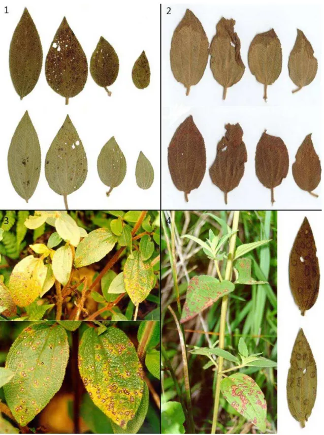
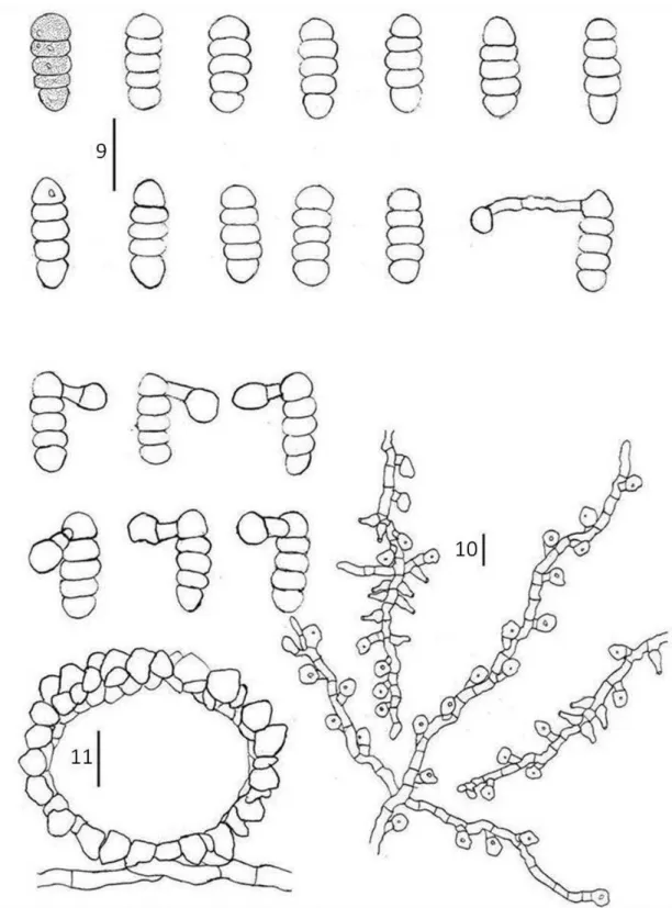
Asteridiella melastomacearum (Speg.) Hansford., The Meliolineae. A Monograph. Sydowia Beih 2:154 (1961).
(FIGS 1, 9-11) Colonies on living leaves, predominantly hypophyllous. Hyphae sinuous, slightly undulate, branching alternate or opposite, cells 5–9 μm diam. Appressoria alternate to unilateral, brown to dark brown, straight to slightly curved, 13.5–21 μm long, stalk cells cylindrical, 3–9 x 5–9.5 μm, head cells subglobose to ovate, sometimes angulose, 9.5–15 x 10–14 μm. Phialides separate, opposite or alternate, brown, ampuliform, 13–20.5 x 6.5–10 μm. Perithecia black, scattered, globose, with crenate to crenulate surface, 43.5–173.5 μm diam. Asci evanescent. Ascospores brown, oblong, obtuse, 4-septate, slightly constricted at septae, 32.5–40 x 11–15 μm, smooth.
Habitat: on living leaves of Tibouchina herbacea.
Known distribution: Minas Gerais and Rio de Janeiro (Brazil).
Material examined: BRAZIL, Minas Gerais, Alvorada de Minas, on living leaves of T. herbacea, 19 April 2008, R. W. Barreto (VIC 30690); BRAZIL,Minas Gerais, Poços de Caldas, Cascata Véu de Noiva, on living leaves of T. herbacea, 8 June 2001, R. W. Barreto (VIC 30639); BRAZIL,Minas Gerais, Ponte Nova, on living leaves of T. herbacea,10 November 1995, R. W. Barreto (VIC 30651); BRAZIL, Minas Gerais, Jequiri, on living leaves of T. herbacea,21 April 1996, R. W. Barreto (VIC 30668); BRAZIL,Rio de Janeiro, Nova Friburgo,on living leaves of T. herbacea, 20 October 1996, R. W. Barreto (VIC 30675)
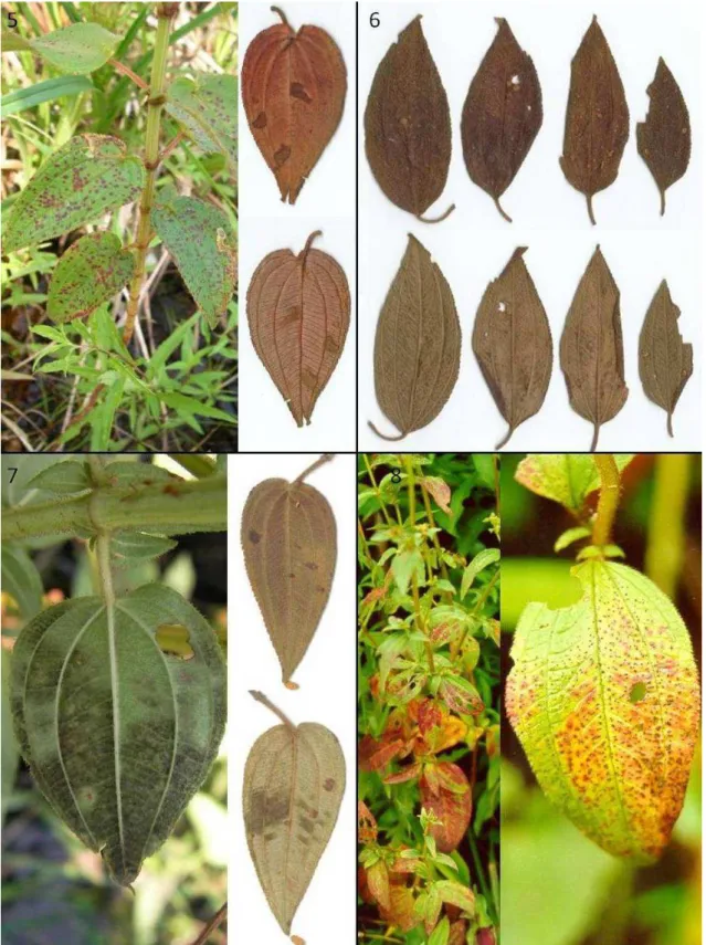
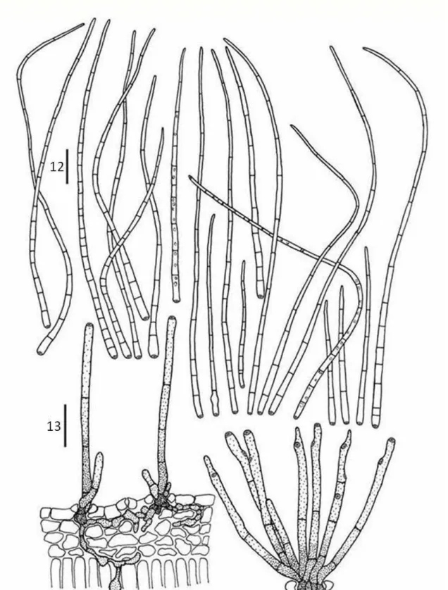
Cercospora apii Fresen. sensu lato, emend. Crous and Braun., Mycosphaerella and its anamorphs: 1. Names published in Cercospora and Passalora CBS Biodiversity Ser. 1: 33-36 (2003)
(FIGS 2, 12-13) Lesions on living leaves, necrotic, initially circular to ellipsoid, later coalescing to form large spots, 3.5–42 x 3–25 mm, with indistinct margins, brow. Internal hyphae 1.5–8 μm diam, branched, septate, brown. External hyphae not observed. Stromata reduced to few cells on substomatal cavity. Conidiophores hypophylous arising through stomata, fasciculate, erect, straight, subcylindrical, 48–175 x 4–5.5 µm, 2–5-septate, unbranched, brown paler at apices, thin-walled, smooth. Conidiogenous cells terminal, integrated, proliferating simpodially, 23.5–73 x 3.5–5 µm, light brown. Conidiogenous loci conspicuous, 1-3 per cell, 2.5–4 µm diam, thickened, darkened. Conidia solitary, ranging from obclavate-cylindrical to (mostly) acicular-filiform, straight to curved, 30–
115 x 2–5 µm, apex subacute, base truncate, 2–7-septate, guttulate, hyaline, thin walled, smooth, hila thickened and darkened.
Teleomorph: not seen.
Habitat: on living leaves of Tibouchina herbacea.
Known distribution: Espirito Santo (Brazil), Minas Gerais (Brazil), Paraná (Brazil) and Rio Grande do Sul (Brazil).
on living leaves of T. herbacea, 19 January 2001, R. W. Barreto (VIC 30696). BRAZIL, Minas Gerais, Viçosa, road to Cajuri on living leaves of T. herbacea, 16 March 1996, R. W. Barreto (VIC 30660). BRAZIL, Minas Gerais, Jequeri on living leaves of T. herbacea, 21 April 1996, R. W. Barreto (VIC 30668). BRAZIL, Minas Gerais, Juiz de Fora, on living leaves of T.herbacea, 06 November 1998, R. W. Barreto (VIC 30673). BRAZIL, Espirito Santo, Venda Nova do Imigrante, entrance of Parque Estadual da Pedra Azul on living leaves of T.herbacea, 09 December 2004, O. L. Pereira (VIC 30682).
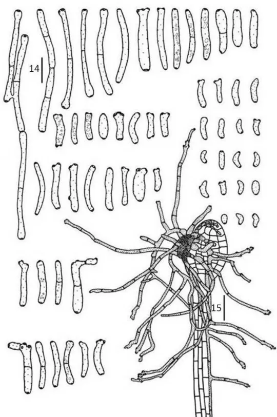
Cladosporium tibouchinensis D.F. Parreira & R.W. Barreto sp. nov.
(FIGS 14-15) Mycobank 512067.
Etymology: named in reference to the host genus.
Differt a Cladosporium hypophyllum conidiophoris longioribus, 15–160.5 x 3.0–5.0 μm, 0–3 septata, conidiis longioribus 4.5–49.5 x 2–5 μm, 0–3 septata.
Colonies formed at the apex of trichomes, partly or completely covering its inflated heads, giving the appearance of burned matches to the trichomes (under the dissecting microscope), connected to neighboring trichomes by external hyphae. Internal hyphae indistinct. External hyphae wrapping the trichomes, 2–3 μm diam,
branched, septate, double layered and smooth walled, brown. Conidiophores
hypophyllous, in loose groups or solitary emerging from external hyphae, cylindrical, straight or flexuous, sometimes branched, 15–160.5 x 3–5 μm, 1–7-septate, double layered, dark brown. Conidiogenous cells terminal, integrated, cylindrical, with simpodial proliferation, 7.5–43.5 x 3–5 µm, brown. Conidiogenous loci conspicuous, 1–
9 per cell, on small lateral shoulders, 1–2(3) µm diam, thickened, darkened. Conidia solitary or catenulate,(in short branched chains), variable in shape, narrowly to broadly ellipsoid, subcylindrical to cylindrical-oblong, subglobose, obovoid or limoniform, straight to slightly curved, curved, 4.5–49.5 x 2–5 µm, 0–3 septate, gutulate, ligth brown, thin-walled, smooth, hila thickened and darkened.
Teleomorph: not seen.
Known distribution: Minas Gerais (Brazil), Paraná (Brazil), Rio Grande do Sul (Brazil) and Santa Catarina (Brazil).
Material examined: BRAZIL, Santa Catarina, Lajes, on living leaves of T.herbacea, 16 January 2001, R. W. Barreto (VIC 30567; holotype). BRAZIL, Minas Gerais, Alvorada de Minas, on living leaves of T.herbacea, 19 April 2008, R. W. Barreto (VIC 30690). BRAZIL, Minas Gerais, Carrancas, on living leaves of T. herbacea, 16 May 2004, O. L. Pereira (VIC 30676). BRAZIL, Minas Gerais, Tiradentes, on living leaves of T.herbacea, 15 May 2004, O. L. Pereira (VIC 30678). BRAZIL, Minas Gerais, Carrancas, on living leaves of T.herbacea, 16 May 2004, O. L. Pereira (VIC 30679). BRAZIL, Paraná, Curutiba, on living leaves of T.herbacea, 31 March 1998, R. W. Barreto (VIC 30619). BRAZIL, Rio Grande do Sul, Nova Petrópolis, on living leaves of T. herbacea, 19 January 2001, R. W. Barreto (VIC 30696).
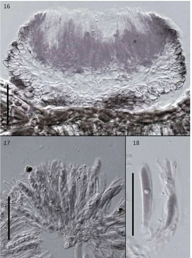
Mollisia tibouchinae D.F. Parreira & R.W. Barreto sp. nov.
(FIGS 3, 16-18) Mycobank XXXXX.
Etymology: named in reference to the host genus.
Differt a Mollisiaparasitica asci minoribus, 52–69 x 6–9 μm, ascosporae minoribus 8.5–12 x 3.5–4 μm.
69 x 6–9 μm, 8-spored,. Ascospores oval to ellipsoid, 8.5–12 x 3.5–4 μm, hyaline, 0-septate, smooth, guttulate.
Anamorph: not observed.
Habitat: on living leaves of Tibouchina herbacea.
Known distribution: Minas Gerais, Paraná, Rio de Janeiro, Santa Catarina, São Paulo, (Brazil).
Material examined: BRAZIL, Minas Gerais, Barbacena, on living leaves of T. herbacea, 14 May 2004, O. L. Pereira (VIC 30677, holotype); BRAZIL, Minas Gerais, Tiradentes, on living leaves of T. herbacea, 15 May 2004, O. L. Pereira (VIC 30678); BRAZIL, Minas Gerais, Carrancas, on living leaves of T. herbacea, 16 May 2004, O. L. Pereira (VIC 30679); BRAZIL, Minas Gerais, Lambari, Parque Nova Baden, on living leaves of T. herbacea, 25 November 1998, R. W. Barreto (VIC 30630); BRAZIL, Minas Gerais, Borda da Mata, on living leaves of T. herbacea, 25 November 1998, R. W. Barreto (VIC 30631); BRAZIL, Minas Gerais, Poços de Caldas, Cascata Véu de Noiva, on living leaves of T. herbacea, 8 June 2001, R. W. Barreto (VIC 30639); BRAZIL, Minas Gerais, Poços de Caldas, Cascata Véu de Noiva, on living leaves of T. herbacea, 5 June 2001, R. W. Barreto (VIC 30640); BRAZIL, Rio de Janeiro, Nova Friburgo, on living leaves of T. herbacea, 19 February 2006, R. W. Barreto (VIC 30642); BRAZIL, Rio de Janeiro, Nova Friburgo, on living leaves of T. herbacea, 2 May 2008, R. W. Barreto (VIC 30644); BRAZIL, Rio de Janeiro, Nova Friburgo, on living leaves of T. herbacea, 08 June 1995, R. W. Barreto (VIC 30645); BRAZIL, Rio de Janeiro, Nova Friburgo, on living leaves of T. herbacea, 08 June 1995, R. W. Barreto (VIC 30646); BRAZIL, Rio de Janeiro, Teresópolis, on living leaves of T. herbacea, 13 October 1995, R. W. Barreto (VIC 30648); BRAZIL, Rio de Janeiro, Petrópolis, Fazenda Inglesa, on living leaves of T. herbacea, 15 October 1995, R. W. Barreto (VIC 30649); BRAZIL, Minas Gerais, Itabirito, on living leaves of T. herbacea, 9 November 1995, R. W. Barreto (VIC 30650); BRAZIL, Rio de Janeiro, Petrópolis, Araras, on living leaves of T. herbacea, 19 December 1995, R. W. Barreto (VIC 30652); BRAZIL, São Paulo, Bananal, Serra da Bocaina,on living leaves of T. herbacea, 28 December 1995, R. W. Barreto (VIC 30654); BRAZIL, São Paulo, Bananal, Serra da Bocaina, on living leaves of T. herbacea, 28 December 1995, R. W. Barreto (VIC 30655); BRAZIL, Minas Gerais, Barão de Cocais, Santuário do Caraça, on living leaves of T. herbacea, 3 May 1996, R. W. Barreto (VIC 30667); BRAZIL, Rio de Janeiro, Nova Friburgo, Alto do Michelis, on living leaves of T. herbacea, 4 April 2000, R. W. Barreto (VIC 30669); BRAZIL, Rio de Janeiro, Nova Friburgo, on living leaves of T. herbacea, 5 April 2000, R. W. Barreto (VIC 30670); BRAZIL, Rio de Janeiro, Nova Friburgo, Alto do Michelis, on living leaves of T. herbacea, 20 October 1996, R. W. Barreto (VIC 30672); BRAZIL, Santa Catarina, São Joaquim, on living leaves of T. herbacea,11 June1997, R. W. Barreto (VIC 30674); BRAZIL, Rio de Janeiro, Nova Friburgo, on living leaves of T. herbacea, 20 October 1996, R. W. Barreto (VIC 30675).
Passalora tibouchinae D.F. Parreira & O.L. Pereira sp. nov.
(FIGS 4, 19-20) Mycobank 512080.
Etymology: named in reference to the host genus.
Maculis in foliis vivis, circularibus, cinereo-brunneis vel atro-brunneis, 1.5-6 mm diam.
Stromatibus amphigenis, parvis vel bene evolutis, atro-brunneis, 6.5–32.5 × 7.5–45 µm. Conidiophoris
fasciculatis, atro-brunneis, apicem versus pallide brunneis, 15.0–76.0 × 2–5 μm, 0–6-septatis. Conidiis
solitariis, acicularibus vel obclavatis, rectis vel leniter curvatis, 30–115 × 2–5 μm, 2–7-septatis, ad apicem acutis, ad basim truncatis, pallide brunneis, levis.
Teleomorph: not seen.
Known distribution: Espirito Santo (Brazil), Minas Gerais (Brazil), Paraná (Brazil), Rio de Janeiro (Brazil), Rio Grande do Sul (Brasil) and São Paulo (Brazil).
Material examined: BRAZIL, Espirito Santo, Ibatiba, on living leaves of T. herbacea, 09 December 2004, O. L. Pereira (VIC 30568; holotype). BRAZIL, Espirito Santo, Venda Nova do Imigrante, entrance of Parque Estadual da Pedra Azul on living leaves of T. herbacea, 09 December 2004, O. L. Pereira (VIC 30680). BRAZIL, Espirito Santo, Venda Nova do Imigrante, entrance of Parque Estadual da Pedra Azul on living leaves of T.herbacea, 09 December 2004, O. L. Pereira (VIC 30681). BRAZIL, Minas Gerais, Lagoa da Prata, on living leaves of T. herbacea, 05 February 2005, O. L. Pereira (VIC 30683). BRAZIL, Paraná, Curitiba, Road Curitiba-Paranaguá Km 51 on living leaves of T. herbacea, 31 March 1998, R. W. Barreto (VIC 30619). BRAZIL, Rio de Janeiro, Nova Friburgo, on living leaves of T.herbacea, 24 february 1998, R. W. Barreto (VIC 30621). BRAZIL, Rio de Janeiro, Vila do Grama, on living leaves of T.herbacea, 24 february 1998, R. W. Barreto (VIC 30625). BRAZIL, Minas Gerais, Rio Pomba, on living leaves of T. herbacea, 19 october 1998, R. W. Barreto (VIC 30628). BRAZIL, Minas Gerais, Santa Barbara do Tugúrio, on living leaves of T. herbacea, 21 november 1998, R. W. Barreto (VIC 30629). BRAZIL, Rio Grande do Sul, Nova Petrópolis, on living leaves of T. herbacea, 19 January 2001, R. W. Barreto (VIC 30696). BRAZIL, Minas Gerais, Coronel Pacheco, on living leaves of T.herbacea, 13 March 2002, R. W. Barreto (VIC 30641). BRAZIL, Rio de Janeiro, Nova Friburgo, on living leaves of T. herbacea, 06 April 2008, R. W. Barreto (VIC 30643). BRAZIL, São Paulo, Bananal, Serra da Bocaina (top) on living leaves of T.herbacea, 28 December 1995, R. W. Barreto (VIC 30656). BRAZIL, Minas Gerais, Araponga, Cachoeira do Estouro on living leaves of T.herbacea, 09 March 1996, R. W. Barreto (VIC 30659). BRAZIL, Minas Gerais, Alto Caparaó, near Hotel Caparaó, river margin on living leaves of T.herbacea, 31 March 1996, R. W. Barreto (VIC 30665). BRAZIL, Rio de Janeiro, Nova Friburgo, on living leaves of T.herbacea, 19 October 1996, R. W. Barreto (VIC 30671).
Perisporiopsis sp.
(FIGS 21-23) Leaf spots absent, pseudothecia randomly distributed on leave surface.. Internal hyphae not observed. External hyphae 3–5 μm diam, branched, septate, brown, smooth. Pseudothecia superficial, epigenous, isolate, unilocular, globose to subglobose, 83.5– 170 x 72.5–160 μm, walls of textura angularis, brown, ornamented with dark brow erect setae on upper half. Hamathecial tissue absent. Asci bitunicate, fasciculate, obclavate to cylindrical, sessile to short pedicellate, straight to slightly curved, 37.5–76 x 14–29.55 μm, 8-spored. Ascospores fusiform, 17.5–28.5 x 5.5–8.5 μm, hyaline, 3-septate, guttulate, smooth.
Anamorph: not observed.
Habitat: on living leaves of Tibouchina herbacea.
Known distribution: Espirito Santo, Minas Gerais, Rio de Janeiro, Rio Grande do Sul (Brazil).
Material examined: BRAZIL, Espirito Santo, Venda Nova do Imigrante, Parque Estadual da Pedra Azul, on living leaves of T. herbacea, 09 December 2004, O. L. Pereira (VIC 30682; holotype); BRAZIL, Minas Gerais, Barbacena, on living leaves of T. herbacea, 14 May 2004 O. L. Pereira (VIC 30677); BRAZIL, Minas Gerais, Santa Barbara do Tugúrio, on living leaves of T. herbacea, 27 November 1998, R. W. Barreto (VIC 30629); BRAZIL, Minas Gerais, Lambari, Parque Nova Baden, on living leaves of T. herbacea, 25 November 1998, R. W. Barreto (VIC 30630); BRAZIL, Rio Grande do Sul, Nova Petrópolis, on living leaves of T. herbacea, 18 January 2001, R. W. Barreto (VIC 30636); BRAZIL, Rio Grande do Sul, Nova Petrópolis, on living leaves of T. herbacea, 19 January 2001, R. W. Barreto (VIC 30637); BRAZIL, Rio Grande do Sul, Nova Petrópolis, on living leaves of T. herbacea, 19 January 2001, R. W. Barreto (VIC 30696); BRAZIL, Rio Grande do Sul, Road Nova Petrópolçis-Caxias do Sul Km 179, on living leaves of T. herbacea, 19 January 2001, R. W. Barreto (VIC 30638); BRAZIL, Rio de Janeiro, Nova Friburgo, road Rio de Janeiro-Nova Friburgo Km 60, on living leaves of T. herbacea, 2 May 2008, R. W. Barreto (VIC 30644); BRAZIL, Rio de Janeiro, Petrópolis, Araras, on living leaves of T. herbacea, 19 December 1995, R. W. Barreto (VIC 30652).
Pseudocercospora subsinematosa D.F. Parreira & D.J. Soares sp. nov.
Mycobank 512071.
Etymology: named in reference to the conidiophores aggregated in short synemata-like structures.
Differt a Pseudocercospora tibouchinae conidiophoris longioribus, 21–76 μm longis, 0–5 septata, conidiis 54–145.5 μm longis, 4–15 septata.
Leaf spots circular to elliptic, 2–7 mm, initially as small spots surrounded by a reddish-brown margin, later developing into a large spot, light brown in center, with a reddish-brown margin, vein-delimited. Internal hyphae thin-walled, 1.5–3.5 μm diam., branched, septate, ligth brown. External hyphae absent. Stromata epiphyllous, well-developed, immersed to erumpent, 21–55 x 16–50 μm, composed of dark brown textura angularis. Conidiophores epigenous, aggregated in dense fascicles, as short synnemata-like structures, erect, straight to slightly sinuose, subcylindrical, 21–76 x 3–5 µm, 1–5 septate, unbranched, brown becoming paler at apice, thin-walled, smooth. Conidiogenous cells terminal, integrated, cylindrical, 8.0–28.5 x 2.5–5.0 µm, light brown, smooth. Conidiogenous loci protruding, 1-2 per cell, 1.5–3 µm diam, unthickened, not darkened. Conidia solitary, obclavate-cylindrical to subcylindrical attenuating gradually towards the apex, straight to slightly curved, 45–145.5 x 2–4 μm, apex subacute to occasionally obtuse, base subtruncate or sometimes endend in a strongly protruding hilum, 4–15-septate, guttulate, pale olivaceous to pale brown, smooth, thin-walled, hila unthickened, not darkened.
Teleomorph: not seen.
Known distribution: Minas Gerais (Brazil).
Material examined: BRAZIL, Minas Gerais, Tabuleiro, on living leaves of T. herbacea, 15 October 2007, D. F. Parreira and D.J. Soares (VIC 30565; holotype). BRAZIL, Minas Gerais, Tabuleiro, on living leaves of T.herbacea, 15 October 2007, D.F. Parreira and D.J. Soares (VIC 30687).
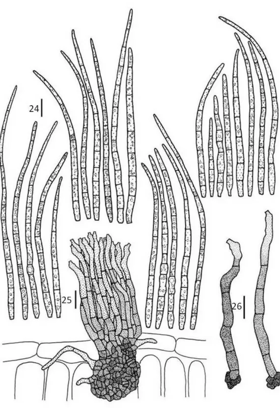
Pseudocercospora tibouchinensis D.F. Parreira & R.W. Barreto sp. nov.
(FIGS 6, 27-28) Mycobank 512076.
Etymology: named in reference to the host genus.
Differt a Pseudocercospora tamoneae, conidiis ad 202.0 μm longis, 3–12 septata.
Leaf spot very similar to those caused by P. subsinematosa, but larger in size 1.5–19 mm diam. Internal hyphae indistinct. External hyphae absent. Stromata subglobose to irregular, well-developed, sub-immersed to erumpent, 17.5–45 x 20–62.5
μm, composed of dark brown, textura angularis. Conidiophores aggregated in sporodochia, erect, straight to slightly curved, subcylindrical, 8.5–40.5 x 2–4.5 µm, 0–4 septate, unbranched, light brown, thin-walled, smooth. Conidiogenous cells terminal, integrated, 5.5–20 x 2–3 µm, light brown. Conidiogenous loci inconspicuous, 1–2 per cell, 1–3 µm diam, unthickened, not darkened. Conidia solitary, cylindrical to acicular, straight to slightly curved, sometimes geniculate, 55–202 x 2–3 µm, apex obtuse or subacute, base truncate, 3–11 septate, guttulate, pale olivaceous, thin-walled, smooth, hila unthickened and not darkened.
Teleomorph: not seen.
Known distribution: Cordillera Oriental (Dominican Republic), Minas Gerais (Brazil), Paraná (Brazil), Rio de Janeiro (Brazil) and Rio Grande do Sul (Brazil).
herbacea, 04 December 1998, R. W. Barreto (VIC 30632). DOMINICAN REPUBLIC, Cordillera Oriental, Miches, on living leaves of T. herbacea, 27 December 1998, R. W. Barreto (VIC 30633). BRAZIL, Rio Grande do Sul, Nova Petrópolis, on living leaves of T. herbacea, 18 January 2001, R. W. Barreto (VIC 30636). BRAZIL, Rio de Janeiro, Nova Friburgo, on living leaves of T. herbacea, 19 February 2006, R. W. Barreto (VIC 30642). BRAZIL, Minas Gerais, Manhuaçu, road Belo Horizonte-Vitória on living leaves of T.herbacea, 30 March 1996, R. W. Barreto (VIC 30661). BRAZIL, Minas Gerais, Manhuaçu, road Belo Horizonte-Vitória (between Realeza and Manhuaçu) on living leaves of T. herbacea, 30 March 1996, R. W. Barreto (VIC 30662). BRAZIL, Minas Gerais, Alto do Caparaó, boundary Alto Jequitibá on living leaves of T. herbacea. 31 March 1996, R. W. Barreto (VIC 30663). BRAZIL, Minas Gerais, Alto do Caparaó, near the entrance park on living leaves of T. herbacea, 31 March 1996 R. W. Barreto (VIC 30664). BRAZIL, Minas Gerais, Caeté, Serra da Piedade on living leaves of T. herbacea, 03 May 1996, R. W. Barreto (VIC 30697). BRAZIL, Minas Gerais, Barão de Cocais, Santuário do Caraça on living leaves of T.herbacea, 03 May 1996, R. W. Barreto (VIC 30667). BRAZIL, Rio de Janeiro, Nova Friburgo, on living leaves of T.herbacea, 05 April 2000, R. W. Barreto (VIC 30670).
Pseudocercospora tibouchinicola D.F. Parreira & D.J. Soares sp. nov.
(FIGS 7, 29-30) Mycobank 512079.
Etymology: named in reference to the host genus.
Differt a Pseudocercospora tibouchinae stromatibus nullis, conidiophoris longioribus, 11.0-53.0
μm, conidiis 16.5-195.0 μm longis, 1–16 septata.
Colonies on living leaves, not forming conspicuous leaf spots, growing mainly
abaxially forming a dense olivaceous mat of conidia and conidiophorores, colonies
somewhat vein-delimited abaxially; adaxially colonies are sparse and less commonly formed;, on senescent leaves green islands formed at colonized areas whereas the rest of the lamina becomes red or brow. Internal hyphae 1.5–3 μm diam., branched, septate, hyaline, thin-walled. External hyphae absent. Stromata absent. Conidiophores arising from stomata, sparsely fascitulate, erect, straight, subcylindrical, 11–53 x 3–5 µm, 1– 4-septate, mostly unbranched, ligth-brow, thin-walled, smooth. Conidiogenous cells terminal, integrated, 5–22.5 x 3–5 µm, pale olivaceous, smooth. Conidiogenous loci protruding, 1–2 per cell, 1–2 µm diam, unthickened, not darkened. Conidia solitary, obclavate-cylindrical to subcylindrical, straight to curved, sometimes geniculate, 16.5–
195 x 2.0–3.5 µm, apex subacute, occasionally obtuse, base endend in a strongly protruding hilum, 1–16-septate, guttulate, pale olivaceous, thin-walled, smooth, hila unthickened not darkened.
Teleomorph: not seen.
Known distribution: Minas Gerais (Brazil).
Material examined: BRAZIL, Minas Gerais, Tabuleiro, on living leaves of T. herbacea, 15 October 2007, D. F. Parreira and D.J. Soares (VIC 30564; holotype).
Septoria tibouchinensis D.F. Parreira & R.W. Barreto sp. nov.
(FIGS 8, 31-33) Mycobank XXXXX.
Etymology: named in reference to the host genus.
Differt a Septoria melastomatis, conidia longioribus, 44.5–101.5 × 1.5–2 μm, 2–4 septata.
of the conidiomata, 0–1-septate, hyaline, smooth, subcylindrical, 6–18 × 2–3.5 μm. Conidiogenous cells holoblastic, terminal, hyaline, smooth, 6–14 × 2–3.5 μm. Conidia solitary, cylindrical, straight to sinuous, 44.5–101.5 × 1.5–2 μm, 2–4-septate, hyaline, smooth, strongly guttulate.
Habitat: on living leaves of Tibouchina herbacea.
Known distribution: Minas Gerais, Paraná, Rio de Janeiro, Rio Grande do Sul, Santa Catarina, São Paulo (Brazil).
Material examined: BRAZIL, Minas Gerais, Alvorada de Minas, on living leaves of T. herbacea, 19 April 2008, R.W. Barreto. (VIC 30690; holotipe); BRAZIL, Minas Gerais, Tiradentes, on living leaves of T. herbacea, 15 May 2004, O L. Pereira (VIC 30678); BRAZIL, Minas Gerais, Carrancas,on living leaves of T. herbacea, 16 May 2004,O. L. Pereira (VIC 30679); BRAZIL, Minas Gerais, São Roque de Minas, on living leaves of T. herbacea, 7 April 2004,O. L. Pereira (VIC 30684); BRAZIL, São Paulo, Eldorado Paulista, Parque Estadual de Jacupiranga, Caverna do Diabo, on living leaves of T. herbacea, 16 April 2005,O. L. Pereira (VIC 30685); BRAZIL, Minas Gerais, Borda da Mata, on living leaves of T. herbacea, 25 November 1998, R. W. Barreto (VIC 30631); BRAZIL, Rio de Janeiro, Parati,on living leaves of T. herbacea, 16 January 2001, R. W. Barreto (VIC 30635); BRAZIL, Rio Grande do Sul, Nova Petrópolis, on living leaves of T. herbacea, 18 January 2001, R. W. Barreto (VIC 30636); BRAZIL, Rio Grande do Sul, Nova Petrópolis,on living leaves of T. herbacea, 19 January 2001, R. W. Barreto (VIC 30637); BRAZIL,Rio Grande do Sul, Nova Petrópolis,on living leaves of T. herbacea, 19 January 2001, R. W. Barreto (VIC 30696); BRAZIL, Rio Grande do Sul, Road Nova Petrópolçis-Caxias do Sul Km 179,on living leaves of T. herbacea, 19 January 2001, R. W. Barreto (VIC 30638); BRAZIL, Rio de Janeiro, Nova Friburgo, on living leaves of T. herbacea, 19 February 2006, R. W. Barreto (VIC 30642); BRAZIL,Rio de Janeiro, Nova Friburgo, road Rio de Janeiro-Nova Friburgo Km 60, on living leaves of T. herbacea, 2 May 2008, R. W. Barreto (VIC 30644); BRAZIL, Rio de Janeiro, Nova Friburgo, on living leaves of T. herbacea, 8 June 1995, R. W. Barreto (VIC 30646); BRAZIL, Minas Gerais, Viçosa,on living leaves of T. herbacea, 24 September 1995, R. W. Barreto (VIC 30647); BRAZIL, São Paulo, Bananal, Serra da Bocaina, on living leaves of T. herbacea, 28 December 1995, R. W. Barreto (VIC 30654); BRAZIL, São Paulo, Bananal, Serra da Bocaina,on living leaves of T. herbacea, 28 December 1995, R. W. Barreto (VIC 30655); BRAZIL, São Paulo, Bananal, Serra da Bocaina,on living leaves of T. herbacea, 28 December 1995, R. W. Barreto (VIC 30656); BRAZIL, Rio de Janeiro, Nova Friburgo, on living leaves of T. herbacea, 05 February 1996, R. W. Barreto (VIC 30657); BRAZIL, Minas Gerais, Araponga, Estevão Araújo,on living leaves of T. herbacea, 9 March 1996, R. W. Barreto (VIC 30658); BRAZIL, Minas Gerais, Alto Caparaó, on living leaves of T. herbacea, 31 March 1996, R. W. Barreto (VIC 30663); BRAZIL, Minas Gerais, Alto Caparaó, on living leaves of T. herbacea, 31 March 1996, R. W. Barreto (VIC 30644); BRAZIL, Minas Gerais, Alto Caparaó, on living leaves of T. herbacea, 31 March 1996, R. W. Barreto (VIC 30665); BRAZIL, Minas Gerais, Caeté,on living leaves of T. herbacea, 3 May 1996, R. W. Barreto (VIC 30666); BRAZIL, Rio de Janeiro, Nova Friburgo, on living leaves of T. herbacea, 5 April 2000, R. W. Barreto (VIC 30670); BRAZIL, Minas Gerais, Juiz de Fora,on living leaves of T. herbacea, 6 December 1998, R. W. Barreto (VIC 30673); BRAZIL,Santa Catarina, São Joaquim,on living leaves of T. herbacea,11 June1997, R. W. Barreto (VIC 30674).
Discussion
Asteridiella melastomacearum is the only species in the group of fungi collected on T. herbacea and studied in the present publication that was readily recognized as previously known taxon. It was originally described from Tibouchina longifolia (Vahl) Baill. ex Cogn. by Hansford (1961). Tibouchina herbacea is the third host in genus Tibouchina, found for this fungal species.
many unresolved issues regarding taxonomic delimitation at the species or infra-specific level within the C. apii complex as revealed by later studies (Groenewald et al. 2006). Under the new concept for this group of species T.herbacea appears to represent a new host for C. apii. This represents the first report of C. apii on a member of the Melastomataceae.
Cladosporium tibouchinensis had conidiophores pigmented and well differenciated, conidia euseptate and conidiogenus loci coronated, therefore it is a typical Cladosporium s. str. (Crous et al, 2007). In the key to biotrophic Cladosporium species provided by Shubert (2005), C. tibouchinensis appears as similar to C. hypophyllum Fuckel, differing from that species by having conidiophores and conidia of a different size. Additionally Cladosporium tibouchinensis was only found growing on the apex of trichomes, differently from C. hypophyllum. It was, therefore recognized and described as a new species.
Mollisia tibouchinae belongs to a genus containing a majority of saprophytic species. Mollisia parasitica (G. Winter) Sacc. is an exception. This species is a known parasite a member of Melastomataceae which has been recorded on Tibouchinapulchra Cogn. (Viégas, 1961). Mollisiatibouchinae can be readily separated from M. parasitica by ascus and ascospore sizes: asci are 52–69 x 6–9 μm, and ascospore 8.5–12 x 3.5–4
μm in M. tibouchinae whereas in M. parasitica asci are 75-78 × 16 µm and ascospores are 14-7 µm.
There is no previous report of a fungus in the genus Passalora on a member of the Melastomataceae. Passaloratibouchinae is clearly a new species.
The fungus identified herein as Perisporiopsis sp. has bitunicate asci, pragmospores that are constricted at septae and fits well within the genus Perisporiopsis (Sivanesan, 1984). Nevertheless, for a complete description allowing for a comparison with other fungi in this genus it is still necessary to find the anamorphic stage of this species, which was apparently absent from all specimens that were collected in this study. For the moment we preferred to keep it identified only at the generic level.
There are at least 14 Pseudocercospora species recorded on members of the Melastomataceae, but only one (P. tibouchinae) is known in association with plants in the genus Tibouchina. All the three new species of Pseudocercospora introduced here have clear morphological differences that allow its distinction to all Pseudocercopora previously described on this host family (Table 1). Pseudocercospora tibouchinicola is similar to P. dissotidis having no stromata and forming indistinct leaf spots. All other Pseudocercospora species on family Melastomataceae are associated to leaf spots. Conidiophores of P. tibouchinicola are smaller and conidia are longer and narrower than those of P. dissotidis. Pseudocercospora subsinematosa is similar to P. leandrae in stromata, conidiophores and conidia size, but the new species is easily distinguished from P. leandrae as well as all other Pseudocercospora on Melastomatace by its conidiophore arrangment in loosely synnemata. Pseudocercospora tibouchinensis is the sole species of Pseudocercospora previously described in association with the genus Tibouchina. Although it causes leave spots that are similar to those formed by P. subsinematosa, conidiophores and conidia which have a similar size to those of P.tibouchinicola, it differs from all new species described in this publication by having conidia with a truncate base whereas P. subsinematosa had conidia with somewhat conical base and P. tibouchinicola has conidia that are ended in a strongly protruding hilum.
longer than in S. miconiae (which is up to 26 µm long as compared to 44.5–101.5 µm long in S. tibouchinae ). The literature contains yet another species of Septoria associated to a member of the Melastomataceae, wich is Septoriamelastomatum (Lév.) Berl. & Voglino (Saccardo, 1892). Nevertheless, this is an obscure species for which no adequate description was found in the literature that might allow for a proper comparison with the new species proposed herein.
Among the species of fungi described in this study, three appear to have potential for use as biological control agents to be used against T. herbaceae because of being associated to severe disease symptoms. These are: Septoria tibouchinensis, Passalora tibouchinensis and Mollisia tibouchinae. Although the specificity of these fungi has not been tested yet, S. tibouchinensis and P. tibouchinensis belong to genera that include species regardes as being rather specific. Asteridiellamelastomacaerum has no potential as a biological control agent, as it causes only minor damage to the infected foliage. Cercospora apii is regarded as a polyphagous pathogen, with a broad host range, although some host-specific populations are known to occur. It is difficult to evaluate its potential at this stage. Cladosporium tibouchinensis and Perisporiopsis sp. are of dubious pathogenic status and, in case they are capable of infecting healthy tissue they are only weak pathogens of no relevance for biological control. Although clearly pathogenic to T. herbacea the three new species of Pseudocercospora should be given a lower priority in investigations for biocontrol as the level of damage to the host, as observed in the field was always lower than that observed for S. tibouchinensis, P. tibouchinensis and M.tibouchinae.
Other fungi that were found associated to T. herbacea were preliminarly identified as: Asterina, Chaetophiophoma, Gnomonia, Hainesia, Leptosphaeria, Pestalotiopsis. They will be described and discussed in a separate publication.
The significant number of 16 fungal species, including seven new taxa, found associated with T. herbacea during the surveys follows the pattern observed during surveys of fungi on other native Brazilian weeds such as Chromolaena odorata (L.) R.M.King & H.Rob (Barreto and Evans, 1994). Lantana camara L. (Barreto et al. 1995; Pereira and Barreto, 2000), Miconia calvescens DC (Seixas et al. 2007) and Mitracarpus hirtus (L.) DC (Pereira & Barreto, 2005). These other surveys also yielded a plethora of fungi, including records of novel fungal-host associations and also new fungal taxa. It is likely that an expansion of the survey to other areas of within the wide range of native distribution of T. herbacea and related species occurrence will expand the list of fungi associated with this species even further.
Acknowledgements
This work forms part of a research project submitted as a M.Sc. dissertation to the Departamento de Fitopatologia/Universidade Federal de Viçosa by D. F. Parreira. This study was supported by USGS BRD Pacific Islands Ecosystem Research Center, National Park Service and the Research Corporation University of Hawaii. The authors also thanks the Cordenação de Aperfeiçoamento de Pessoal de Nível Superior (CAPES) and de Conselho Nacional do Desenvolvimento Científico e Tecnológico (CNPq) for financial support.
References
Almasi, K.N. (2000). A non-native perennial invades a native forest. Biological Invasions 2: 219-230.
Barreto, R.W. and Evans, H.C. (1994). The mycobiota of the weed Chromolaena odorata in southern Brazil with particular reference to fungal pathogens for biological control. Mycological Research 98: 1107-1116.
Barreto, R.W., Evans, H.C. and Ellison, C. (1995). The mycobiota of the weed Lantana camara in Brazil, with particular reference to biological control. Mycological Research 99: 407-409.
Braun, U. (1999). Taxonomic notes on some species of the Cercospora complex. Schlechtendalia 2:1-28.
Braun, U. and Hill, C.F. (2002). Some new micromycetes from New Zealand. Mycological Progress 1:19-20.
Braun, U., Crous, P.W. and Pons, N. (2002). Annotated list of Cercospora species (epithets a-b) described by C. Chupp. Feddes Repertorium Specierum Novarum Regni Vegetabilis 113:112-127
Braun, U., David, J. and Freire F.d.C.O. (1999).Some cercosporoid hyphomycetes from Brazil. Cryptogamie Mycologie 20(2): 95-106.
Chupp, C. (1954). A monograph of the fungus genus Cercospora. Published by the author, Ithaca.
Cronk, Q.C.B. and Fuller, J.L. (1995). Plant Invaders: the Threat to Natural Ecosystems. Chapman & Hall, New York.
Crous, P.W. and Braun, U. (1996). Cercosporoid fungi from South Africa. Mycotaxon 57:233-321.
Crous, P.W. and Braun, U. (2003). Mycosphaerella and its anamorphs: 1. Names published in Cercospora
and Passalora. CBS Biodiversity Ser.
Crous, P.W., Alfenas, A.C. and Barreto, R.W. (1997). Cercosporoid fungi from Brazil. Mycotaxon 64: 405-430.
Crous, P.W., Braun, U., Schubert, K. and Groenewald, J.Z. (2007). The genus Cladosporium and similar dematiaceous hyphomycetes. Studies in Mycology 58:51-55.
Culliney, T.W., Nagamine, W.T. and Teramoto, K.K. (2003). Introductions for Biological Control in Hawaii 1997–2001. Proceedings Hawaiian Entomological Society 36: 145-153.
Culliney, T.W., Nagamine, W.T. and Teramoto, K.K. (2003). Introductions for Biological Control in Hawaii 1997–2001. Proceedings Hawaiian Entomological Society 36: 145-153.
Deighton, F.C. (1987). New species of Pseudocercospora and Mycovellosiella, and new combinations into Pseudocercospora and Phaeoramularia. Transactions of the British Mycological Society 88: 365-391.
DeWalt, J.S., Denslow, J.S. and Ickes, K. (2003). Natural-enemy release facilitates habitat expansion of the invasive tropical shrub Clidemia hirta.Ecology 85: 471-483.
Dornelo-Silva, D., Pereira-Carvalho, R.C., Dianese, J.C. (2007). New Stenella and Parastenella species from the Brazilian Cerrado. Mycologia 99: 753-764.
Groenewald, M., Groenewald, J.Z., Braun, U. and Crous, P.W. (2006). Host range of Cercospora apii and
C. beticola and description of C. apiicola, a novel species from cerely. Mycologia 98: 275-285.
Hansford, C.G. (1961). The Meliolineae. A Monograph. Sydowia Beih 2: 1-806.
Julien, M.H. and Griffiths, M.W. (1998). Biological Control of Weeds: AWorld Catalogue of Agents and Their Target Weeds. 4th edition. CABI Publishing, Wallingford, Oxon., U.K.
Killgore, E.M., Sugyama, L.S. and Barreto, R.W. (1997). Prospective biological control of Miconia calvescens in Hawai’i with a non-indigenous fungus Colletotrichum gloeosporioides (Penz.) Sacc. f.sp. miconiae. Proceedings of the First Conference on Miconia Control.
Motooka, P., Castro, L., Nelson, D., Nagay, G. and Ching, L (2003). Weeds of Hawai‘i’s Pastures and Natural AreasAn Identification and Management Guide. CTAHR Publication, 184 pp.
Pereira, J.M. and Barreto, R.W. (2000). Additions to the mycobiota of the weed Lantanacamara
(Verbenaceae) in Southeastern Brazil.Mycophatologia 151: 71-80.
Pereira, O.L., Barreto, R.W., Cavalazzi, J.R.P. and Braun, U. (2007). The micobiota of the cactus weed
Pereskia aculeata in Brazil, with comments on the life-cicle of Uromyces perskiae. Fungal Diversity 25: 167-180.
Pereira, O.L. and Barreto, R.W. (2005). The mycobiota of the weed Mitracarpus hirtus in Minas Gerais (Brazil), with particular reference to fungal pathogens for biological control. Australasian plant pathology 34: 41-50.
Rocha, F.B., Soares, D.J. and Barreto R.W. (2008) Pseudocercospora species on Piperaceae from Viçosa, Minas Gerais, Brazil. Mycological Progress. doi10.1007/s11557-008-0566-0.
Saccardo, P.A. (1889). Sylloge Fungorum 8: 329
Saccardo, P.A. (1892). Sylloge Fungorum. 10: 355.
Schubert, K. (2005). Morphotaxonomic revision of foliicolus Cladosporium species (hyphomycetes). Ph D. Thesis. Martin-Luther-University Halle, Germany.
Seixas, C.D.S., Barreto, R.W. and Killgore E. (2007). Fungal pathogens of Miconia calvescens
(Melastomataceae) from Brazil, with reference to classical biological control. Mycologia 99: 99-111.
Seixas, C.D.S., Barreto, R.W., Freitas, L.G., Maffia, L.A. and Monteiro, F.T. (2004). Ditylenchus drepanocercus (Nematoda), a potential biological control agent for Miconia calvescens
(Melastomataceae): host-specificity and epidemiology. Biological Control 31: 29-37.
Sivanesan, A. (1984). The Bitunicate Ascomycetes and Their Anamorphs. J. Cramer, Vaduz, Germany.
Soares, D.J. and Barreto, R.W. (2008). Fungal pathogens of the invasive riparian weed Hedychium coronarium from Brazil and their potential for biological control. Fungal Diversity 28: 85-96.
Viégas, A.P. (1961). Índice de fungos da América do Sul. IAC, Campinas.
Wagner, W.L., Herbst, D.R. and Sohmer, S.H. (1999). Manual of the flowering plants of Hawaii. Revised edition. Bernice P. Bishop Museum special publication. University of Hawai‘i Press/Bishop Museum Press, Honolulu.