Retinitis pigmentosa in pantothenate
kinase-associated neurodegeneration
Retinose pigmentar na neurodegeneração associada à pantotenato quinase
José Luiz Pedroso
1, Priscilla Proveti
1, Luiz Fernando Teixeira
2, Juliana Maria Ferraz Sallum
2,
Orlando G. P. Barsottini
1A 16-year-old boy presented to our hospital with
4-year-history of generalized dystonia (predominantly
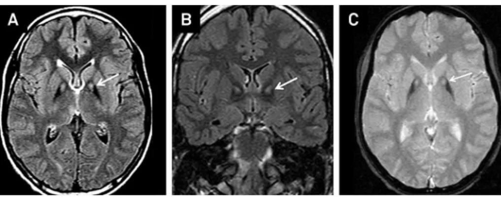
cranio-cervical and upper limbs) (Figure 1) and visual loss. Brain
MRI revealed globus pallidus hypointensity with central
hyperintense signal (eye-of-the-tiger) (Figure 2). Retinitis
pigmentosa was observed in ophthalmologic evaluation
(Figure 3). Genetic test confirmed mutation in PANK2
gene.
Pantothenate kinase-associated neurodegeneration (PKAN)
is classically characterized by early-onset dystonia and pyramidal
signs but other features may include parkinsonism,
choreoathe-tosis and dementia
1. Brain MRI typically depicts the
eye-of-the-tiger pattern
1. When retinitis pigmentosa, an unusual finding
3,
is observed in the clinical spectrum of PKAN, we must consider
variants
2,3,4, such as HARP syndrome (hypoprebetalipoproteinemia,
acanthocytosis, retinitis pigmentosa, and pallidal degeneration).
1Departamento de Neurologia, Universidade Federal de São Paulo, Sao Paulo SP, Brazil;
2Departamento de Oftalmologia, Universidade Federal de São Paulo, Sao Paulo SP, Brazil.
Correspondence:José Luiz Pedroso; Rua Botucatu, 740; 04023-900 São Paulo SP, Brasil; E-mail: jlpedroso.neuro@gmail.com Conflict of interest:There is no conflict of interest to declare.
Received 29 April 2014; Received in final form 01 July 2014; Accepted 21 July 2014.
Figure 1.
Dystonia in upper limbs (A). Note marked dystonia involving cranio-cervical segment and facial
“
grimacing
”
(B and C).
Figure 2.
Axial FLAIR (A), coronal FLAIR (B) and spin echo (C) sequences brain MRI disclose marked hypointense signal of the
globus pallidus with central hyperintense signal (eye-of-the tiger appearance) (arrows).
DOI:10.1590/0004-282X20140122
IMAGES IN NEUROLOGY
References
1. Schneider SA, Hardy J, Bhatia KP. Syndromes of neurodegeneration with brain iron accumulation (NBIA): an update on clinical presentations, histological and genetic underpinnings, and treatment considerations. Mov Disord. 2012;27(1):42-53. http://dx.doi.org/10.1002/mds.23971
2. Orrell RW, Amrolia PJ, Heald A, et al. Acanthocytosis, retinitis pigmentosa, and pallidal degeneration: a report of three patients, including the second reported case with hypoprebetalipoproteinemia (HARP syndrome). Neurology. 1995;45(3):487-92. http://dx.doi.org/10.1212/wnl.45.3.487
3. Ching KH, Westaway SK, Gitschier J, Higgins JJ, Hayflick SJ. HARP syndrome is allelic with pantothenate kinase-associated neurode-generation. Neurology. 2002;58(11):1673-4. http://dx.doi.org/10.1212/ wnl.58.11.1673
4. Houlden H, Lincoln S, Farrer M, Cleland PG, Hardy J, Orrell RW. Com-pound heterozygous PANK2 mutations confirm HARP and Hallervorden-Spatz syndromes are allelic. Neurology. 2003;61(10):1423-6. http://dx.doi. org/10.1212/01.wnl.0000094120.09977.92