Submitted11 July 2016 Accepted 8 October 2016 Published9 November 2016
Corresponding authors Jerson Lima Silva, [email protected], [email protected] Andréa Cheble de Oliveira, [email protected]
Academic editor Vladimir Uversky
Additional Information and Declarations can be found on page 17
DOI10.7717/peerj.2670
Copyright 2016 De Souza et al.
Distributed under
Creative Commons CC-BY 4.0
OPEN ACCESS
Charge neutralization as the major factor
for the assembly of nucleocapsid-like
particles from C-terminal truncated
hepatitis C virus core protein
Theo Luiz Ferraz de Souza1,2,*, Sheila Maria Barbosa de Lima3,*,
Vanessa L. de Azevedo Braga2,4, David S. Peabody5, Davis Fernandes Ferreira2,6,
M. Lucia Bianconi4, Andre Marco de Oliveira Gomes2,4, Jerson Lima Silva2,4and
Andréa Cheble de Oliveira2,4
1Faculdade de Farmácia, Universidade Federal do Rio de Janeiro, Rio de Janeiro, Brazil
2Instituto Nacional de Ciência e Tecnologia de Biologia Estrutural e Bioimagem, Universidade Federal do Rio
de Janeiro, Rio de Janeiro, Brazil
3Bio-Manguinhos, Funda¸cão Oswaldo Cruz, Rio de Janeiro, Brazil
4Programa de Biologia Estrutural, Instituto de Bioquímica Médica Leopoldo de Meis, Universidade Federal do
Rio de Janeiro, Rio de Janeiro, Brazil
5Department of Molecular Genetics and Microbiology and Cancer Research and Treatment Center, University
of New Mexico, Albuquerque, United States
6Instituto de Microbiologia Paulo de Góes, Universidade Federal do Rio de Janeiro, Rio de Janeiro, Brazil
*These authors contributed equally to this work.
ABSTRACT
Background. Hepatitis C virus (HCV) core protein, in addition to its structural role to form the nucleocapsid assembly, plays a critical role in HCV pathogenesis by interfering in several cellular processes, including microRNA and mRNA homeostasis. The C-terminal truncated HCV core protein (C124) is intrinsically unstructured in solution and is able to interact with unspecific nucleic acids, in the micromolar range, and to assemble into nucleocapsid-like particles (NLPs) in vitro. The specificity and propensity of C124 to the assembly and its implications on HCV pathogenesis are not well understood.
Methods. Spectroscopic techniques, transmission electron microscopy and calorimetry were used to better understand the propensity of C124 to fold or to multimerize into NLPs when subjected to different conditions or in the presence of unspecific nucleic acids of equivalent size to cellular microRNAs.
Results. The structural analysis indicated that C124 has low propensity to self-folding. On the other hand, for the first time, we show that C124, in the absence of nucleic acids, multimerizes into empty NLPs when subjected to a pH close to its isoelectric point (pH≈12), indicating that assembly is mainly driven by charge neutralization. Isothermal calorimetry data showed that the assembly of NLPs promoted by nucleic acids is enthalpy driven. Additionally, data obtained from fluorescence correlation spectroscopy show that C124, in nanomolar range, was able to interact and to sequester a large number of short unspecific nucleic acids into NLPs.
unspecific cellular polyanions, which may correspond to microRNAs and mRNAs in a host cell infected by HCV, triggering their confinement into infectious particles.
SubjectsBiochemistry, Biophysics, Virology
Keywords HCV core protein, Capsid assembly, Circular dichroism, Fluorescence spectroscopy, Structural biology
INTRODUCTION
Hepatitis C Virus (HCV) is a member of theFlaviviridaefamily, genusHepacivirus, which causes acute and chronic liver diseases. It is estimated that about 170–200 million people worldwide are infected with HCV (Pol et al., 2012). Chronic hepatitis C can lead to cirrhosis and subsequent complications such as hepatocellular carcinoma (HCC). Although in remarkable development, current therapies against HCV are still unsatisfactory and several researchers seek a better understanding of processes associated to the HCV infection and pathogenesis in order to favor the development of novel alternative treatments (Vermehren & Sarrazin, 2011;Wendt et al., 2014;Douam, Ding & Ploss, 2016).
HCV contains one copy of a positive single-stranded RNA genome which encodes a single polyprotein with approximately 3,000 amino acids. This polyprotein is processed by both cellular and viral proteases at the level of the endoplasmic reticulum (ER) into at least 10 mature proteins (Grakoui et al., 1993). The structural proteins include the core protein and the envelope glycoproteins E1 and E2. The nonstructural proteins NS3, NS4A, NS4B, NS5A and NS5B are components of the viral replicase complex, and p7 and NS2 proteins are dispensable for replication but necessary for the production of progeny viruses (Moradpour, Penin & Rice, 2007;Jones et al., 2007;Gentzsch et al., 2013).
2006), which involves the three basic clusters of N-terminal (Sharma et al., 2010). Different truncated hepatitis C virus core proteins can induce proapoptotic and pronecrotic effects (Yan et al., 2005), which might play an important role in the pathogenesis of HCV persistent infection and HCC.
The first cleavage of the polyprotein forms an immature core protein of 191 amino acids residues (Lauer & Walker, 2001). During the following cleavage, the core protein remains anchored to the ER through a C-terminal hydrophobic region (Grakoui et al., 1993) being further processed at its C-terminus by a signal peptide peptidase resulting in the mature protein (Liu et al., 1997;Yasui et al., 1998). The C-terminus of mature core protein is not precisely elucidated but probably lies between residues 170 and 182 (Kopp et al., 2010;
Oehler et al., 2012). Although core protein has been shown to possess a nuclear localization signal (Suzuki et al., 1995;Yan et al., 2005), it is located exclusively in the cytoplasm in cells infected with cell culture-derived HCV, consistent with the cytoplasmic life cycle of HCV (Lindenbach & Rice, 2001).
The mature core protein consists of a N-terminal two-thirds hydrophilic domain of 120 aa or so and a C-terminal one-third hydrophobic domain of 50 aa or so (McLauchlan, 2000). Similar to the core proteins of HCV-related flaviviruses, the HCV core protein is essentially composed ofα-helices (50%). In the presence of mild detergents, it is a dimeric alpha-helical protein, but it forms heterogeneous soluble micelle-like aggregates of high molecular weight in the absence of detergents. In contrast, the C-terminal truncated form (117 residues) is soluble, monodispersed, and unfolded in the absence of detergent. The 117–169 hydropho-bic domain contains the structural determinants for the HCV core protein binding to cellular membranes and is essential for the folding of the highly basic domain. Therefore, the hydrophobic C-terminal domain is responsible for core association with lipid droplets in mammalian cells and with endoplasmic reticulum membranes, and the N-terminal domain that includes numerous positively charged amino acids is mainly involved in RNA binding (Boulant et al., 2005).
The focus of our research was to seek new structural and thermodynamic information to better understand the molecular aspects of the N-terminal region of core protein from C-terminal truncated HCV core protein (C124). Our data indicate that C124 has a low propen-sity for overall folding. In contrast, by electron microscopy, we show an unusual capacity of C124, at low concentration and in the absence of nucleic acids, to naturally multimerize into empty nucleocapsid-like particles (NLPs) when subjected to a pH close to its isoelectric point. Moreover, our data indicate that C124 is able to sequester a great number of unspe-cific nucleic acids of molecular size equivalent to the cellular microRNAs into NLPs in the nanomolar range. Our findings reveal features that can be related to the multiplicity of func-tions of HCV core protein, such as gene regulation, and explain why thein vitroformation of NLPs does not require high specificity, being mainly driven by neutralization of basic residues, which correspond to approximately 20% of the C-terminal truncated HCV core protein. Implications in virus-host interaction and HCV pathogenesis are discussed.
MATERIALS & METHODS
ChemicalsAll reagents were of analytical grade. Distilled water was filtered and deionized through a Millipore water purification system. The probe bis-8-anilinonaphthalene-1-sulfonate (bis-ANS) was purchased from Invitrogen. All experiments were performed at 20◦C using
the standard buffer: 10 mM phosphate (pH 7.0) with 100 mM NaCl.
Nucleic acid samples
High pressure liquid chromatography-purified synthetic single-stranded RNA fragment 43–59 of SAF93 aptamer (SAF9343–59-5′-GGA UGC AAU CUC CAU
CCC-3′) (Rhie et al., 2003) was obtained from Integrated DNA Technologies, Inc.
(Coralville, IA, USA). Synthetic RNA samples were maintained lyophilized at −20 ◦C and used in RNase-free water. Double-stranded oligonucleotides were prepared by mixing equimolar amounts of the complementary single-stranded oligonucleotides, poly(GC) DNA (5′ ATAATTGCGCGCGCGCGCAGGAAA3′)
(purchased from DNAgency, Malvern, PA) or consensus DNA (5′
TTTCCTAGACATGC-CTAATTA 3′) (purchased from Invitrogen, Carlsbad, CA, USA), in 50 mM Tris–HCl, pH
7.2, containing 250 mM NaCl. This mixture was incubated at 96 ◦C for 5 min, and the
temperature was slowly reduced to 25◦C.
Cloning and expression of the C-terminal truncated HCV core protein We amplified the HCV core sequence by PCR from pCV-H77C, an infectious cDNA clone of type 1a (from J Bukh, NIH) (Yanagi et al., 1997), using a 5′ primer with the
sequence 5′-GCGCCATATGAGCACGAATCCTAAACCT-3′ a 3′ primer of sequence 5′
-GCGGATCCTCAGGCTGAAGCGGGCACAGTCAG-3′, and Vent DNA polymerase (New
ease the purification process on a nickel affinity column (Qiagen)) cleaved with the same enzymes, Biolabs. The C124 was propagated to midlog phase (OD600=0.8) inEscherichia
colistrain BL21(DE3) at 25◦C. Expression of C124 fused to a histidine tail was induced with
1 mM IPTG. Three hours after induction the cells were centrifuged (5,500 g for 20 min) at 4◦C and frozen at−20◦C overnight.
Purification of the C-terminal truncated HCV core protein
After thawing, the cells were ressuspended in lysis buffer (25 mM NaH2PO4, 250 mM
NaCl, 8 M urea, 2 mM EDTA and 2 mM DTT, pH 7.0), and were sonicated. The cell debris was pelleted by centrifugation (13,500 g for 20 min). The clarified lysate containing the core protein was applied to a cation-exchange column (SP Sepharose) equilibrated with denaturing cation buffer (25 mM Hepes, pH 7.0, 50 mM NaCl, 8 M urea). The core protein was eluted with a linear NaCl gradient (0.05–1.0 M). Fractions containing core protein were subsequently applied to a nickel-NTA agarose resin at pH 7.0, and to promote the elution we utilized the buffer (10 mM Tris, 100 mM NaCl) at pH 3.0. To the measurements in neutral conditions, the protein in high concentration was diluted in the same buffer at pH 7.0. The homogeneity of purified core protein was determined by 15% sodium dodecyl sulfate-polyacrylamide gel electrophoresis (SDS-PAGE) stained with Comassie Brilliant Blue. Protein concentration was determined by UV spectrophotometry at a wavelength of 280 nm in 6 M guanidine hydrochloride solution, using an extinction coefficient of 31,970 M−1determined using ExPASy program (Appel, Bairoch & Hochstrasser, 1994).
Fluorescence spectroscopy
Fluorescence spectra were recorded in an ISSK2 spectrofluorometer (ISS Inc., Champaign, IL, USA). The protein was excited at 280 nm and emission was observed from 300 to 420 nm. For experiments in the presence of bis-ANS, the excitation wavelength was 360 nm and emission was collected from 400 nm to 600 nm.
Light scattering
Light scattering (LS) measurements were performed in an ISSK2 spectrofluorometer (ISS Inc., Champaign, IL, USA). Scattered light was collected at an angle of 90◦to the incident
light. The samples were illuminated at 320 nm and the light was collected in the range of 315 to 325 nm. This wavelength was chosen because neither protein nor nucleic acid absorbs at 320 nm. In each pH, the LS values were obtained from the area under LS curve.
Circular dichroism
Circular dichroism (CD) spectra of C124 were recorded on a Jasco J-715 (1505 model) spec-tropolarimeter (Jasco Corporation, Tokyo, Japan) with 0.02 cm circular path length cells at 20◦C. Wavelength range: 260–190 nm. The spectra were averaged from three scans that
Temporal evolution studies of in vitroassembly reactions
For temporal evolution studies, the optical density was monitored at 350 nm in a spectrophotometer at 20◦C. For analysis of NLPs formation promoted by unspecific nucleic
acids, fixed amount of DNA (100 µL), to the final concentration of 5µM, was added to different concentrations of C124 to a final volume of 600µL. Optical density was recorded by Swift II software every 2 s for 5 min. The maximum value represents the maximum optical density obtained during this 5 min analysis.
Transmission electron microscopy
After specific temporal studies the same samples were adsorbed to carbon coated Cu grids, 400 mesh, for 10 min. Grids were washed three times with filtered, distilled water and stained for 2 min with 2% uranyl acetate, and the NLPs were examined on a Morgani electron microscope (FEI Co., Hillsboro, OR, USA) operated at 100 kV. After scanning the negatives, images were processed with Adobe Photoshop (Adobe Systems Inc., Mountain View, CA).
Isothermal titration calorimetry (ITC)
ITC measurements were performed using a VP-ITC calorimeter from MicroCal, Llc (Northampton, MA, USA). The titration of 20µM HCV core protein with unspecific nucleic acids (RNA or DNA) involved 13 injections (1×1µL and 12×20µL) of 0.1 mM nucleic acid solution at 5 min intervals, with constant stirring at 317 rpm. The temperature was set at 37 ◦C. The C124 solutions were degassed under vacuum before the titrations,
and the reference cell was filled with Milli-Q water. The heat of dilution of nucleic acids into the buffer was subtracted from the raw data obtained using C124. Three independent experiments were analyzed separately.
Fluorescence correlation spectroscopy (FCS) measurements
FCS measurements were carried out in an ALBA fluorescence correlation spectrometer (ISS, Champaign, IL, USA) using a Nikon TE2000-U inverted microscope with a two-photon excitation regime. A Ti:Sa Tsunami laser, pumped by a Millenia Pro 15sJ (Spectra Physics, Mountain View, CA, USA), was focused on the sample with a water immersion 63_objective, 1.2 NA. A wavelength of 780 nm was used for excitation. The nucleic acids were FITC or Alexa-488-labeled. Fluorescence was collected through the same lens and separated from excitation by a dichroic mirror 700dcxru (Chroma, VT, USA). After passing through the dichroic mirror, the beam was split by a 50/50 beam splitter, and the resulting beams were focused to two APD detectors. Recorded fluctuation traces were processed in Vista ISS software for autocorrelation.
RESULTS
HCV C124 protein structure and the effect of alcohols
Figure 1 HCV core protein.(A) Primary sequence of HCV core protein. Hydrophobic residues (L, I, V, F, W and M) are shown in green, acidic residues in red and basic residues in blue. (B) Scheme of HCV core protein domains and their respective functions are highlighted.
shown in green, being important to the cellular membrane interaction (Fig. 1A), and the signal peptide direct hepatitis C virus polyprotein processing in the endoplasmic reticulum (Fig. 1B).
In order to analyze the C124 structure, we probed the intrinsic fluorescence of the six tryptophan residues, located at positions 76, 83, 93, 96, 107 and 113 of the protein. Our data showed that these residues were extremely solvent-exposed (Fig. 1A) as evidenced by a center of spectral mass of 352 nm, reflecting characteristics of an disordered protein. The far-UV CD spectrum of C124 exhibited a negative signal at 200 nm, in agreement with a non-structured protein (Fig. 1B).
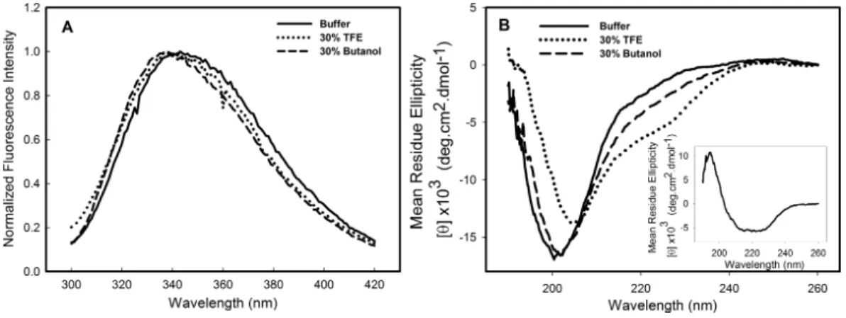
Structural studies in the presence of different solvents can provide information about the stability, folding pathway and intermolecular interactions of a protein. Alcohols are known to modulate the interactions of the polypeptide chain, and trifluorethanol (TFE) has been used to induce the formation of helical structure in protein fragments and peptides (Dyson & Wright, 1993;Hamada et al., 2000) and in many other instances is known to transform proteins into molten globule-like intermediates (Konno, Iwashita & Nagayama, 2000) and sometimes to stabilize intermediate structures (Luo & Baldwin, 1998). To investigate the effects of alcohols on the C124 structure, the samples were incubated in the presence of 10–50% of either butanol or its fluorinated derivative, TFE. The most significant effects were observed at 30% of both alcohols and are shown inFig. 2. The intrinsic fluorescence spectrum (Fig. 2A—dotted line, TFE, dashed line, butanol) underwent a slight blue shift in the presence of the alcohols, indicating little change in the tertiary structure. The spectral center of mass of the C124 fluorescence spectrum changed from 343 nm to 341 nm in the presence of 30% TFE and to 337 nm in the presence of 30% butanol. Although small changes in tryptophan environment were observed, the results showed that TFE was able to promote significant alterations in the secondary structure of C124, as indicated by the effects on the circular dichroism spectrum (Fig. 2B, dotted line).
Analysis of bis-ANS binding to C124
Figure 2 Structural changes of HCV C124 induced by TFE and butanol.Changes on the tertiary struc-ture (A) and secondary strucstruc-ture (B) of C124 (25µM) in the presence of 30% butanol or 30% TFE. Inset shows the resulting CD spectrum obtained after subtracting the C124 CD spectrum in the presence of TFE from that in the absence of TFE.
in proteins, especially in proximity to positive charges (Rosen & Weber, 1969;Silva et al., 1992). Since its binding is followed by a large increase in its fluorescence quantum yield, this probe has been frequently used to investigate the presence of hydrophobic sites, conformational changes, and to detect the presence of molten globule states of proteins (Silva et al., 1992). Additionally, the effects of this fluorescent probe on protein conformation and stability have been studied (Shi, Palleros & Fink, 1994;Foguel et al., 1996;
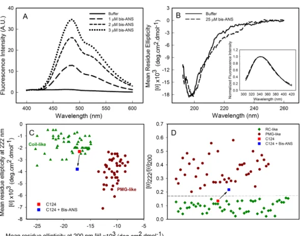
Lima et al., 2006). Here, we show that bis-ANS was able to bind to C124, as verified by the increase on bis-ANS fluorescence emission intensity (Fig. 3A). Nevertheless, binding of the probe did not significantly change either the CD (Fig. 3B) or the intrinsic fluorescence spectra (Inset, Fig. 3B), indicating no significant alterations in the secondary structure or in the tryptophan environment of C124 (Fig. 3B). We were not able to observe any increase in turbidity or in light scattering in the presence of bis-ANS or alcohols (data not shown). IDPs CD spectra may be derived of at least three different disordered equilibrium conformations, molten globule (MG), premolten globule (PMG), and random coil (RC) (Uversky, 2002;Habchi et al., 2014). The PMG-like and RD-like forms can be subdivided from the correlation between θ222 andθ200values, as described byUversky (2002)(Fig.
3C). This analysis shows that C124 is a random coil in solution (Fig. 3C) as confirmed by the analysis of the ratio between theθ222 andθ200 (Fig. 3D), as described byBlocquel et al. (2012). However, the ratioθ222/θ200of C124 CD spectrum in the presence of bis-ANS
changed to 0.217 suggesting that bis-ANS binding promotes a transition of a RC-like to a PMG-like form (Fig. 3D). Theθ222/θ200analysis has the advantage since it undergoes smaller
interference of errors in estimations of protein concentrations (Blocquel et al., 2012).
Changes in pH induce the formation of empty nucleocapsid-like particles (NLPs)
Figure 3 Analysis of binding of bis-ANS to C124.(A) Analysis of bis-ANS binding to C124 (1µM) by the increase of fluorescence intensity of bis-ANS spectra. (B) Changes on the secondary structure observed by Far-UV CD of C124 (25µM) in the presence of the probe (25µM). Inset: intrinsic fluorescence mea-surements of C124 in the absence and in the presence of bis-ANS at room temperature. (C) Double wave-length plot, [θ]222versus [θ]200, modified fromUversky (2002), of a set of well-characterized unfolded, RC-like (dark green diamonds) or PMG-like proteins (dark red circles), and of the C124 in the absence or presence of Bis-ANS (25µM) that the positions are highlighted (red and blue squares, respectively). (D) Plot of the ratio between the ellipticity at 222 nm and the ellipticity at 200 nm ([θ]222/[θ]200) of the same set of well-characterized RC-like or PMG-like proteins shown in (C). The position in the plot of C124 in the absence or presence of bis-ANS is highlighted (red and blue squares, respectively). The arrows in (C) and (D) are indicating the changes in the C124 position promoted by bis-ANS binding.
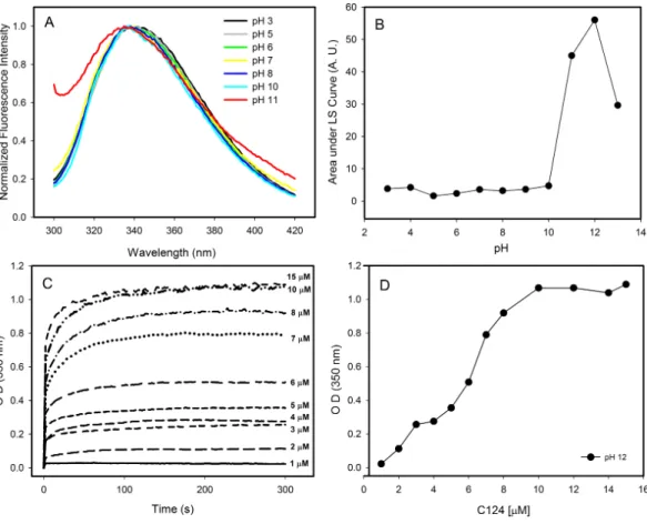
Figure 4 Effects of pH on the structure of C124.(A) Changes in the intrinsic fluorescence induced by high pH values. (B) Light scattering of C124 at different pH values. (C) Temporal evolution analysis ofin vitroassembly of NLPs at different concentrations of C124 triggered by the pH 12. (D) Plot of the maxi-mum O.D. values at 350 nm as derived from the curves in (C) at different C124 concentrations.
concentration correlated with an increase of optical density, as expected to protein self-assembly process (Fig. 4D).
Images obtained by transmission electron microscopy confirmed the formation of NLPs at pH 12 (Fig. 5). The micrographs showed the formation of NLPs with heterogeneous particle size at pH 12, as already observed for NLP assembly induced by some nucleic acids (Kunkel et al., 2001).
The process of NLPs assembly is triggered by short unspecific nucleic acids
The formation of NLPs triggered by addition of nucleic acids has been extensively characterized (Fromentin et al., 2007; Kunkel et al., 2001). Here, we used other
Figure 5 Electron micrographs of negatively stained nucleocapsid-like particles (NLPs) produced from truncated HCV core protein at pH 12.The protein concentration was 20µM. Bars: 100 nm (A) and 300 nm (B and C).
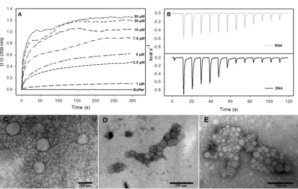
Figure 6 Interaction of HCV core protein with unspecific nucleic acids and NLPs formation.(A)
Tem-poral evolution studies ofin vitroassembly of NLPs at different concentrations of C124 triggered by the addition of 5µM DNA poly(GC). (B) Heat flux profile associated with injections of 5µM nucleic acid (poly(GC) DNA or RNA (SAF9343–59)) in the calorimetric cell containing C124 (20µM) at 37 ◦C. (C–
E) Electron micrograph of negatively stained nucleocapsid-like particles (NLPs) produced from C124 at 2.5µM (C), 10µM (D) and 50µM (E). To each protein concentration was added 5µM poly(GC) DNA. Bars: (C)—100 nm and (D, E)—300 nm.
The process of NLPs assembly triggered by nucleic acids is enthalpy driven
Isothermal titration calorimetry (ITC) is often used to measure the heat absorbed or released during a reaction and, thus, can provide a universal means to follow biological processes (Ladbury, 2004). We used ITC to investigate the energetics of NLP assembly induced by the interaction of C124 with nonspecific nucleic acids. As shown in the calorimetric traces inFig. 6B, each injection of nucleic acids (DNA or RNA) into the protein solution resulted in an exothermic reaction, showing that nucleic acid-triggered capsid assembly in solution is enthalpically driven at 37◦C (Fig. 6B). These data indicate an important role
of non-hydrophobic interactions, such as the electrostatic interactions between basic residues of the protein and phosphate groups on the nucleic acids, and suggest that charge neutralization plays an important role in the particle assembly process. This is also consistent with our results showing capsid assembly at pH=12. These may be the main interactions driving the assembly processin vitro.
Fluorescence correlation spectroscopy analysis indicates capsid assembly with unspecific nucleic acid in nanomolar range
Although the capsid assembly promoted by unspecific nucleic acids has been studied (Fromentin et al., 2007;Kunkel et al., 2001), the concentrations of both protein and nucleic acid used in most of that work were in the micromolar range. In the experiments described here, we applied fluorescence correlation spectroscopy (FCS) to gain new information on the interaction between nucleic acids and the C-terminal truncated HCV core protein in the nanomolar range, by using DNA or RNA labeled with Alexa-488.
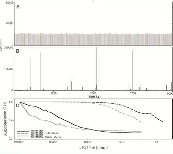
Observing the raw data from FCS measurements we verified that the fluorescence fluc-tuation from the free RNA is very homogeneous (Fig. 7A). The results were similar to RNA (Fig. 7A) and DNA (Fig. S2). On the other hand, in the presence of higher concentrations of HCV core protein we observed rare fluorescent species with very high fluorescence intensity (Fig. 7B). In addition, the signal of the free nucleic acids basically disappeared, presumably due to RNA oligomerization promoted by capsid assembly (Fig. 7B). These data suggest that the assembly is a highly cooperative process since stable intermediates were not observed. For comparison of the diffusion times in DNA or RNA in the absence or in the presence of C124, the normalized autocorrelation curves are shown inFig. 7C. The protein promoted a great increase in the diffusion time of both RNA and DNA, consistent with the formation of NLPs.
DISCUSSION
Figure 7 C-terminal truncated HCV core protein and unspecific nucleic acids (DNA or RNA) interac-tion analyzed by fluorescence correlainterac-tion spectroscopy.Fluctuation of the fluorescence intensity of RNA labeled with Alexa-488 in the absence (A) or in the presence (B) of C-terminal truncated HCV core pro-tein, and the normalized autocorrelation curves of free DNA or RNA at 100 nM and in the presence of 1 µM or 500 nM of C124 (C). The buffer used was 10 mM phosphate (pH 7.4) with 100 mM NaCl.
Figure 8 Free energy diagram representing the energy barrier between the disordered state of C124 to oligomeric state (empty capsid or nucleic acid loaded capsid).
Although bis-ANS was able to induce only subtle changes of protein secondary structure, the binding verified by the increase of fluorescence intensity indicated the presence of structured hydrophobic regions in the C-terminal truncated HCV core protein (Fig. 3A). Bis-ANS binds noncovalently to nonpolar segments in proteins, especially in proximity to positive charges (Rosen & Weber, 1969;Silva et al., 1992). Proteins that consist of disordered regions interspersed with short structured regions are typically intrinsically disordered proteins. There are at least three different disordered equilibrium conformations, molten globule, premolten globule, and randon coil in IDPs (Uversky, 2002;Habchi et al., 2014). Analysis of the correlation betweenθ200 andθ222 values (Uversky, 2002) and
ratio betweenθ222andθ200(Blocquel et al., 2012) indicates that C124 is RC-like in solution
(Figs. 3Cand3D). Additionally, the change ofθ222/θ200=0.135 toθ222/θ200=0.217 indicates that bis-ANS binding promotes the transition of a RC-like to a PMG-like form possibly by folding intermediate stabilization. Additionally, consistent with this finding, previous studies described that hydrophobic fluorescence probe ANS also is able to interact with premolten globule state in proteins (Uversky & Ptitsyn, 1996;Uversky, 2002). These structured regions are known to be important in specific ligand binding in some cases (Fuxreiter et al., 2004;Mohan et al., 2006;Habchi et al., 2014). The stabilization of PMG-like form in the C124 structure, as indicated by bis-ANS binding analysis, may also account for the ability of binding to different cellular targets.
Energetic aspects of the capsid assembly are important to the understanding of the bio-logical process. The assembly of Hepatitis B Virus capsids is driven by weak protein-protein interactions and is characterized by positive enthalpy and entropy (Ceres & Zlotnick, 2002). This assembly process is entropy-driven, and is characterized largely by hydrophobic con-tacts. On the other hand, the binding of intrinsically disordered proteins, which are usually highly hydrophilic, involves an entropic cost associated with the disorder-to-order transi-tion (Dyson & Wright, 2005;Habchi et al., 2014). Here, we verified that a negative enthalpic contribution is the key thermodynamic driving force for NLP assembly by C124 (Fig. 6B). It is likely that the negative enthalpy offsets the entropic cost, giving a good example of enthalpy-entropy compensation associated to assembly process. The heat released in the assembly process is probably mainly due to the neutralization of basic residues as they interact electrostatically with nucleic acids.
and length of the DNA used, suggesting that the neutralization of positive charges might be the main event driving the assembly process (López et al., 2009). The fact that all these studies were performed in micromolar range and the binding affinities are not well characterized must be given due importance.
of miR-122 targets over the human transcriptome favoring the unbalance liver homeostasis and the development of liver cancers (Luna et al., 2015). We suggest that core protein could contribute to the deregulation of microRNA homeostasis from encapsulation of unspecific microRNAs or mRNA into infectious particles.
Recent studies have demonstrated that, besides the viral genome, host RNAs, such as mRNA and non-coding RNAs, are encapsidated by authentic flock house virus virions and virus-like particles. This important finding showed that, although there is high specificity for the viral RNA, a variable genetic content may also be packed by nonenveloped RNA viruses (Routh, Domitrovic & Johnson, 2012a;Routh, Domitrovic & Johnson, 2012b). One site containing three conserved phenylalanine residues at the C-terminal of the coat protein alpha of Flock House Virus has been identified as essential for the specific encapsidation of viral RNA. Deletion of all three residues almost totally abolishes viral RNA encapsidation, resulting in particles that primarily package cellular RNA (Schneemann & Marshall, 1998). These findings are comparable to the unspecific RNA packaging by the C-terminal truncated HCV core protein described here. Therefore, we speculate that similar events could occur during the production of HCV virions.
CONCLUSIONS
The present study shows that the N-terminal of HCV core protein has low propensity to overall folding. However, in the absence of nucleic acids, it multimerizes itself into empty NLPs when submitted to pH values close to the isoelectric point (pH≈12). This finding suggests that electrostatic repulsion among the positive charges of the basic residues of the C124, which represents about 20% of their residues, is the only energy barrier that avoids protein multimerization, and that unspecific polyanions could promote the formation of NLPs. Our observations suggest that C124 can physically interact with different polyanions, triggering their confinement into NLPs. It also explains why thein vitroNLP formation does not require high specificity. Based on these new findings, we speculate that this process could also happen in a host cell infected by HCV, promoting the confinement of microRNAs and mRNAs into infectious particles. We believe that the new data here obtained provide advances in the understanding of the molecular basis of some secondary effects of the core protein on the HCV pathogenesis.
ACKNOWLEDGEMENTS
We gratefully acknowledge Emerson R. Gon¸calves and Ana Carolina Q. Vaz for competent technical assistance.
ADDITIONAL INFORMATION AND DECLARATIONS
Funding
Janeiro (FAPERJ), Funda¸cão Universitária José Bonifácio (FUJB), Instituto Milênio de Biologia Estrutural em Biomedicina e Biotecnologia (IMBEBB), Instituto Nacional de Ciência e Tecnologia de Biologia Estrutural e Bioimagem (INBEB), and Programa de Apoio a Núcleos de Excelência (PRONEX) to JLS, AMOG, and ACO. The funders had no role in study design, data collection and analysis, decision to publish, or preparation of the manuscript.
Grant Disclosures
The following grant information was disclosed by the authors:
Conselho Nacional de Desenvolvimento Científico e Tecnológico (CNPq). Coordena¸cão de Aperfei¸coamento de Pessoal de Nível Superior (CAPES).
Funda¸cão Carlos Chagas Filho de Amparo à Pesquisa do Estado do Rio de Janeiro (FAPERJ). Funda¸cão Universitária José Bonifácio (FUJB).
Instituto Milênio de Biologia Estrutural em Biomedicina e Biotecnologia (IMBEBB). Instituto Nacional de Ciência e Tecnologia de Biologia Estrutural e Bioimagem (INBEB). Programa de Apoio a Núcleos de Excelência (PRONEX).
Competing Interests
Jerson Lima Silva is an Academic Editor for PeerJ.
Author Contributions
• Theo Luiz Ferraz de Souza and Sheila Maria Barbosa de Lima conceived and designed the experiments, performed the experiments, analyzed the data, wrote the paper, prepared figures and/or tables, reviewed drafts of the paper.
• Vanessa L. de Azevedo Braga conceived and designed the experiments, performed the experiments, analyzed the data, prepared figures and/or tables, reviewed drafts of the paper.
• David S. Peabody conceived and designed the experiments, performed the experiments, analyzed the data, contributed reagents/materials/analysis tools, reviewed drafts of the paper.
• Davis Fernandes Ferreira performed the experiments, analyzed the data, reviewed drafts of the paper.
• M. Lucia Bianconi analyzed the data, reviewed drafts of the paper.
• Andre Marco de Oliveira Gomes conceived and designed the experiments, performed the experiments, analyzed the data, contributed reagents/materials/analysis tools, wrote the paper, reviewed drafts of the paper.
• Jerson Lima Silva analyzed the data, contributed reagents/materials/analysis tools, wrote the paper, reviewed drafts of the paper.
• Andréa Cheble de Oliveira conceived and designed the experiments, performed the experiments, analyzed the data, contributed reagents/materials/analysis tools, wrote the paper, prepared figures and/or tables, reviewed drafts of the paper.
Data Availability
Supplemental Information
Supplemental information for this article can be found online athttp://dx.doi.org/10.7717/ peerj.2670#supplemental-information.
REFERENCES
Acosta-Rivero N, Rodrigues A, Mussachio A, Poutu J, Falcon V, Torres D, Aguillar C, Linares M, Alonso M, Perez A, Menezes I, Morales-Grillo J, Marques G, Dueñas-Carrera S. 2005.A C-terminal truncated hepatitis C virus core protein variant assembles into virus-like particlesin vitroin the absence of structured nucleic acids. Biochemical and Biophysical Research Communications334:901–906
DOI 10.1016/j.bbrc.2005.06.185.
Ahmad E, Rahman SK, Khan JM, Varshney A, Khan RH. 2010.Phytolacca America lectin (Pa-2; pokeweed mitogen): an intrinsically unordered protein and its conver-sion into partial order at low pH.Bioscience Reports30:125–134
DOI 10.1042/BSR20090035.
Appel RD, Bairoch A, Hochstrasser DF. 1994.A new generation of information retrieval tools for biologists: the example of the ExPASy WWW server.Trends in Biochemical Sciences19:258–260 DOI 10.1016/0968-0004(94)90153-8.
Bartenschlager R, Penin F, Lohmann V, André P. 2011.Assembly of infectious hepatitis C virus particles.Trends in Microbiology 19(2):95–103
DOI 10.1016/j.tim.2010.11.005.
Blocquel D, Habchi J, Gruet A, Blangy S, Longhi S. 2012.Compaction and binding properties of the intrinsically disordered C-terminal domain of Henipavirus nucleoprotein as unveiled by deletion studies.Molecular BioSystems8:392–410 DOI 10.1039/C1MB05401E.
Boson B, Granio O, Bartenschlager R, Cosset F. 2011.A concerted action of hepatitis C virus P7 and nonstructural protein 2 regulates core localization at the endoplasmic reticulum and virus assembly.PLoS Pathogens7(7):e1002144
DOI 10.1371/journal.ppat.1002144.
Boulant S, Vanbelle C, Ebel C, Penin F, Lavergne J. 2005.Hepatitis C virus core protein is a dimeric alpha-helical protein exhibiting membrane protein features.Journal of Virology79(17):11353–11365DOI 10.1128/JVI.79.17.11353-11365.2005.
Ceres P, Zlotnick A. 2002.Weak protein–protein interactions are sufficient to drive assembly of hepatitis B virus capsids.Biochemistry41:11525–11531
DOI 10.1021/bi0261645.
Chen X. 2009.MicroRNA signatures in liver diseases.World Journal of Gastroenterology
15(14):1665–1672DOI 10.3748/wjg.15.1665.
Conrad KD, Niepmann M. 2014.The role of microRNAs in hepatitis C virus RNA replication.Archives of Virology159(5):849–862 DOI 10.1007/s00705-013-1883-4.
Cristofari G, Ivanyi-Nagy R, Gabus C, Boulant S, Lavergne JP, Penin F, Darlix JL. 2004.The hepatitis C virus Core protein is a potent nucleic acid chaperone that directs dimerization of the viral (+) strand RNAin vitro.Nucleic Acids Research
32:2623–2631DOI 10.1093/nar/gkh579.
De Giorgi V, Monaco A, Worchech A, Rornesello M, Izzo F, Buonaguro L, Marincola FM, Wang E, Buonaguro FM. 2009.Gene profiling, biomarkers and pathways characterizing HCV-related hepatocellular carcinoma.Journal of Translational Medicine7: Article 85DOI 10.1186/1479-5876-7-85.
Douam F, Ding Q, Ploss A. 2016.Recent advances in understanding hepatitis C. F1000Research5(F1000 Faculty Rev): Article 131
DOI 10.12688/f1000research.7354.1.
Dunker AK, Babu MM, Barbar E, Blackledge M, Bondos SE, Dosztánvi Z, Dyson HJ, Forman-Kay J, Fuxreiter M, Gsponer J, Han K, Jones DT, Longhi S, Metallo SJ, Nishikawa K, Nussinov R, Obradovic Z, Pappu RV, Rost B, Selenko P, Subrama-niam V, Sussman JL, Tompa P, Uversky VN. 2013.What’s in a name? Why these proteins are intrinsically disordered.Intrinsically Disordered Proteins1(1):e24157 DOI 10.4161/idp.24157.
Duvignaud JB, Leclerc D, Gagné SM. 2010.Structure and dynamics changes induced by 2,2,2-trifluoro-ethanol (TFE) on the N-terminal half of hepatitis C virus core protein.Biochemistry and Cell Biology88(2):315–323DOI 10.1139/O09-155.
Duvignaud JB, Savard C, Fromentin R, Majeau N, Leclerc D, Gagné SM. 2009.Structure and dynamics of the N-terminal half of hepatitis C virus core protein: an intrinsically unstructured protein.Biochemical and Biophysical Research Communications
378(1):27–31DOI 10.1016/j.bbrc.2008.10.141.
Dyson HJ, Wright PE. 1993.Peptide conformation and protein folding.Current Opinion in Structural Biology3:60–65DOI 10.1016/0959-440X(93)90203-W.
Dyson HJ, Wright PE. 2005.Intrinsically unstructured proteins and their functions. Nature Reviews6:19–208DOI 10.1038/nrm1589.
Fan Z, Yang QR, Twu JS, Sherker AH. 1999.Specificin Vitroassociation between the hepatitis C viral genome and core protein.Journal of Medical Virology59:131–134 DOI 10.1002/(SICI)1096-9071(199910)59:2<131::AID-JMV1>3.0.CO;2-C.
Foguel D, Suarez MC, Barbosa C, Rodrigues Jr JJ, Sorenson MM, Smillie LB, Silva JL. 1996.Mimicry of the calcium-induced conformational state of troponin C by low temperature under pressure.Proceedings of the National Academy of Sciences of the United States of America93(20):10642–10646DOI 10.1073/pnas.93.20.10642.
Fromentin R, Majeau N, Laliberté Gagné ME, Boivin A, Duvignaud J, Leclerc D. 2007.
A method forin vitroassembly of hepatitis C virus core protein and for screening of inhibitors.Analytical Biochemistry 366:37–45DOI 10.1016/j.ab.2007.03.033.
Fuxreiter M, Simon I, Friedrich P, Tompa P. 2004.Preformed structural elements feature in partner recognition by intrinsically unstructured proteins.Journal of Molecular Biology 338:1015–1026DOI 10.1016/j.jmb.2004.03.017.
capsid assembly and envelopment.PLoS Pathogens9(5):e1003355 DOI 10.1371/journal.ppat.1003355.
Grakoui A, Wychowski C, Lin C, Feinstone SM, Rice C. 1993.Expression and iden-tification of hepatitis C virus polyprotein cleavage products.Journal of Virology
67:1385–1395.
Habchi J, Tompa P, Longhi S, Uversky VN. 2014.Introducing protein intrinsic disorder. Chemical Reviews114(13):6561–6588DOI 10.1021/cr400514h.
Hamada D, Chiti F, Guijarro JL, Katoaba M, Taddei N, Dobson CM. 2000.Evidence concerning rate-limiting steps in protein folding from the effects of trifluoroethanol. Nature Structural & Molecular Biology7:58–61DOI 10.1038/71259.
Irshad M, Dhar I. 2006.Hepatitis C virus core protein: an update on its molecular biology, cellular functions and clinical implications.Medical Principles and Practice
15(6):405–416DOI 10.1159/000095485.
Ivanyi-Nagy R, Kanevsky I, Gabus C, Lavergne JP, Ficheux D, Penin F, Fosse P, Darlix JL. 2006.Analysis of hepatitis C virus RNA dimerization and core-RNA interactions. Nucleic Acids Research34:2618–2633DOI 10.1093/nar/gkl240.
Jones CT, Murray CL, Eastman DK, Tassello J, Rice CM. 2007.Hepatitis C virus p7 and NS2 proteins are essential for production of infectious virus.Journal of Virology
81:8374–8383DOI 10.1128/JVI.00690-07.
Jopling CL, Yi M, Lancaster AM, Lemon SM, Sarnow P. 2005.Modulation of hepatitis C virus RNA abundance by a liver-specific MicroRNA.Science309:1577–1581 DOI 10.1126/science.1113329.
Kao CC, Yi G, Huang H. 2016.The core of hepatitis C virus pathogenesis.Current Opinion in Virology17:66–73DOI 10.1016/j.coviro.2016.01.009.
Kasprzak A, Adamek A. 2008.Role of hepatitis C virus proteins (C, NS3, NS5A) in hep-atic oncogenesis.Hepatology Research38:1–26
DOI 10.1111/j.1872-034X.2007.00261.x.
Konno T, Iwashita J, Nagayama K. 2000.Fluorinated alcohol, the group of cosolvents that stabilize the molten-globule state relative to a highly denatured state of cy-tochrome c.Protein Science9:564–569 DOI 10.1110/ps.9.3.564.
Kopp M, Murray CL, Jones CT, Rice CM. 2010.Genetic analysis of the carboxy-terminal region of the hepatitis c virus core protein.Journal of Virology84(4):1666–1673 DOI 10.1128/JVI.02043-09.
Kunkel M, Lorinczi M, Rijnbrand R, Lemon SM, Watowich SJ. 2001.Self-assembly of nucleocapsid-like particles from recombinant hepatitis C virus core protein.Journal of Virology75:2119–2129DOI 10.1128/JVI.75.5.2119-2129.2001.
Kunkel M, Watowich SJ. 2004.Biophysical characterization of hepatitis C virus core protein: implications for interactions within the virus and host.FEBS Letters
557:174–180DOI 10.1016/S0014-5793(03)01486-8.
Ladbury JE. 2004.Application of isothermal titration calorimetry in the biological sciences: things are heating up!BioTechniques37(6):885–887.
Lima S, Vaz A, Souza T, Peabody SD, Silva JL, Oliveira AC. 2006.Dissecting the role of protein-protein and protein-nucleic acids interactions on MS2 Bacteriophage stabil-ity.The FEBS Journal273(7):1463–1475DOI 10.1111/j.1742-4658.2006.05167.x.
Lindenbach BD, Rice CM. 2001. Flaviviridae: the viruses and their replication. In: Knipe DM, Howley PM, eds.Fields virology. Philadelphia: Lippincott Williams and Wilkins, 991–1042.
Liu Q, Tackney C, Bhat R, Prince A, Zhang P. 1997.Regulated processing of hepatitis C virus core protein is linked to subcellular localization.Journal of Virology7:657–662.
Liu X, Wang T, Wakita T, Yang W. 2010.Systematic identification of microRNA and messenger RNA profiles in hepatitis C virus-infected human hepatoma cells.Virology
398:57–67DOI 10.1016/j.virol.2009.11.036.
López C, Gil L, Lazo L, Menéndez I, Marcos E, Sánchez J, Valdés I, Falcón V, De la Rosa MC, Márquez G, Guillén G, Hermida L. 2009.In vitroassembly of nucleocapsid-like particles from purified recombinant capsid protein of dengue-2 virus.Archives of Virology154:695–698DOI 10.1007/s00705-009-0350-8.
Lorenzo LJ, Dueñas-Carrera S, Falcón V, Acosta Rivero N, González E, De la Rosa MC, Menéndez I, Morales J. 2001.Assembly of truncated hcv core antigen into virus-like particles inEscherichia coli.Biochemical and Biophysical Research Communications
281:962–965DOI 10.1006/bbrc.2001.4449.
Luna JM, Scheel TK, Danino T, Shaw KS, Mele A, Fak JJ, Nishiuchi E, Takacs CN, Catanese MT, Jong YP, Jacobson IM, Rice CM, Darnell RB. 2015.Hepatitis C virus RNA functionally sequesters miR-122.Cell160(6):1099–1110
DOI 10.1016/j.cell.2015.02.025.
Luo P, Baldwin RL. 1998.Trifluoroethanol stabilizes the pH 4 folding intermediate of sperm whale apomyoglobin.Journal of Molecular Biology 279:49–57
DOI 10.1006/jmbi.1998.1774.
Majeau N, Gagne V, Boivin A, Bolduc M, Majeau J, Ouellet D, Leclerc D. 2004.The N-terminal half of the core protein of hepatitis C virus is sufficient for nucleocapsid formation.Journal of General Virology85:971–981DOI 10.1099/vir.0.79775-0.
McGivern DR, Lemon SM. 2008.Tumor suppressors, chromosomal instability, and hepatitis c virus-associated liver cancer.Annual Review of Pathology4:399–415 DOI 10.1146/annurev.pathol.4.110807.092202.
McLauchlan J. 2000.Properties of the hepatitis C virus core protein: a structural protein that modulates cellular processes.Journal of Viral Hepatitis7(1):2–14 DOI 10.1046/j.1365-2893.2000.00201.x.
Mohan A, Oldfield CJ, Radivojac P, Vacic V, Cortese MS, Dunker KA, Uversky VN. 2006.Analysis of molecular recognition features (MoRFs).Journal of Molecular Biology362:1043–1059DOI 10.1016/j.jmb.2006.07.087.
Moradpour D, Penin F, Rice CM. 2007.Replication of hepatitis C virus.Nature Reviews Microbiology5:453–463DOI 10.1038/nrmicro1645.
Oehler V, Filipe A, Montserret R, Costa D, Brown G, Penin F, McLauchlana J. 2012.
Structural analysis of hepatitis C virus core-E1 signal peptide and requirements for cleavage of the genotype 3a signal sequence by signal peptide peptidase.Journal of Virology86(15):7818–7828DOI 10.1128/JVI.00457-12.
Peng X, Li Y, Walters KA, Rosenzweig ER, Lederer SL, Aicher LD, Proll S, Katze MG. 2009.Computational identification of hepatitis C virus associated micro-RNA-mRNA regulatory modules in human livers.BMC Genomics10:373
DOI 10.1186/1471-2164-10-373.
Pol S, Vallet-Pichard A, Corouge M, Mallet VO. 2012.Hepatitis C: epidemiology, diagnosis, natural history and therapy.Contributions to Nephrology176:1–9 DOI 10.1159/000332374.
Polyak SJ, Klein KC, Shoji I, Miyamura T, Lingappa JR. 2006. Assemble and interact: pleiotropic functions of the HCV core protein. In:Hepatitis C viruses: genome and molecular biology. Norwich: Horizon Scientific Press.
Rhie A, Kirby L, Sayer N, Wellesley R, Disterer P, Sylvester I, Gill A, Hope J, James W, Tahiri-Alaoui A. 2003.Characterization of 2’-fluoro-RNA aptamers that bind preferentially to disease-associated conformations of prion protein and inhibit conversion.Journal of Biological Chemistry 278:39697–39705 DOI 10.1074/jbc.M305297200.
Rodríguez-Casado A, Molina M, Carmona P. 2006.Conformational features of trun-cated hepatitis c virus core protein in virus-like particles.Biopolymers82:334–338 DOI 10.1002/bip.20474.
Rodríguez-Casado A, Molina M, Carmona P. 2007.Spectroscopy study of confor-mational changes accompanying self-assembly of HCV core protein.Proteins
66:110–117DOI 10.1002/prot.21192.
Rosen CG, Weber G. 1969.Dimer formation from 1-anilino-8-naphthalenesulfonate catalyzed by bovine serum albumin. A new fluorescent molecule with exceptional binding properties.Biochemistry8:3915–3919DOI 10.1021/bi00838a006.
Routh A, Domitrovic T, Johnson JE. 2012a.Host RNAs, including transposons, are encapsidated by a eukaryotic single-stranded RNA virus.Proceedings of the National Academy of Sciences of the United States of America109(6):1907–1912 DOI 10.1073/pnas.1116168109.
Routh A, Domitrovic T, Johnson JE. 2012b.Packaging host RNAs in small RNA viruses: an inevitable consequence of an error-prone polymerase?Cell Cycle
11(20):3713–3714DOI 10.4161/cc.22112.
Ruan K, Fang X, Ouyang G. 2009.MicroRNAs: novel regulators in the hallmarks of human cancer.Cancer Letters285:116–126DOI 10.1016/j.canlet.2009.04.031.
Schneemann A, Marshall D. 1998.Specific encapsidation of Nodavirus RNAs is medi-ated through the C terminus of capsid precursor protein alpha.Journal of Virology
72(11):8738–8746.
of the Hepatitis C virus core protein.Nucleic Acids Research38(11):3632–3642 DOI 10.1093/nar/gkq094.
Shi L, Palleros DR, Fink AL. 1994.Protein conformational changes induced by 1,1-Bis(4-anilino-5-napthalenesulfonic acid): Preferential binding to the molten globule of DnaK.Biochemistry33:7536–7546DOI 10.1021/bi00190a006.
Siber A, Podgornik R. 2007.Role of electrostatic interactions in the assembly of empty spherical viral capsids.Physical Review E76: Article 061906
DOI 10.1103/PhysRevE.76.061906.
Silva JL, Silveira CF, Correia Jr A, Pontes L. 1992.Dissociation of a native dimer to a molten globule monomer. Effects of pressure and dilution on the associa-tion equilibrium of arc repressor.Journal of Molecular Biology 223(2):545–555 DOI 10.1016/0022-2836(92)90669-B.
Steuerwald NM, Parsons JC, Bennett K, Bates TC, Bonkovsky HL. 2010.Parallel microRNA and mRNA expression profiling of (genotype 1b) human hepatoma cells expressing hepatitis C virus.Liver International30(10):1490–1504
DOI 10.1111/j.1478-3231.2010.02321.x.
Suzuki T. 2011.Assembly of hepatitis C virus particles.Microbiology and Immunology
55(1):12–18DOI 10.1111/j.1348-0421.2010.00274.x.
Suzuki R, Matsuura Y, Suzuki T, Ando A, Chiba J, Harada S, Saito I, Miyamura T. 1995.Nuclear localization of the truncated hepatitis C virus core protein with its hydrophobic C terminus deleted.Journal of General Virology76:53–61
DOI 10.1099/0022-1317-76-1-53.
Tanaka Y, Shimoike T, Ishii K, Suzuki R, Suzuki T, Ushijim H, Matsuura Y, Miyamura T. 2000.Selective binding of hepatitis C virus core protein to synthetic oligonu-cleotides corresponding to the 59 untranslated region of the viral genome.Virology
270:229–236DOI 10.1006/viro.2000.0252.
Tang H, Grisé H. 2009.Cellular and molecular biology of HCV infection and hepatitis. Clinical Science117:49–65DOI 10.1042/CS20080631.
Tompa P. 2005.The interplay between structure and function in intrinsically unstruc-tured proteins.FEBS Letters59:3346–3354DOI 10.1016/j.febslet.2005.03.072.
Tran G. 2008.The role of hepatitis C virus in the pathogenesis of hepatocellular carci-noma.Bio Horizons1(2):167–175DOI 10.1093/biohorizons/hzn020.
Uversky VN. 1999.A multiparametric approach to studies of self-organization of globular proteins.Biochemistry64:250–266.
Uversky VN. 2002.Natively unfolded proteins: a point where biology waits for physics. Protein Science11(4):739–756.
Uversky VN, Ptitsyn OB. 1996.Further evidence on the equilibrium pre-molten globule state: four-state GdmCl-induced unfolding of carbonic anhydrase B at low temperature.Journal of Molecular Biology255:215–228DOI 10.1006/jmbi.1996.0018.
Wendt A, Adhoute X, Castellani P, Oules V, Ansaldi C, Benali S, Bourlière M. 2014.
Chronic hepatitis C: future treatment.Clinical Pharmacology6:1–17 DOI 10.2147/CPAA.S30338.
Yan XB, Chen Z, Luo DH, Xu XY, Wu W, Zhou LF. 2005.Proapoptotic and pronecrosis effect of different truncated hepatitis C virus core proteins.Journal of Zhejiang University-Science6(4):295–300DOI 10.1631/jzus.2005.B0295.
Yanagi M, Purcell RH, Emerson SU, Bukh J. 1997.Transcripts from a single full-length cDNA clone of hepatitis C virus are infectious when directly transfected into the liver of a chimpanzee.Proceedings of the National Academy of Sciences of the United States of America94:8738–8743DOI 10.1073/pnas.94.16.8738.
Yasui K, Wakita T, Tsukiyama-Kohara K, Funahashi SI, Ichikawa M, Kajita T,